Liquid Medium For Preventing Charge-Up in Electron Microscope and Method of Observing Sample Using The Same

a technology of electron microscope and liquid medium, which is applied in the direction of instruments, non-metal conductors, conductors, etc., can solve the problems of complicated steps, measurement becomes complicated, and no complete prevention of charge-up is possible, and achieves the effect of easy releas

Active Publication Date: 2009-07-09
HITACHI HIGH-TECH CORP
View PDF5 Cites 42 Cited by
  • Summary
  • Abstract
  • Description
  • Claims
  • Application Information

AI Technical Summary

Benefits of technology

[0028]According to the charge-up preventing liquid medium for electron microscope, according to the present invention, the charges built up on the sample surface can be easily released so as to solve the problem of charge-up. Further, according to the observation method using the liquid medium according to the present invention, even when a sample impregnated or coated with an ionic medium is placed under vacuum, the ionic liquid is not volatilized from the sample, and hence, in particular, when a biological sample is observed, the shape of the biological sample free from contraction or the like can be observed as it is.
[0029]Additionally, according to the specimen and the preparation method thereof according to the present invention, the specimen is impregnated or coated with an ionic liquid, and hence the charge-up can be prevented in the same manner as described above, and the shape of the sample can be observed as it is.
[0030]Further, according to the sample cell and the electron microscope according to the present invention, a nonvolatile ionic liquid is injected into the cell and the ionic liquid expels the electrons to outside the sample, and hence the sample can be observed, similarly as above, in a manner free from the problem of charge-up.

Problems solved by technology

However, in the observation with a SEM, for a method in which the charge-up of the sample surface is prevented by vapor deposition of carbon (C), aluminum (Al), platinum (Pt) or the like on the surface of the sample made up of an insulating material, a separate vapor deposition step is required to be conducted before the observation of the sample, and hence the steps become complicated and a vapor deposition apparatus and the like are required to be prepared.
Additionally, as described in Patent Document 1, in a SEM in which the sample surface is irradiated with primary electrons at an acceleration voltage at which the secondary electron emission gain of 1 is attained, although the secondary electron emission gain is maintained at 1 when the portion irradiated with the electron beam is a portion made up of a certain material, the secondary electron emission gain is varied when the portion irradiated with the electron beam is moved to a portion made up of a different material, and accordingly, no complete prevention of the charge-up is possible.
At the instant when the portion irradiated with the electron beam is moved to the portion made up of the different material, a revision may be made so as to render the secondary electron emission gain to be 1; however, for that purpose, a revision technique capable of conducting such a revision is needed to be provided, and hence there has been a problem that the measurement becomes complicated.
Further, for the SEM in which the back side opposite to the sample surface is irradiated with an ion shower and thus the sample surface negatively charged by the electron irradiation is neutralized by the ion shower (Patent Document 2), there has been a problem that the sample surface is irradiated with the ion shower, and hence the sample itself is processed by the ion shower to be damaged.
Additionally, both in the SEM observation and in the TEM observation, the water content in a biological sample is required to be completely evaporated to be dried, and hence there has been a problem that the shape or the like as it is naturally possessed by a biological object cannot be observed.
Accordingly, there have been made attempts in which a sample is dried by freeze drying or critical point drying while the shape possessed by a sample is maintained as much as possible; however, there has been a problem that it is difficult to prepare a sample while the shape of the sample is completely maintained and such a preparation takes time and labor.

Method used


Image

  • Liquid Medium For Preventing Charge-Up in Electron Microscope and Method of Observing Sample Using The Same
  • Liquid Medium For Preventing Charge-Up in Electron Microscope and Method of Observing Sample Using The Same
  • Liquid Medium For Preventing Charge-Up in Electron Microscope and Method of Observing Sample Using The Same

Examples


example 1

[0123]Hereinafter, Examples according to the present invention are described. The following Examples are for the purpose of illustration and it is understood that the present invention is not to be limited to following Examples.

[0124]In Example 1, hydrophilic 1-butyl-3-methylimidazolium tetrafluoroborate (BMI-BF4) was used.

[0125]First, a dried specimen of wakame (Undaria pinnatifida) was rehydrated with water. Thereafter, the rehydrated specimen of wakame was immersed in hydrophilic BMI-BF4 for 2 hours, the water contained in the specimen of wakame was slowly replaced with BMI-BF4. Thereafter, the specimen of wakame was placed under near vacuum (2 mmHg or less) for 30 minutes and vacuum dried. FIG. 9 is a SEM micrograph (300×) of a specimen of wakame impregnated with BMI-BF4, observed with a SEM. An observation method according to the present invention was able to observe a rehydrated specimen of wakame, namely, a wet specimen of wakame.

[0126]FIG. 10 is a SEM micrograph (300×) obtai...

example 2

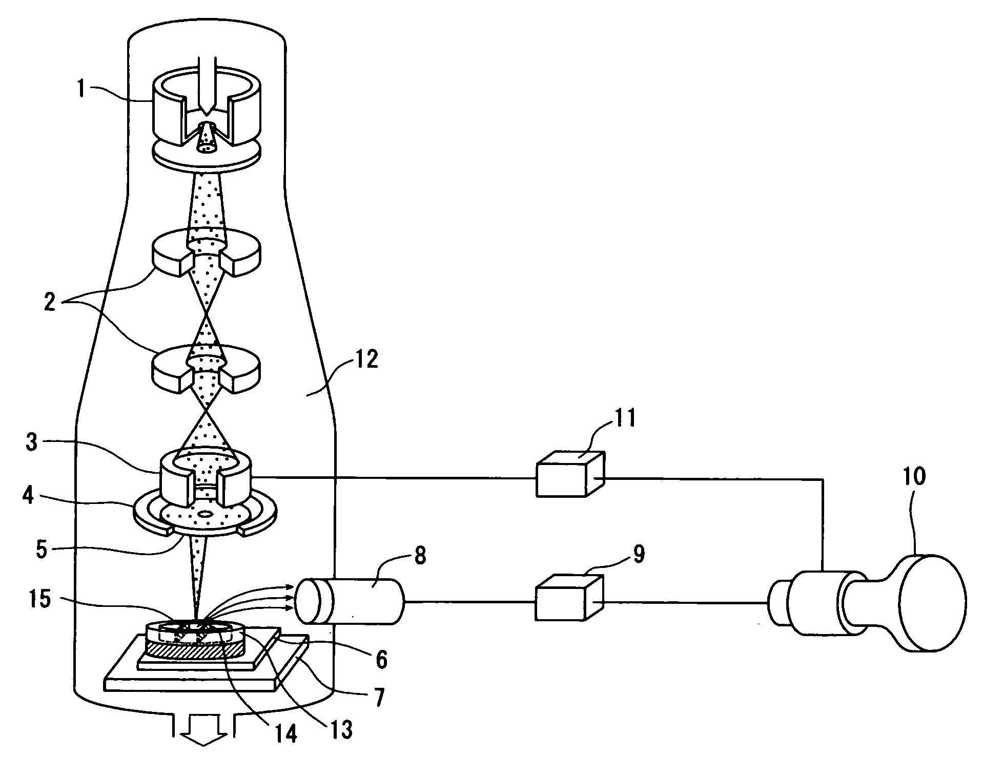
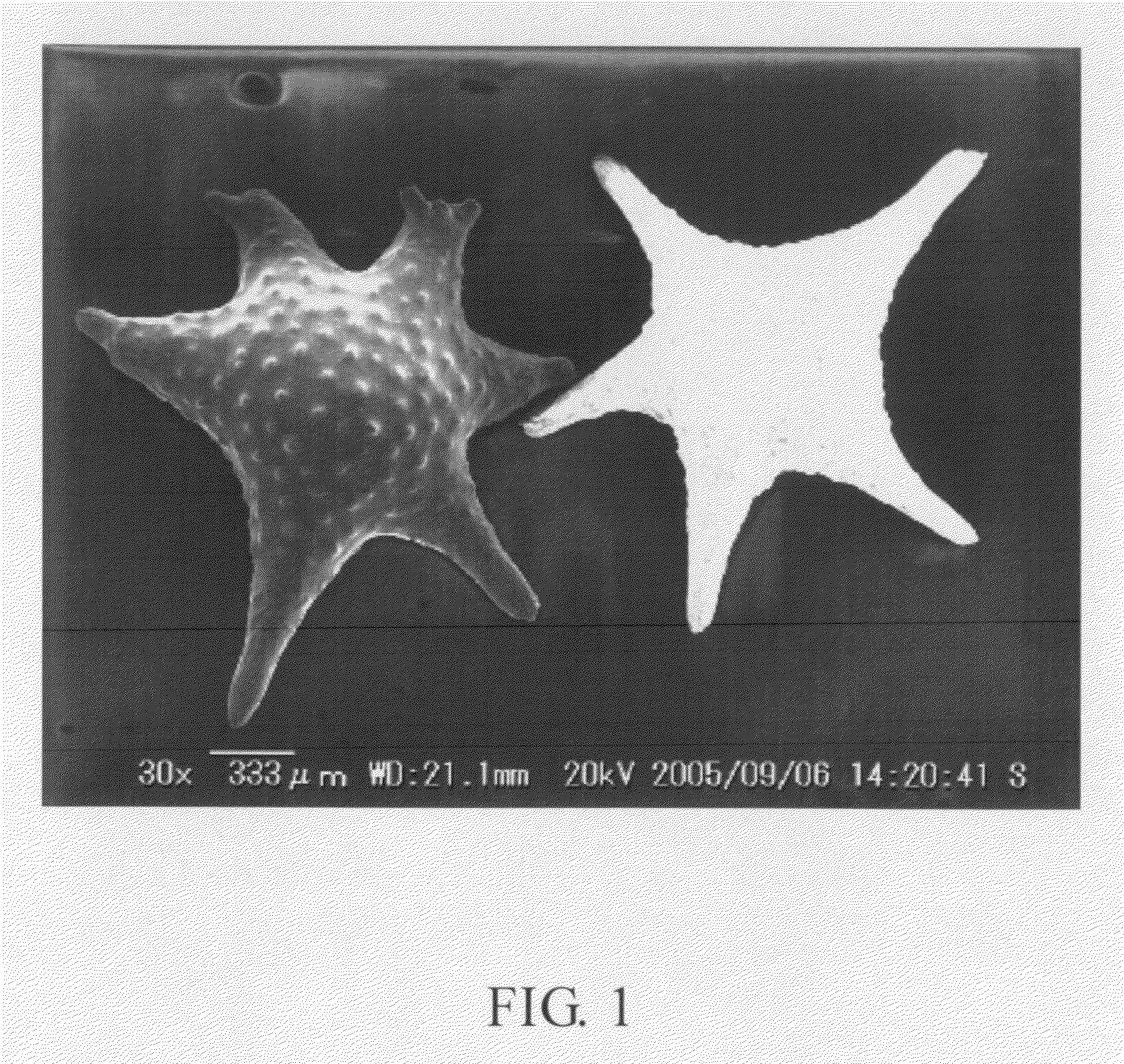
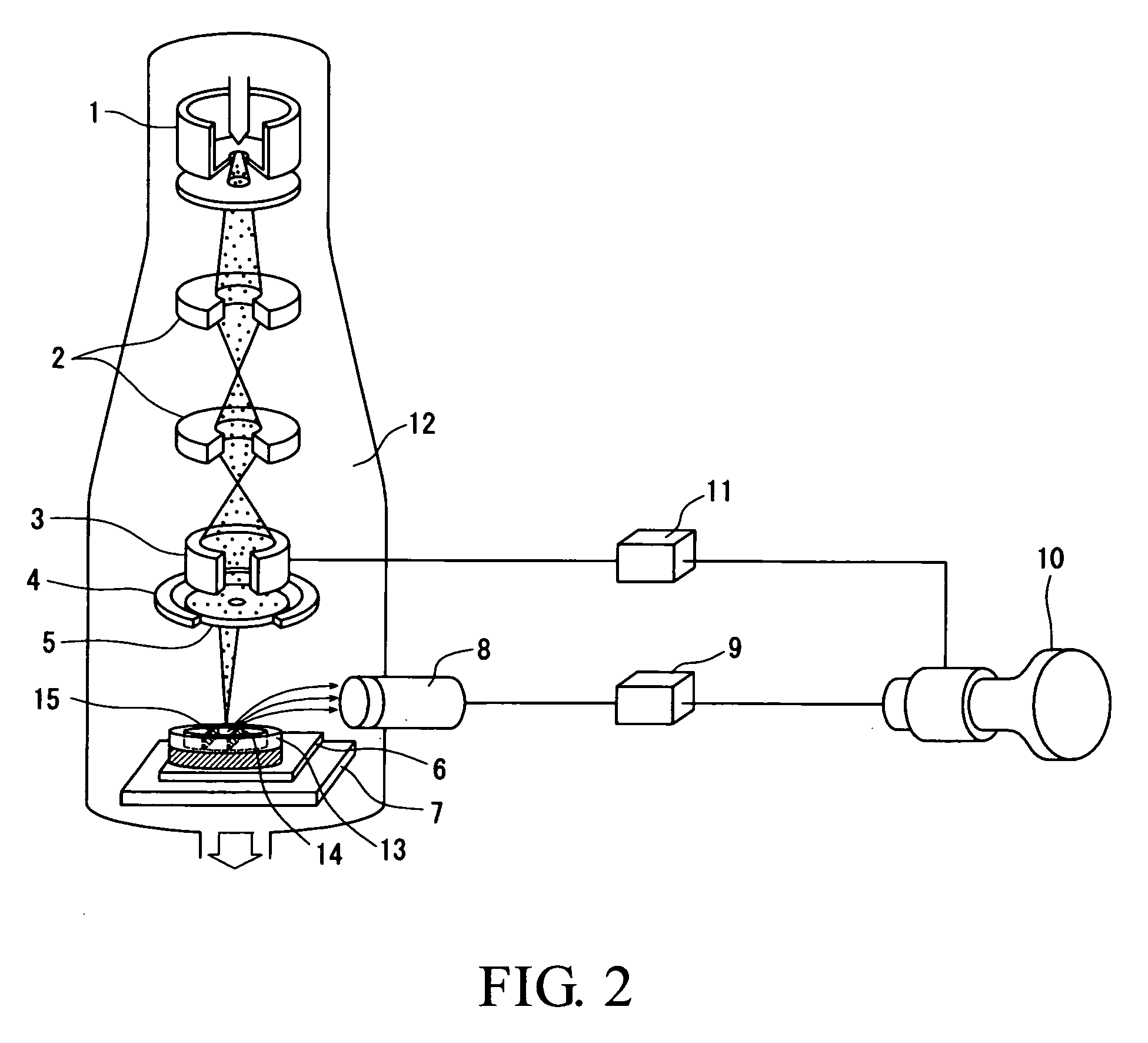
[0129]In Example 2, 1-ethyl-3-methylimidazolium tetrafluoroborate was used as an ionic liquid, and this ionic liquid was impregnated into a sample of star sand and the thus impregnated sample of star sand was observed with a SEM system. As the ionic liquid, 1-ethyl-3-methylimidazolium tetrafluoroborate manufactured by Lancaster Inc. was used. First, the contamination and the like attached to the sample of star sand were removed with a cleaning liquid, and thereafter the sample (star sand) was adhered and fixed to the SEM sample base with an adhesive, and successively the sample was impregnated with 1-ethyl-3-methylimidazolium tetrafluoroborate. The setting of the SEM sample base was conducted in the SEM system. Thereafter, the electron microscope sample chamber was evacuated to vacuum. The SEM acceleration voltage was set at 20 kV and the working distance (WD) was set at 21.1 mm. The star sand was observed under the above-described conditions and an image of the star sand was able t...

example 3

[0131]In Example 3, 1-butyl-3-methylimidazolium tetrafluoroborate was used as an ionic liquid, and this ionic liquid was impregnated into a sample of star sand and the thus impregnated sample of star sand was observed with a SEM system. In present Example 3, the operations were conducted in the same manner as in Example 2 except that 1-butyl-3-methylimidazolium tetrafluoroborate was used in place of 1-ethyl-3-methylimidazolium tetrafluoroborate. Also in this Example 3, a satisfactory observation was enabled in the same manner as described above.


PUM

PropertyMeasurementUnit
thicknessaaaaaaaaaa
SEM acceleration voltageaaaaaaaaaa
constant currentaaaaaaaaaa

Abstract

An object of the present invention is to provide a medium; a specimen; a method for preparing the specimen; a method for observing the specimen; a sample cell; and an electron microscope capable of easily solving the problem of charge-up and further capable of observing a real shape or the like of a sample with a SEM, a TEM or the like. For the purpose of achieving the above-described object, the present invention uses an electrical conductivity-imparting liquid medium, for use in a microscope, which includes an ionic liquid as an essential component thereof and is impregnated into the entirety of a SEM or TEM sample or applied to the observation surface of a SEM or TEM sample to impart electrical conductivity at least to the observation surface of the sample. According to the present invention, the charge built up on the sample surface can be released simply by impregnating or coating the sample with the ionic liquid, and hence the problem of charge-up can be easily solved. Further, even when a sample impregnated or coated with the ionic liquid is placed under vacuum, the ionic liquid is not evaporated from the sample, and hence a biological sample can be observed as it is in an original shape.

Description

TECHNICAL FIELD[0001]The present invention relates to a liquid medium that is used, in the observation of a sample with an electron microscope such as a scanning electron microscope or a transmission electron microscope, to prevent the charge-up of the observation surface of the sample, and a method of observing a sample by using the liquid medium.BACKGROUND ART[0002]In these days, high-precision observation of nano-level or micro-level microstructures has been requested, and in general, a scanning electron microscope (hereinafter referred to as SEM) is used as an apparatus for observing microstructures or the like.[0003]Here, it may be noted that a SEM is a type of electron microscope that can display a three-dimensional image of an observation sample wherein the observation sample is irradiated with an electron beam while the electron beam is scanning the sample; secondary electrons, reflected electrons, characteristic X-rays, fluorescence and the like are generated by the collisi...

Claims


Application Information

Patent Timeline
Patent Type & Authority Applications(United States)
IPC IPC(8): G01N23/00H01B1/12B05D1/18
CPCG01N1/30G01N23/225H01J2237/2004H01J2237/0044H01J37/20
Inventor KUWABATA, SUSUMUTORIMOTO, TSUKASA
Owner HITACHI HIGH-TECH CORP
Why Eureka
Try Eureka