

Systems, methods and computer-accessible medium for providing spectral-domain optical coherence phase microscopy for cell and deep tissue imaging
a technology of optical coherence phase and cell, applied in the field of system, method and computer-accessible medium for providing spectral-domain optical coherence phase microscopy for cell and deep tissue imaging, can solve the problems of low light from the reference surface that is likely too low to generate interference with the light scattered from the focal volume, and the configuration of the separate beam interferometer is likely to have at least some phase instability. to achieve the effect of facilitating high-resolution imaging and high-sensitivity measuremen
- Summary
- Abstract
- Description
- Claims
- Application Information
AI Technical Summary
Benefits of technology
Problems solved by technology
Method used
Image

Examples
Embodiment Construction
[0010]To address and / or overcome such deficiencies, exemplary embodiments of the present invention can be provided.
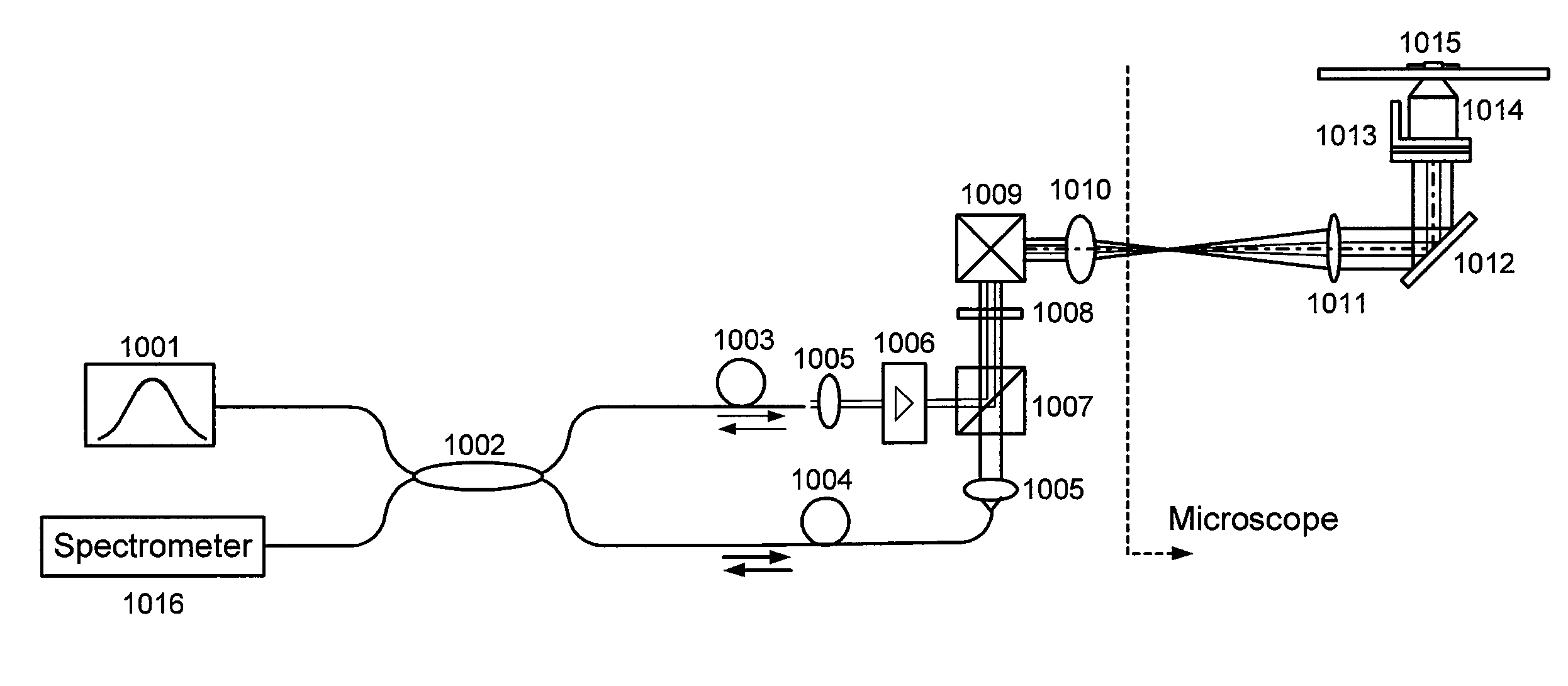
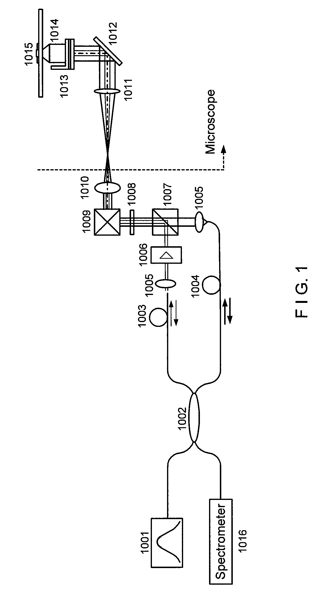
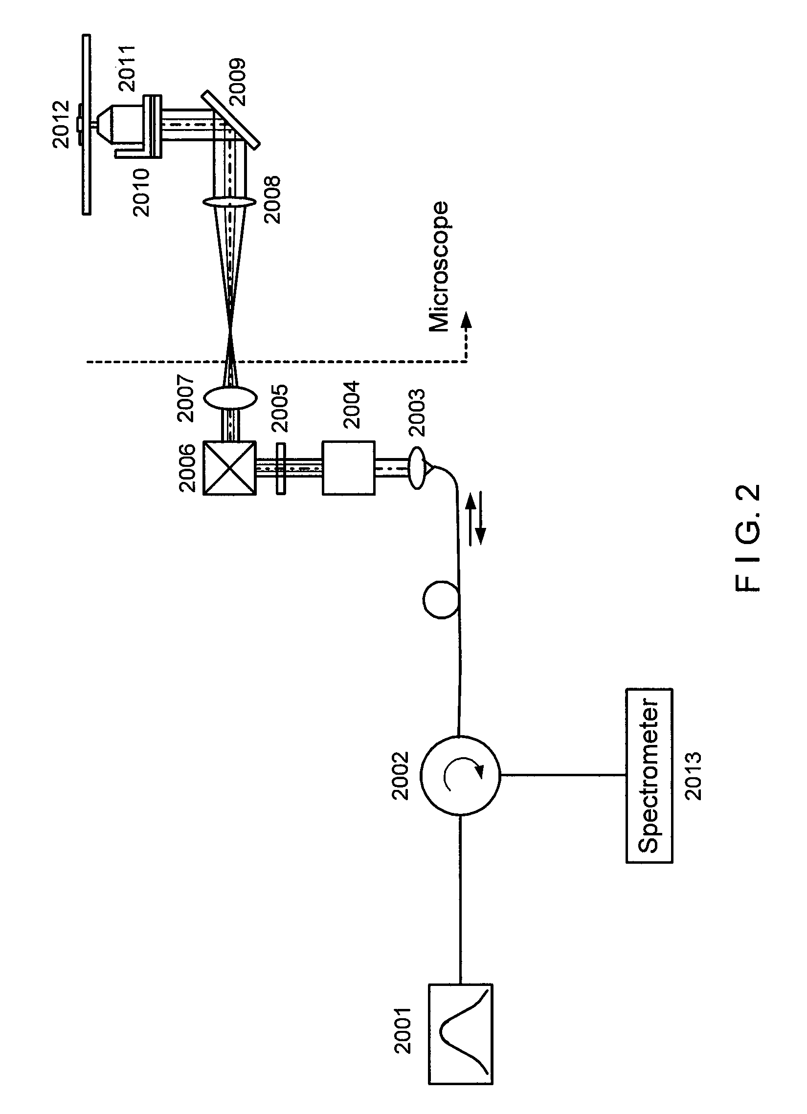
[0011]In general, certain exemplary embodiments of the systems, methods and computer-accessible medium according to the present invention can facilitate high-resolution imaging based on the interferometric detection of scattered light from the sample.
[0012]For example, exemplary embodiments of the present invention can provide the systems, methods and computer-accessible medium which can facilitate high-sensitive measurement and imaging of structural variations deep in the biological sample based on phase-stable low coherence interferometer. Such exemplary embodiments can be applied to functional implementations associated with the motion of structures at a particular depth location. Moreover, by scanning the beam in a volumetric space, the exemplary embodiments of the present invention can generate three-dimensional intensity, phase, and diffusive property images of bi...
PUM
 Abstract
Description
Claims
Application Information