
Fluorescence detection system

a fluorescence detection and detection system technology, applied in the field of intraoperative systems for detecting cancer, can solve the problems of preoperative approach that does not provide the oncologic surgeon with real-time, dynamic intraoperative information, handling and disposal of radioactive-labeled materials, etc., and achieves enhanced illumination and fluorescence collection efficiency, and facilitates intraoperative detection

Inactive Publication Date: 2009-09-17
THE OHIO STATE UNIV RES FOUND
View PDF17 Cites 63 Cited by
  • Summary
  • Abstract
  • Description
  • Claims
  • Application Information

AI Technical Summary

Benefits of technology

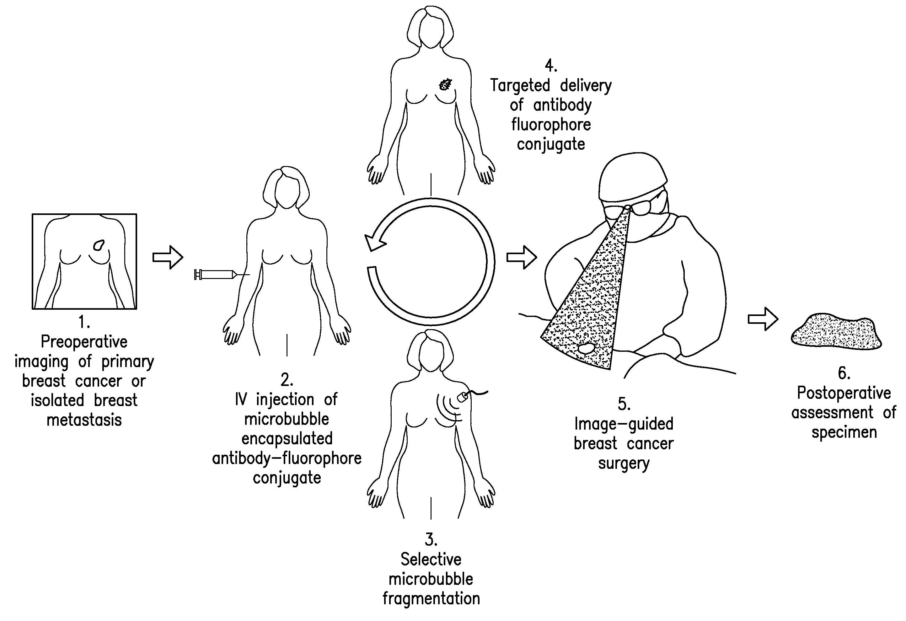
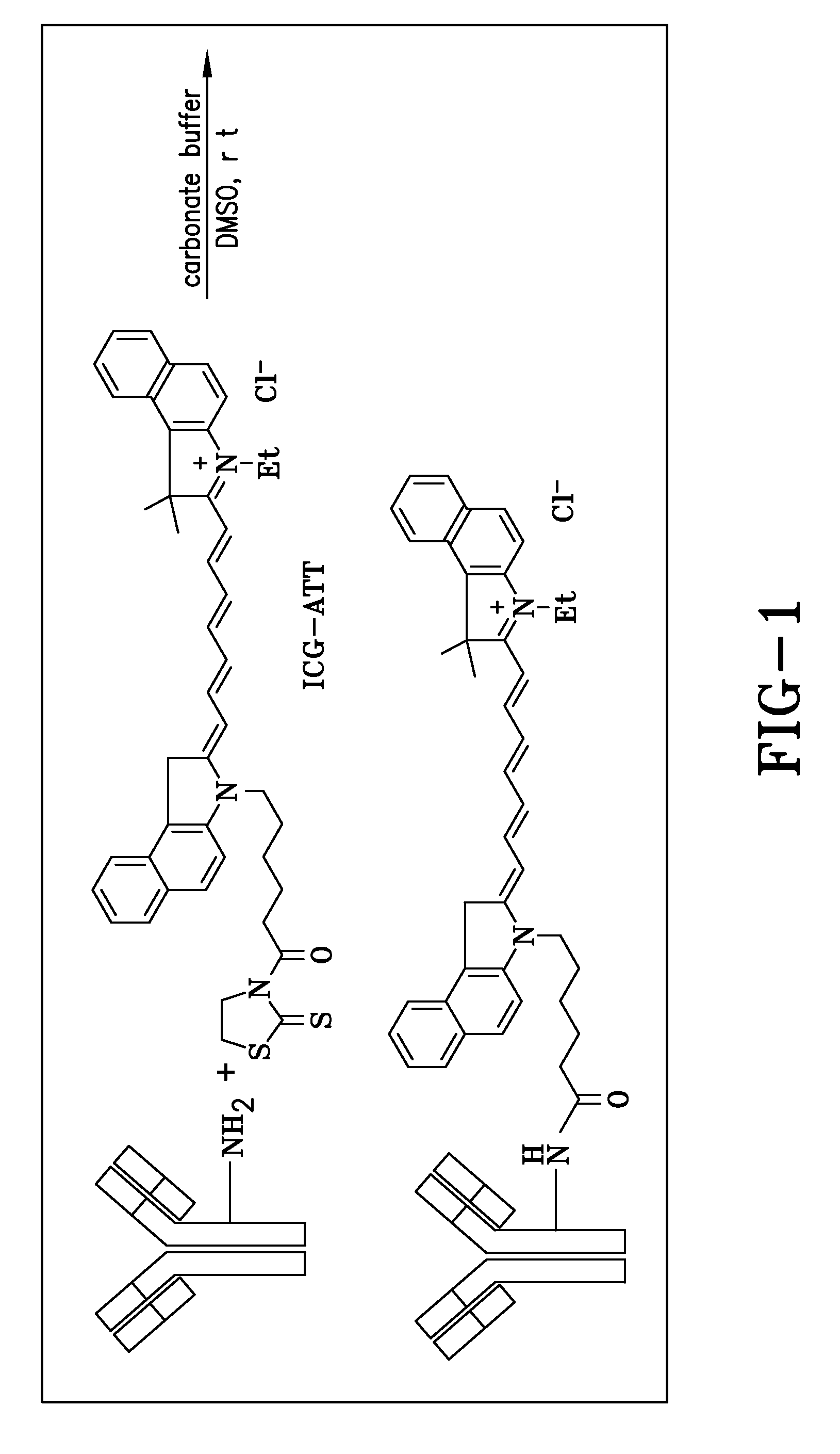
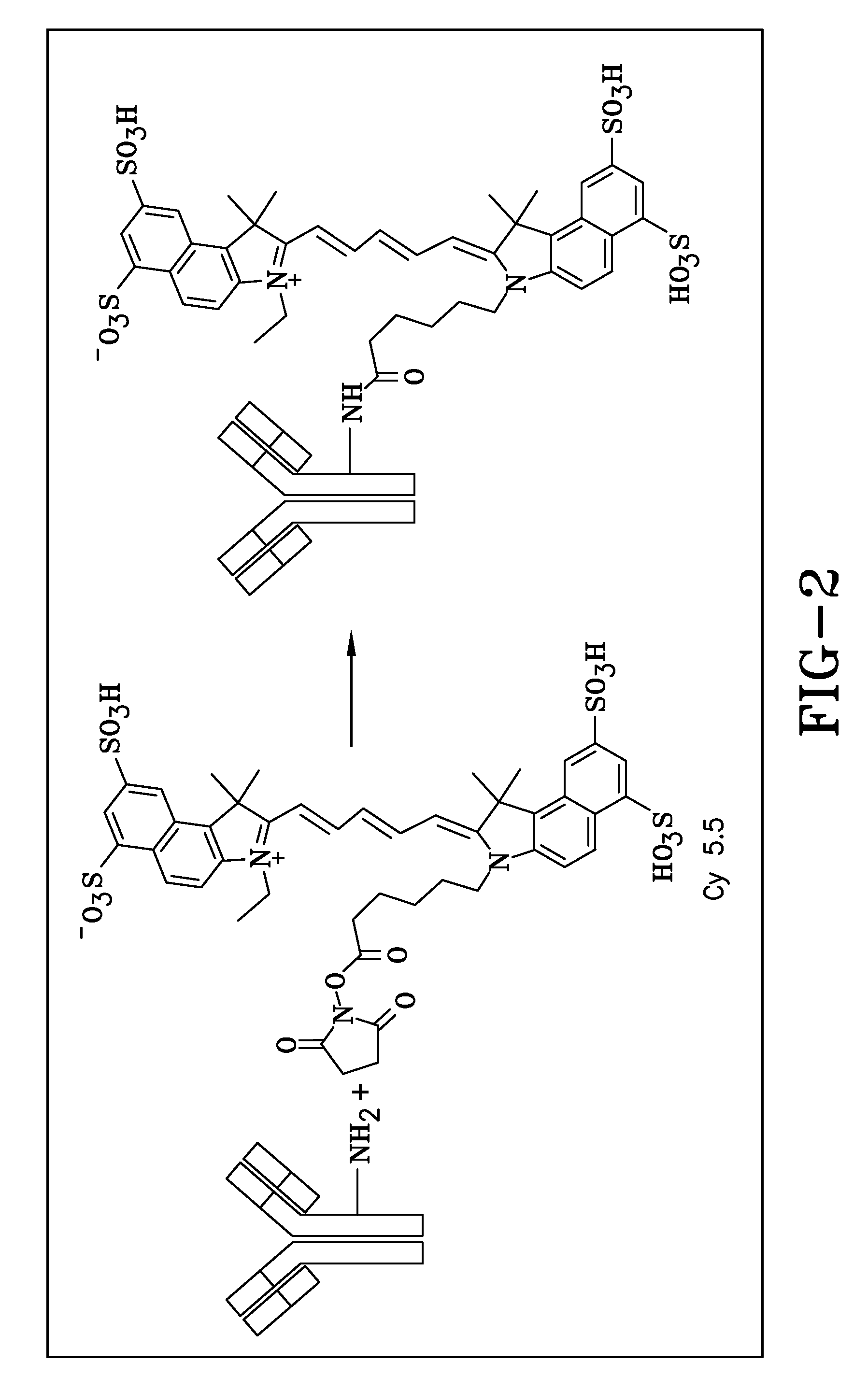
[0012]An exemplary embodiment provides novel fluorescent tumor targeting agents that comprise a molecular targeting unit coupled to a fluorescent unit. In particular, the embodiment provides targeting units comprising at least one motif that is capable of locating both tumor endothelium and tumor cell mass. Before detection, the targeting unit will be associated with at least one fluorescent species. The phrase “operably linked” is used to refer broadly to any useful coupling between a targeting unit and the fluorophore. Such fluorescent targeting units are therapeutically and diagnostically useful, especially in the treatment and diagnosis of cancer, including metastases. More specifically, the fluorescent targeting agents of an exemplary embodiment, when used in conjunction with the appropriate fluorescence detection equipment provide the user with real-time information to more precisely locate occult disease within adjacent peritumoral tissues and within regional lymph node basins, as well as improve the assessment of surgical resection margins.
[0018]To facilitate intraoperative detection, a hand-held optical instrument and a control unit for the detection of fluorescence from the above described novel fluorescent molecular targeting agent system are disclosed. Preferably, the optical detection instrument is equipped with a light source to provide the fluorescence excitation radiation that is capable of illuminating a relatively large area of tissue. The excitation light may also be provided by an external source. The handheld sensor head of an exemplary embodiment may be specifically designed for enhanced illumination and fluorescence collection efficiency.
[0021]The fluorescence detection apparatus of an exemplary embodiment has a large acceptance angle and high efficiency for fluorescence collection. In an exemplary embodiment, the apparatus is able to reject the ambient noise and the background excitation light. Optionally, the enclosure of the hand-held instrument may be made from biocompatible materials. Preferably, the instrument can be sterilized to prevent infections.
[0022]In operation, the targeting agents are administered to a patient in an effective amount. The agent specifically binds a marker produced by or associated with neoplastic tissue, e.g. cancer. Time is then permitted to elapse following the administration for the fluorophore-labeled targeting agent to preferentially concentrate the agent in any neoplastic tissue. Given sufficient time, unbound fluorophore-labeled targeting agent may be cleared so as to increase the ratio of photon emissions from neoplastic tissue to background photon emissions in the patient. After the time has elapsed, the patient is accessed with a fluorescence detection instrument for determining sites exhibiting accretion of the fluorophore locator by detecting with the instrument elevated levels of fluorescence emission at the neoplastic sites. Tissue exhibiting elevated levels of fluorescent emissions may be removed and subjected to gross visual analysis. Such sites may alternatively or additionally be subjected to histological analysis.
[0023]This system will provide the surgeon with intra-operative, real-time cancer-specific information to accurately assess surgical resection margins and precisely locate occult metastatic disease. Such a cancer-specific system offers the potential for use in the detection of malignancies in a wide range of organ systems.

Problems solved by technology

However, such a preoperative approach fails to provide the oncologic surgeon with real-time, dynamic intraoperative information that may significantly impact on critical decision-making within the operating room.
Despite the success of RIGS in such clinical trials, the handling and disposal of the radioactive-labeled material, specifically I125, has remained a major stumbling block to the widespread acceptance and implementation of RIGS technology.

Method used


Image

  • Fluorescence detection system
  • Fluorescence detection system
  • Fluorescence detection system

Examples


examples

[0068]The following examples are included to demonstrate exemplary embodiments of the invention. It should be appreciated by those skilled in the art that the techniques disclosed in the examples which follow represent techniques discovered by the inventors to function well in the practice of the invention, and thus can be considered to constitute preferred modes for its practice. However, those of skill in the art should, in light of the present disclosure, appreciate that many changes can be made in the specific embodiments which are disclosed and still obtain a like or similar result without departing from the concept, spirit and scope of the invention. More specifically, it will be apparent that certain agents that are both chemically and physiologically related may be substituted for the agents described herein while the same or similar results would be achieved. All such similar substitutes and modifications apparent to those skilled in the art are deemed to be within the spir...

example

Imaging Results in Small Animal Model

[0116]A simplified goggle prototype is shown in FIG. 12. An exemplary goggle system may comprise a camera with fluorescence filter, a multi-wavelength excitation light source, a head mount display (HMD), a laptop computer, and other imaging processing accessories, as shown in FIG. 12. A Labview program may be used to fuse the background image and the fluorescence emission captured by the camera, and project to HMD for real-time image guidance.

[0117]As demonstrated in FIG. 13, useful fluorescence images can be visualized after an IV injection of MuCC49-Cy7 conjugate on a LS174T colon cancer xenograft nude mouse. The fluorescence imaging system used to obtain the images shown in FIG. 13 is similar to the exemplary system described in FIG. 12, except that the images were projected to a computer monitor instead of a HMD. However, the core technique is similar to that of the system described in FIG. 12.

[0118]As is evident from FIG. 13, tumor can be re...


PUM


Abstract

Exemplary embodiments include systems, methods, and compositions for the intra-operative detection of target tissue. At least one embodiment includes a fluorescence detection instrument that may be used for intra-operative detection of a fluorescent targeting agent, its binding site, and its interaction within cancer tissues. An exemplary embodiment is highly sensitive to the local deposition of fluorescence agents even at a low concentration. In at least one embodiment, the system includes a handheld navigation instrument that is usable to excite, detect, and report the fluorescent deposition of the targeting agent in real-time. In alternative embodiments, the system includes a wearable unit to excite, detect, and visually report the fluorescent deposition of the targeting agent to the user. The wearable unit includes eyewear that allow the user to perform image-guided surgery based on the near real-time fluorescence detection of the fluorescent targeting agent.

Description

CROSS-REFERENCE TO RELATED APPLICATIONS[0001]This non-provisional patent application claims the benefit of priority to U.S. Provisional Patent Application No. 61 / 020,345 filed Jan. 10, 2008, which is hereby incorporated by reference in its entirety.[0002]The present invention was made with United States Government support under grant no. R01 CA120023 by the National Institute of Health. The United States Government may have certain rights to this invention under 35 U.S.C. §200 et seq.TECHNICAL FIELD[0003]The disclosed embodiments of the present invention are in the field of intra-operative systems for detecting cancer, particularly in vivo detection systems utilizing fluorescent molecular targeting agents.BACKGROUND OF THE ART[0004]The accurate assessment of surgical resection margins and the recognition of occult disease within adjacent peritumoral tissues and within regional lymph node basins during cancer surgery are important oncologic principles that minimize recurrence rates a...

Claims


Application Information

Patent Timeline
Patent Type & Authority Applications(United States)
IPC IPC(8): A61B6/00A61K49/00
CPCG01N21/6428G01N33/582G01N33/57469G01N21/6456
Inventor MARTIN, EDWARD W.XU, RONALD X.SUN, DUXINPOVOSKI, STEPHEN P.HEREMANS, JOSEPH PIERRELEE, ROBERTSUBRAMANIAM, VISH V.TURRO, CLAUDIA
Owner THE OHIO STATE UNIV RES FOUND
Learn More