Insertable medical devices having microparticulate-associated elastic substrates and methods for drug delivery

Active Publication Date: 2009-10-01
SURMODICS INC
View PDF23 Cites 187 Cited by
  • Summary
  • Abstract
  • Description
  • Claims
  • Application Information

AI Technical Summary

Benefits of technology

[0013]The invention generally relates to insertable medical devices associated with microparticulates containing a bioactive agent. The microparticulates can be released from the device and provide bioa

Problems solved by technology

Such preparations are generally undesirable because the drug can be easily removed from the surface during insertion.
In addition, release of the drug is generally difficult to control following implantation.
For certain medical applications, these polymer systems are not ideal.
For the po

Method used


Image

  • Insertable medical devices having microparticulate-associated elastic substrates and methods for drug delivery
  • Insertable medical devices having microparticulate-associated elastic substrates and methods for drug delivery
  • Insertable medical devices having microparticulate-associated elastic substrates and methods for drug delivery

Examples


Example

Example 1

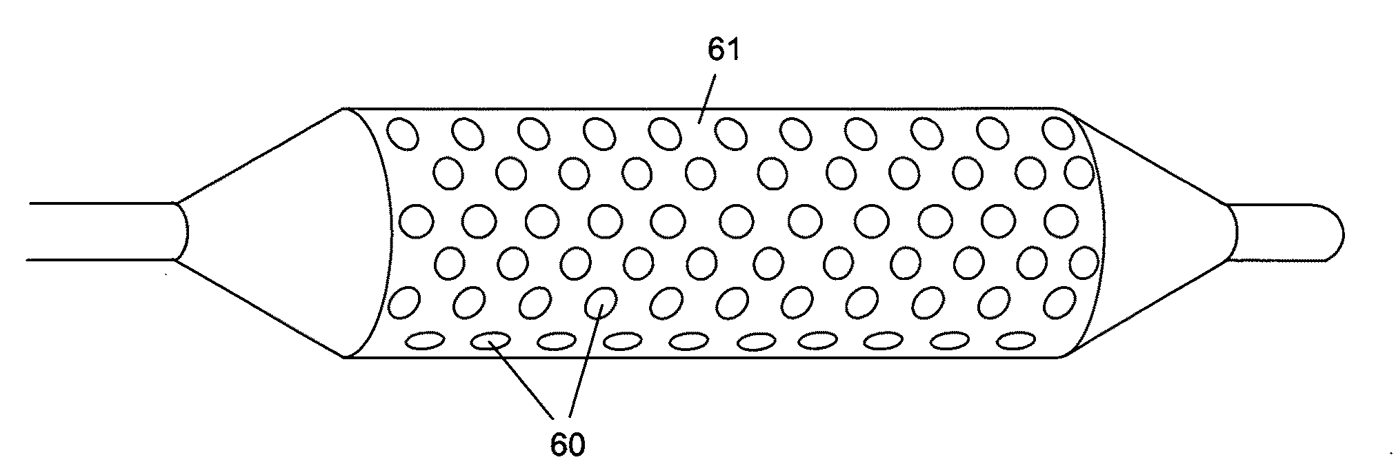
[0194]The elastic surface of the balloon of a balloon catheter was provided with a flexible hydrogel coating with associated paclitaxel microparticulates. The balloon catheter that was used in the coating process was obtained from Minnesota Medtec (Maple Grove, Minn.). The elastic portion of the balloon was made from nylon and has a balloon wall thickness of 5-10 μm.

[0195]A hydrogel coating solution was prepared using photo-polyacrylamide (prepared as described U.S. Pat. No. 6,007,833, Examples 1 & 2), which was weighed and dissolved into a mixture of IPA and water (50% IPA / 50% water (v / v)) at a concentration of 10 mg / mL. The balloon was coated in the photo-polyacrylamide coating solution using a dip process with a withdrawal rate of 0.5 cm / s. After the hydrogel coating solution was applied to the balloon, it was subjected to UV cure. The coated balloon was placed in front of a Dymax 2000-EC Series UV Floodlamb with a 400 Watt metal halide bulb, approximately 20 cm from lig...

Example

Example 2

[0198]The coating process as described in Example 1 was repeated, with the exception of the use of a different coating composition to form the hydrogel coated layer.

[0199]A hydrogel coating solution was prepared using photo-polyacrylamide (Example 1) at 5 mg / mL, photo-poly(vinylpyrrolidone) (prepared as described in Example 4 of U.S. Pat. No. 5,414,075) at 25 mg / mL, poly(vinylpyrrolidone) K90 (BASF) at 10 mg / mL, and 4,5-bis(4-benzoylphenylmethyleneoxy)benzene-1,3-disulfonic acid (prepared as described in U.S. Pat. No. 6,278,018 (Example 1)) at 0.25 mg / mL, dissolved into a mixture of IPA and water (15% IPA / 85% water).

[0200]Dipcoating, UV treatment, and paclitaxel microparticulate coating was performed as described in Example 1.

Example

Example 3

[0201]The elastic surface of the balloon of a balloon catheter was provided with a flexible hydrogel coating and then paclitaxel microparticulates were formed on the hydrogel surface.

[0202]The coating processes as described in Examples 1 and 2 were repeated to provide hydrogel coated layers.

[0203]Next, a microparticulate-forming composition was prepared by dissolving paclitaxel in methanol at a concentration of 30 mg / mL. The composition was then applied to the photo-polymer coated surfaces by pipetting a known volume of drug suspension (typically 10-20 μl). The pipetted droplet was evenly distributed over the balloon surface by rotating the balloon until the solvent was visibly dry.

[0204]FIGS. 12a and 12b are micrographs of the elastic substrate having a hydrogel coating with paclitaxel microparticulates partially embedded in the hydrogel, prepared according to this method.


PUM

PropertyMeasurementUnit
Lengthaaaaaaaaaa
Lengthaaaaaaaaaa
Fractionaaaaaaaaaa

Abstract

The present invention provides insertable medical devices having elastic surfaces associated with bioactive agent-containing microparticulates and a coating material. Upon expansion of the elastic surfaces the microparticulates can be released to a subject.

Description

CROSS-REFERENCE TO RELATED APPLICATIONS[0001]The present non-provisional application claims the benefit of commonly owned provisional application having Ser. No. 61 / 072,234, filed on Mar. 28, 2008, and entitled INSERTABLE MEDICAL DEVICES HAVING MICROPARTICLE-ASSOCIATED ELASTIC SUBSTRATES AND METHODS FOR DRUG DELIVERYFIELD OF THE INVENTION[0002]The present invention relates to the field of drug delivery from insertable medical articles.BACKGROUND OF THE INVENTION[0003]The release of drugs from an implanted medical device has been shown to be beneficial for the function of devices and the treatment of various medical conditions. For example, delivery of a drug from the device surface can prevent cellular responses initiated by the presence of the implantable device. Also, drug released from the device can prevent conditions that would otherwise shorten the functional life of the device following implantation. Drug released from the device may also be directed at treating a diseased ar...

Claims


Application Information

Patent Timeline
IPC IPC(8): A61L27/54A61K31/337A61P9/00
CPCA61L29/085A61L29/145A61L29/16A61L2300/416A61L29/148A61L2300/622C08L3/02A61M2025/1031A61M2025/1075A61K31/337A61M25/1002A61M2025/1004A61M2025/105A61P29/00A61P35/00A61P43/00A61P7/02A61P9/00A61L29/041A61M25/104A61L2300/604A61L2300/606
Inventor ARPS, JAMES HOWARDBACH, ANDREW G.
Owner SURMODICS INC
Why Eureka
Try Eureka