Cerebral Infarction Suppressant

a technology for cerebral infarction and suppressant, which is applied in the direction of antibody medical ingredients, drug compositions, peptides, etc., can solve the problems of limited efficacy and achieve the effect of effective suppression of brain tissue necrosis, low genotoxic side effects, and low side effects

Inactive Publication Date: 2009-10-08
UNIV OKAYAMA +1
View PDF15 Cites 13 Cited by
  • Summary
  • Abstract
  • Description
  • Claims
  • Application Information

AI Technical Summary

Benefits of technology

[0019]The cerebral infarction suppressant of the present invention can effectively suppress brain tissue necrosis resulted from a long term ischemia due to cerebral thrombosis and cerebral embolism or the like. In addition, it is considered that a possibility generating a serious adverse side effect is extremely low based on the relatively safe clinical application of antibody drugs currently used. Therefore, the cerebral infarction suppressant of the present invention is extremely useful as being capable of suppressing the cerebral infarction for which a particularly effective treating means has not been present.

Problems solved by technology

However, a drug having a novel mechanism of action is desired, since the conventional drugs have problems of adverse side effects and limited efficacy.
For example, edaravone which is a cerebral infarction therapeutic being solely approved in Japan has many problems such as adverse side effects, and the efficacy remains a limited level.

Method used


Image

  • Cerebral Infarction Suppressant
  • Cerebral Infarction Suppressant
  • Cerebral Infarction Suppressant

Examples


example 1

Preparation of Anti-HMGB1 Monoclonal Antibody

(a) Immunization of Rat

[0035]Into a 2 ml-glass syringe, was taken 1 mg / mL of a commercially available mixture of bovine thymus-derived HMGB1 and HMGB2 (manufactured by Wako Pure Chemical Industries, Ltd., code No. 080-070741), and an equivalent volume of a complete Freund's adjuvant was taken into another 2 mL-glass syringe. These syringes were connected with a connecting tube. The mixture and the adjuvant were gradually kneaded through the connecting tube to obtain an emulsion. Each 0.1 mL of the obtained emulsion at a total of 0.2 mL was injected to a rat anesthetized with sevoflurane in a hind limb footpads. After 2 weeks, blood was probatively taken from jugular, and the increase of antibody titer was confirmed. Then, an enlarged iliac lymph node was sterilely taken out 5 weeks after the injection administration. From the two lymph nodes obtained, about 6×10⁷ cells could be recovered.

(b) Cell Fusion and Cloning

[0036]The iliac lymph no...

example 2

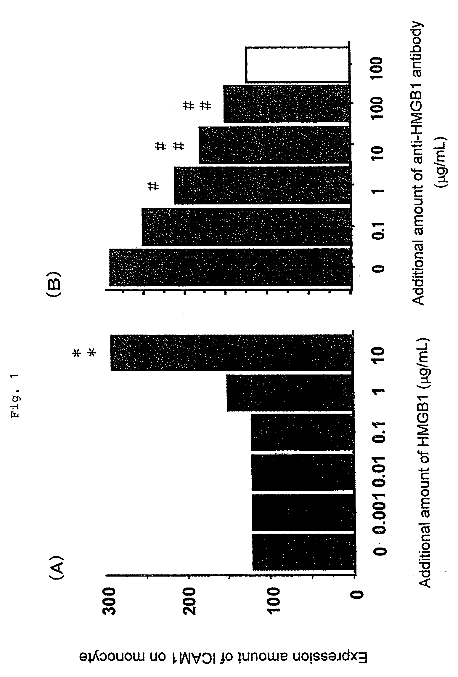
[0038]The neutralization activity of the anti-HMGB1 monoclonal antibody prepared in Example 1 was tested.

[0039]First, 1×10⁶ / mL peripheral blood mononuclear cells were prepared from peripheral blood of a healthy person by a conventional method, and were cultured for 24 hours in a basal medium for culturing an animal cell containing 10% bovine fetal serum (manufactured by Sigma, RPMI1640). Then, bovine HMGB1 purified from a bovine thymus-derived HMGB1 / 2 mixture manufactured by Wako Pure Chemical Industries, Ltd. was added to the medium at a concentration of 0.001 to 10 μg / mL to stimulate the monocytes. Twenty four hours after addition of HMGB1, the cells were collected, and an expression amount of ICAM-1 (intercellular adhesion molecule-1) expressed on the monocytes with HMGB1 was quantitated by a fluorescent antibody method (FACS method). Results are shown in FIG. 1(A). In FIG. 1(A), “**” indicates the case where there was a significant difference at p<0.01 by t-test as compared with...

example 3

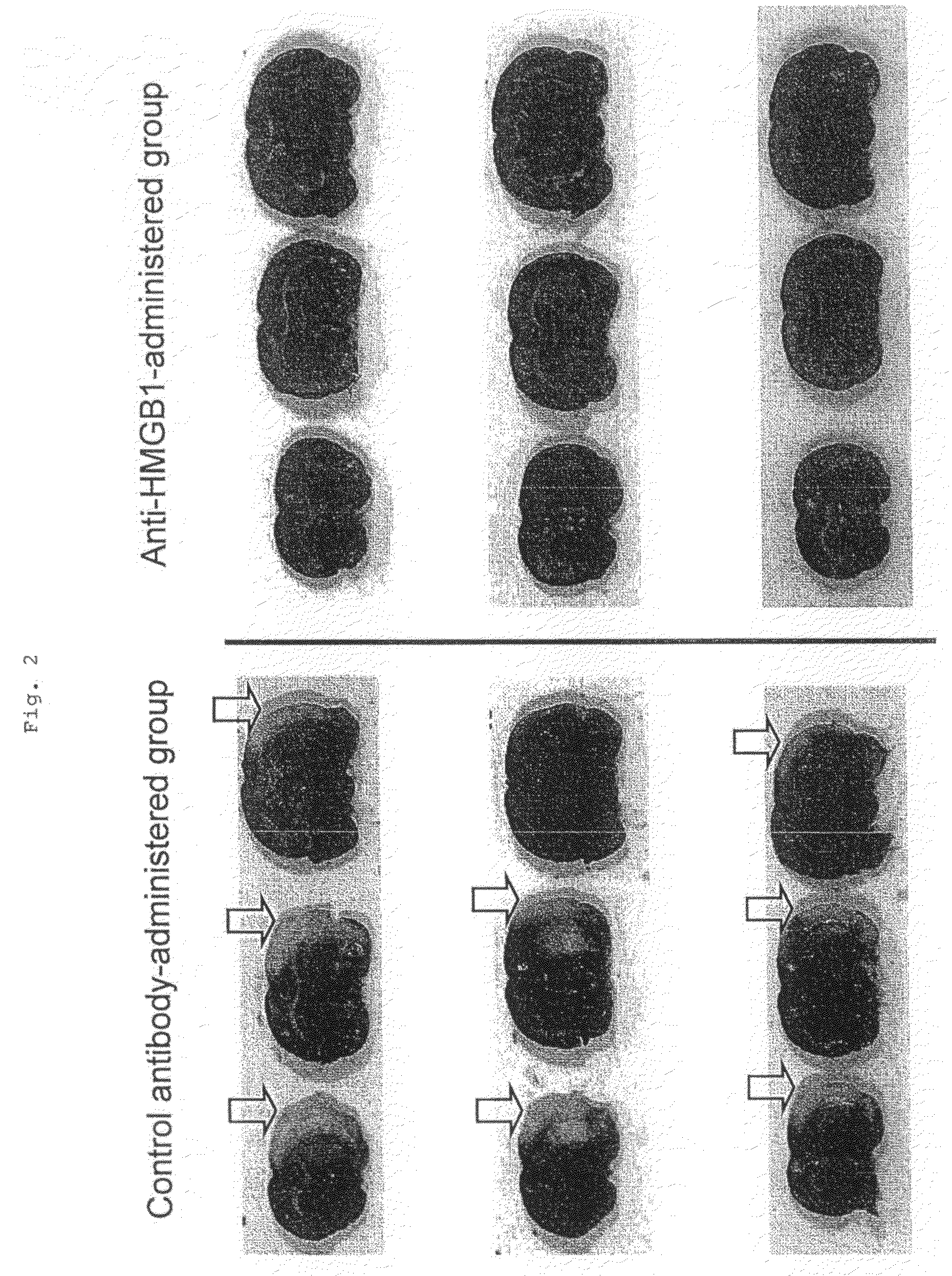
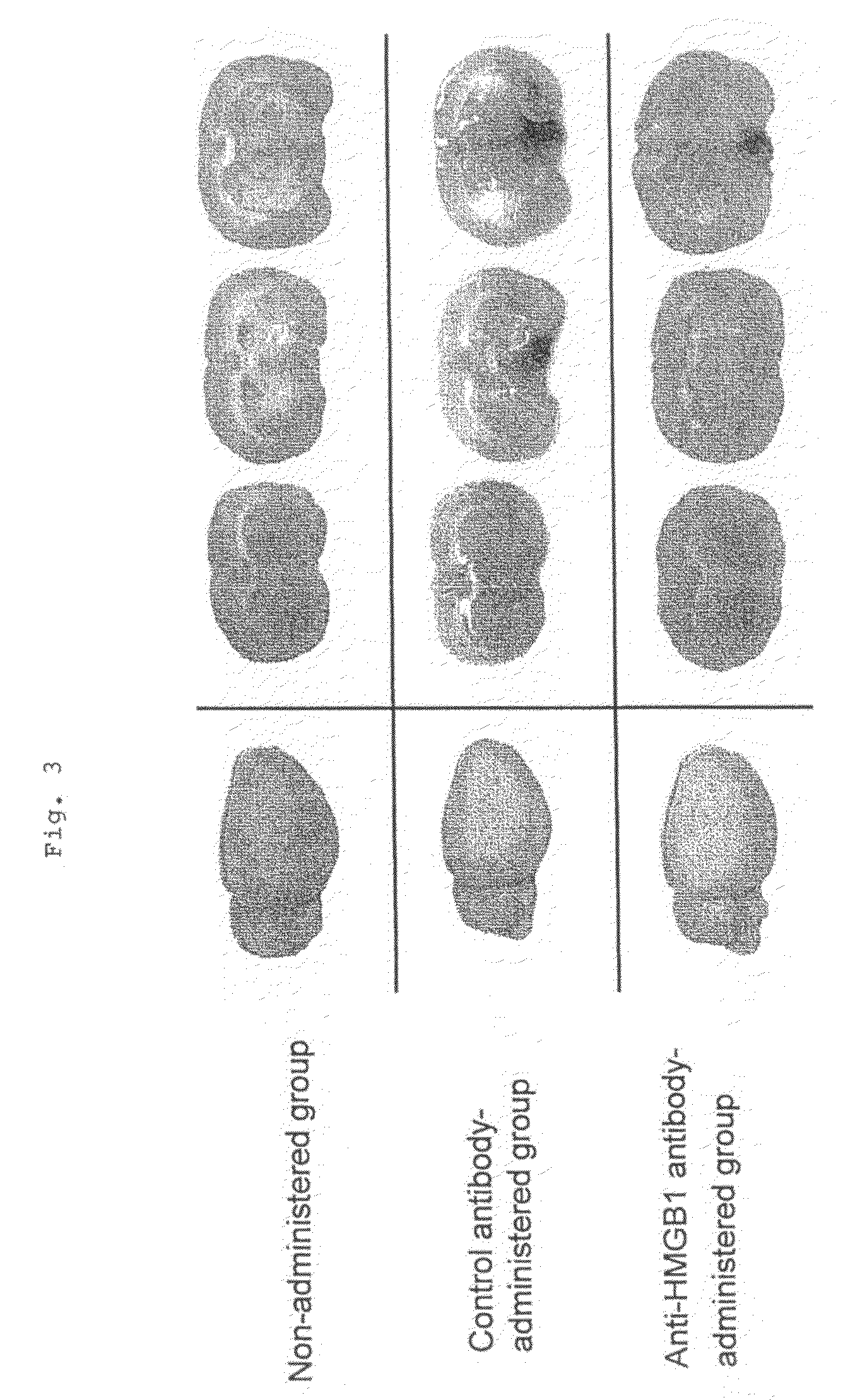
[0041]Fifteen male Wistar rats weighing about 300 g were divided into an anti-HMGB1 monoclonal antibody-administered group of 7 animals and a control antibody-administered group of 8 animals. These rats were anesthetized with a gas mixture of 2% halothane and 50% laughing gas, and kept under spontaneous breathing. Subsequently, a median incision was made in the neck of the rat placed on its back, and the right common carotid artery was exposed. After an intraperitoneal injection of 100 units of heparin, the root of the right middle cerebral artery was occluded by inserting 4.0 nylon thread coated with silicone into the right internal carotid artery from the bifurcation of the internal and external carotid arteries. The tip of the nylon thread was placed 18 mm from the bifurcation. After suture of the incision of the skin, the rats were allowed to recover from anesthesia. During surgery, an electronic thermometer was inserted into the rectum, and the rectum temperature was maintained...


PUM

PropertyMeasurementUnit
concentrationaaaaaaaaaa
timeaaaaaaaaaa
timeaaaaaaaaaa

Abstract

An objective of the present invention is to provide a suppressant for cerebral infarction occurred after long-term ischemia associated with actual cerebral infarction, and has fewer side effects. The cerebral infarction suppressant of the present invention is characterized in comprising an anti-HMGB1 monoclonal antibody as an active ingredient.

Description

TECHNICAL FIELD[0001]The present invention relates to a drug for suppressing cerebral infarction caused by cerebral ischemia.BACKGROUND ART[0002]Cerebral infarction is a disease wherein the brain tissue with impaired blood flow is necrotized due to insufficient-blood flow. The insufficient blood flow is caused by the cerebral blood vessel occluded or narrowed due to various factors, such as atherosclerosis of the cerebral blood vessel and transfer of thrombus formed in an extracerebral blood vessel into the brain. Once cerebral infarction occurs, the brain tissue in which necrosis has developed does not regain its function. Therefore, even if the patient survives, symptoms of dementia, motor weakness, sensory abnormality and language disorder often persist. Meanwhile, in recent years, diseases called lifestyle-related diseases, such as hypertension, cardiac diseases, hyperlipidemia and diabetes, are increasing, and thereby the risks for cerebral infarction are increasing. In our cou...

Claims


Application Information

Patent Timeline
Patent Type & Authority Applications(United States)
IPC IPC(8): A61K39/395A61P9/10
CPCA61K2039/505C07K2316/96C07K16/24C07K2317/76A61P25/00A61P9/10
Inventor NISHIBORI, MASAHIROMORI, SHUJITAKAHASHI, HIDEOTOMONO, YASUKOADACHI, NAOTOLIU, KEYUE
Owner UNIV OKAYAMA
Why Eureka
Try Eureka