

Method to Detect Transmissible Spongiform Encephalopathies Via Electroretinogram
a technology of spongiform encephalopathy and electroretinogram, which is applied in the field of antemortem method for screening transmissible spongiform encephalopathies or neurodegenerative diseases, can solve the problems of inability to reliably detect prpsup>sc /sup>, the investigation of the pathobiology of tses is further complicated, and the sole reliance on prpsup>sc /sup>
- Summary
- Abstract
- Description
- Claims
- Application Information
AI Technical Summary
Benefits of technology
Problems solved by technology
Method used
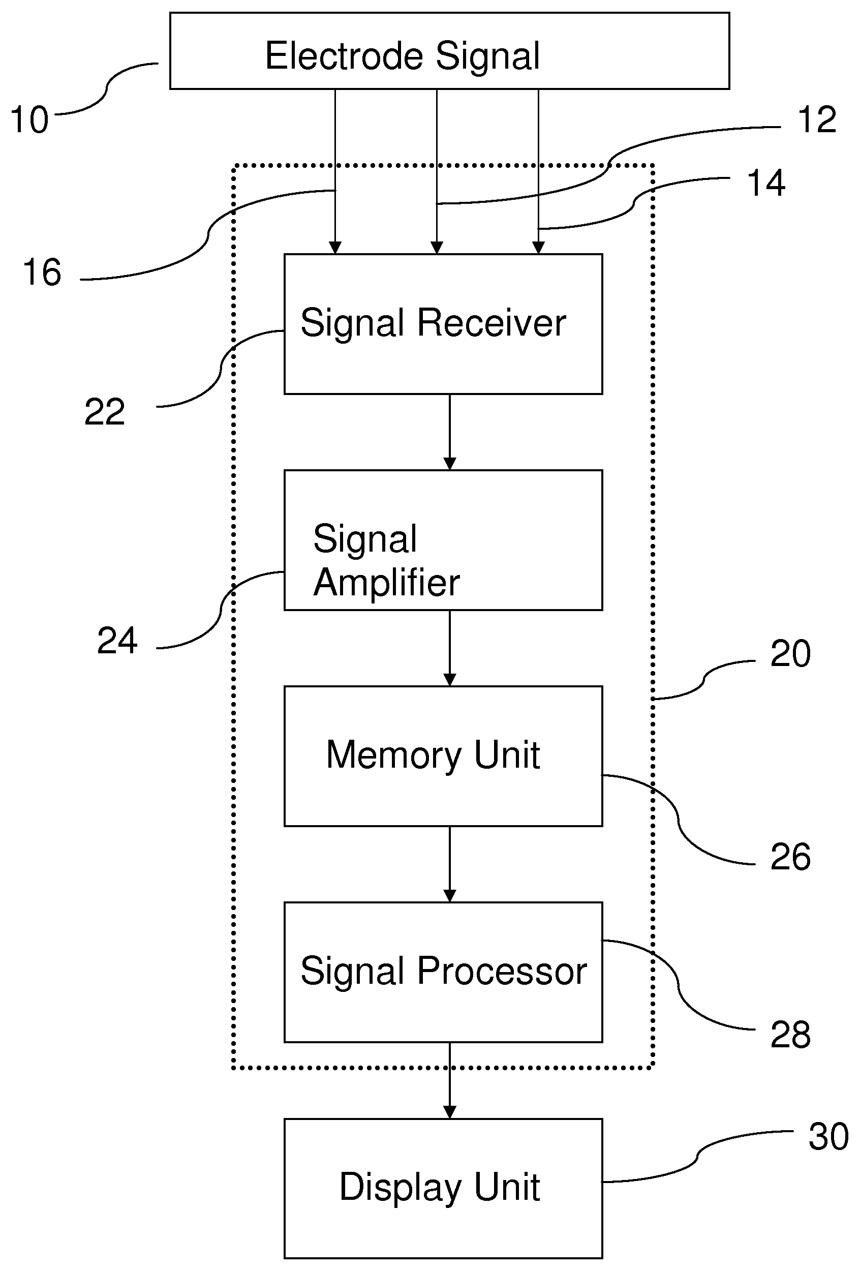
Image

Examples
example 1
ERG of Transmissible Mink Encephalopathy-Inoculated Holstein Cattle
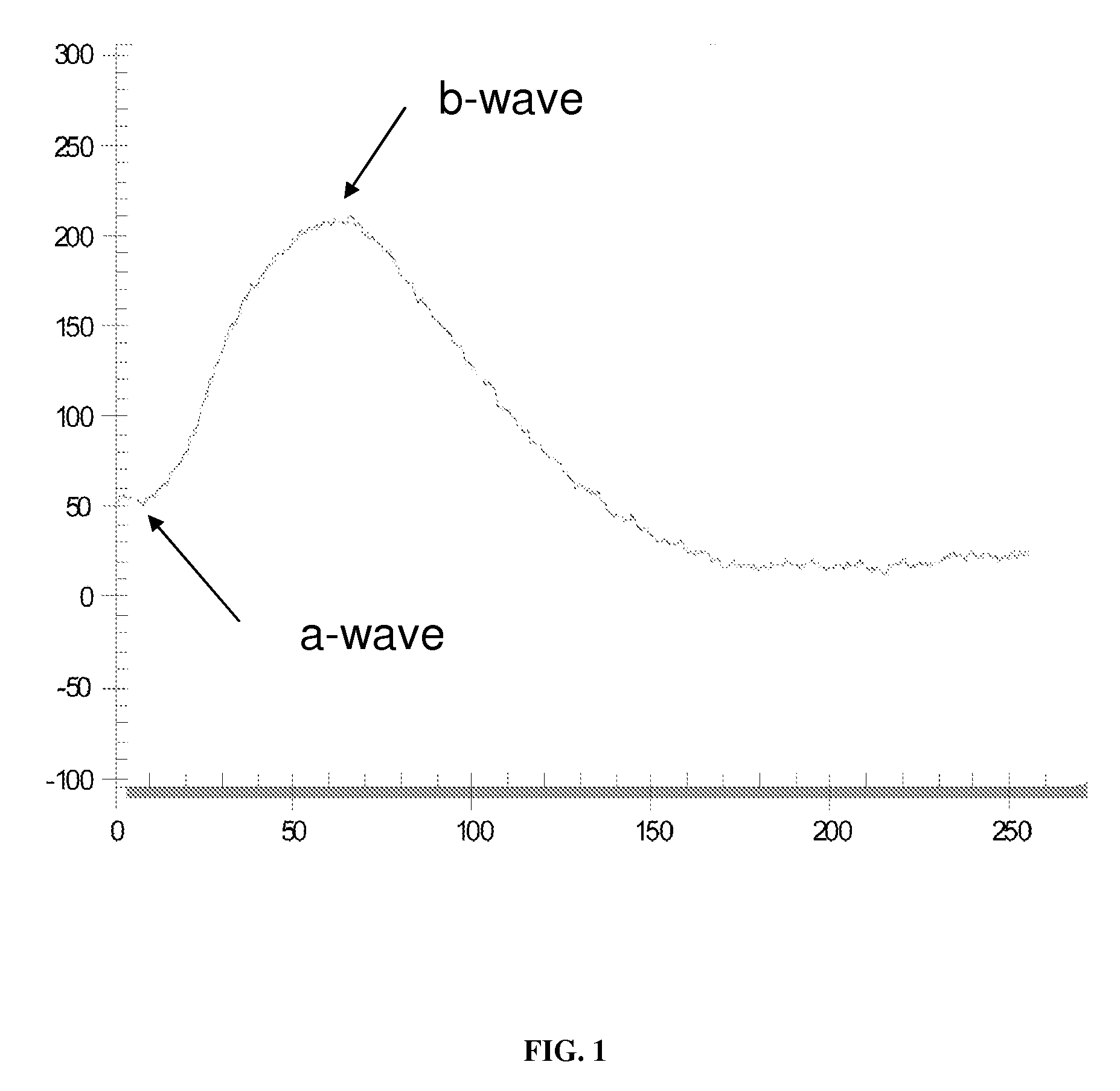
[0029]Five Holstein steers were inoculated intracerebrally with brain homogenate prepared transmissible mink encephalopathy (TME) at 9-months of age and evaluated prior to clinical signs of disease at 12.5, 13.5, and 14.5 months post-inoculation, and at 18.5 months. All cattle developed clinical disease and were euthanized when deemed humanely necessary at between approximately 16.5 to 19 months post-inoculation. Inoculated steers were housed in a Biosafety Level 2 isolation barn (two animals per pen) at the National Animal Disease Center (NADC), Ames, Iowa. They were fed pelleted growth and maintenance rations that contained no ruminant protein, and clean water was available ad libitum. Control steers were housed together in an open shed and fed the pelleted growth ration (without ruminant protein) and alfalfa hay. Personnel wore protective clothing while in the isolation facility and showered before leaving the fac...
example 3
Immunohistochemical Data
[0034]To evaluate the morphologic effects of TSE infection on the retina of cattle, retinas from cattle clinically affected with TME using standard histologic techniques and immunohistochemistry. Immunoreactivity for PrPSc was detected in the retinas of all TME cattle, and was localized primarily to the synaptic layers and the cytoplasm of retinal ganglion cells (FIG. 4B). Despite marked PrPSc accumulation within the retinas of TME-affected cattle, severe pathologic change was not observed on examination of hematoxylin and eosin stained sections (FIG. 4A and FIG. D). However, multifocal distinct, round vacuoles (consistent with spongiform change) were observed within the IPL of TME affected cattle, but not in controls (arrows FIG. 4D). Cell density within the GCL differed between the two groups with controls having an average of 114 nuclei per five 40× fields versus 44 nuclei per five 40× fields in TME-affected cattle. Additionally, the optic nerves of TME-af...
example 4
ERG of Scrapie Inoculated Suffolk Sheep
[0039]A single Suffolk sheep (number 3742) was inoculated intracerebrally with brain homogenate prepared from a fourth-passage scrapie-affected sheep. The original source of inoculum was from 13 scrapie-affected sheep from 7 flocks (designated 13-7). These sheep were verified scrapie positive by immunohistochemistry for PrPSc. The inoculum was ground in a mechanical grinder, gentamicin was added at 10 μg / ml, and the final concentration of 10% (wt / vol) was made with PBS. For subsequent passages the scrapie-infected brain tissue was obtained from the animal with the shortest incubation to terminal disease (survival) time from the previous passage and the inoculum was prepared as described (supra). The inoculum was passaged in 5 generations of lambs. Sheep #3742 was from the 5th generation of lambs (4th passage) and had a 12.2-month survival time.
[0040]The procedure for intracerebral inoculation is similar to cattle. Briefly, the animals were seda...
PUM
 Abstract
Description
Claims
Application Information