
Method and apparatus for measuring cancerous changes from reflectance spectral measurements obtained during endoscopic imaging

a technology of reflectance spectral and imaging, applied in the field of optical spectroscopy, can solve the problems of increasing the cost of detection specificity, increasing the detection sensitivity, and unable to detect about 25 percent of lung cancer,

Inactive Publication Date: 2009-10-29
PERCEPTRONIX MEDICAL +1
View PDF10 Cites 66 Cited by
  • Summary
  • Abstract
  • Description
  • Claims
  • Application Information

AI Technical Summary

Problems solved by technology

Unfortunately, conventional lung endoscopy (bronchoscopy) based on white light reflectance (WLR) imaging, which is used typically to detect the cancer lesions in the central airways of the lung, can only detect about 25 percent of the lung cancers.
Most of these lesions are in the late stage when cancer has progressed and is fatal.
However, such increase in detection sensitivity was at the cost of the decreased detection specificity, which was reduced to 66 percent compared to 90 percent for WLR imaging alone.
The result was increased medical costs related to the enlarged number of biopsies caused by the increased number of false positives.
The development of an in vivo tissue Raman probe, however, is technically challenging due to the weak Raman signal of tissue, interference from tissue fluorescence, and spectral contamination caused by the background Raman and fluorescence signals generated in the fiber itself.
However, the spatial resolution of commercial OCT systems still cannot meet the clinical requirements for accurate in vivo endoscopy diagnosis.
Nevertheless, these LSS measurements are limited since LSS does not allow obtaining quantitative information about the absorption properties of the tissue such as chromophore concentration.
In addition, LSS measurements are difficult if not impossible to perform during endoscopy applications.
However, in most studies quantitative information obtained from DRS measurements was limited to the estimation of the average bulk tissue optical properties (reduced scattering and absorption coefficient) rather than obtaining quantitative information related directly to tissue microstructure and morphology.
This limitation is mainly due to the complex nature of light propagation (multiple scattering) in tissue with such microstructures and morphology.
Therefore, it is difficult to characterize the scattering properties at cellular levels from DRS.
However, for internal organs such as the lung, such set-up would not be practical because of the interferences of the instrument-channel-based fiber probe with biopsy or other therapeutic tools.
Few studies have investigated the potential of diffuse reflectance spectroscopy for detecting tissue cancerous changes.
Despite the fact that employment of the contact probe geometry gives more controlled diffuse reflectance measurements with less measurement artefacts, the limitation of this kind of measurement is that it is awkward and time consuming for in vivo endoscopic imaging of internal organs.

Method used


Image

  • Method and apparatus for measuring cancerous changes from reflectance spectral measurements obtained during endoscopic imaging
  • Method and apparatus for measuring cancerous changes from reflectance spectral measurements obtained during endoscopic imaging
  • Method and apparatus for measuring cancerous changes from reflectance spectral measurements obtained during endoscopic imaging

Examples


Embodiment Construction

[0041]While the invention may be susceptible to embodiments in different forms, there is shown in the drawings, and herein will be described in detail, specific embodiments with the understanding that the present disclosure is to be considered an exemplification of the principles of the invention, and is not intended to limit the invention to that as illustrated and described herein.

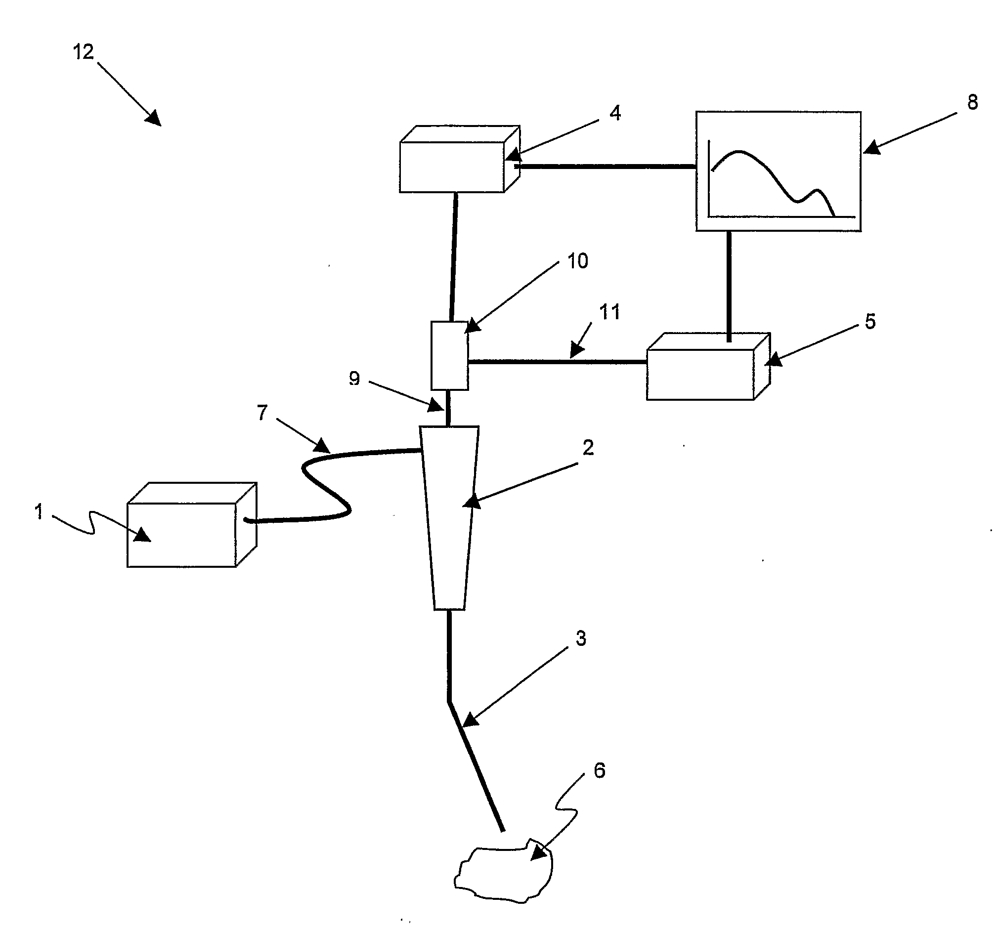
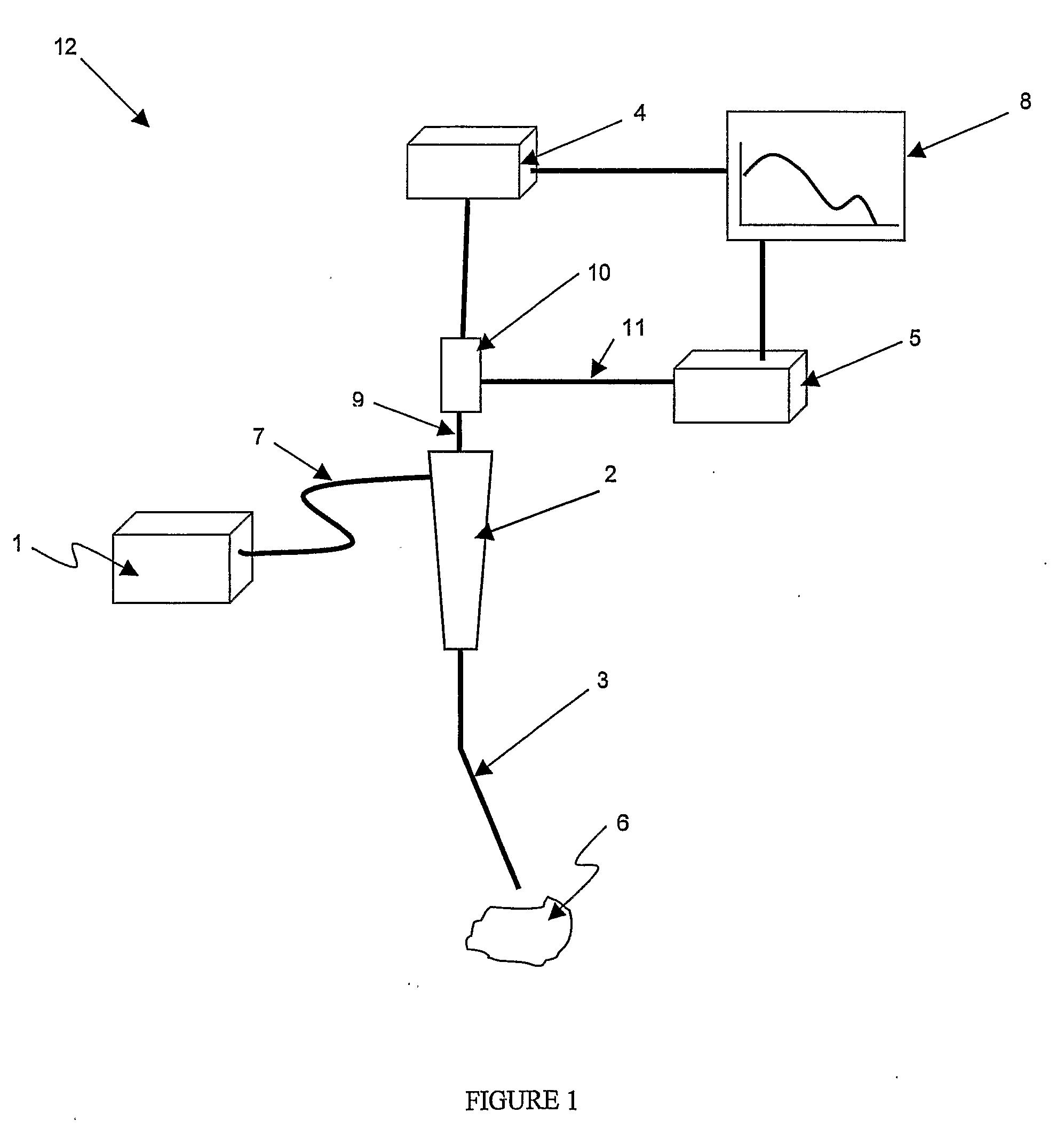
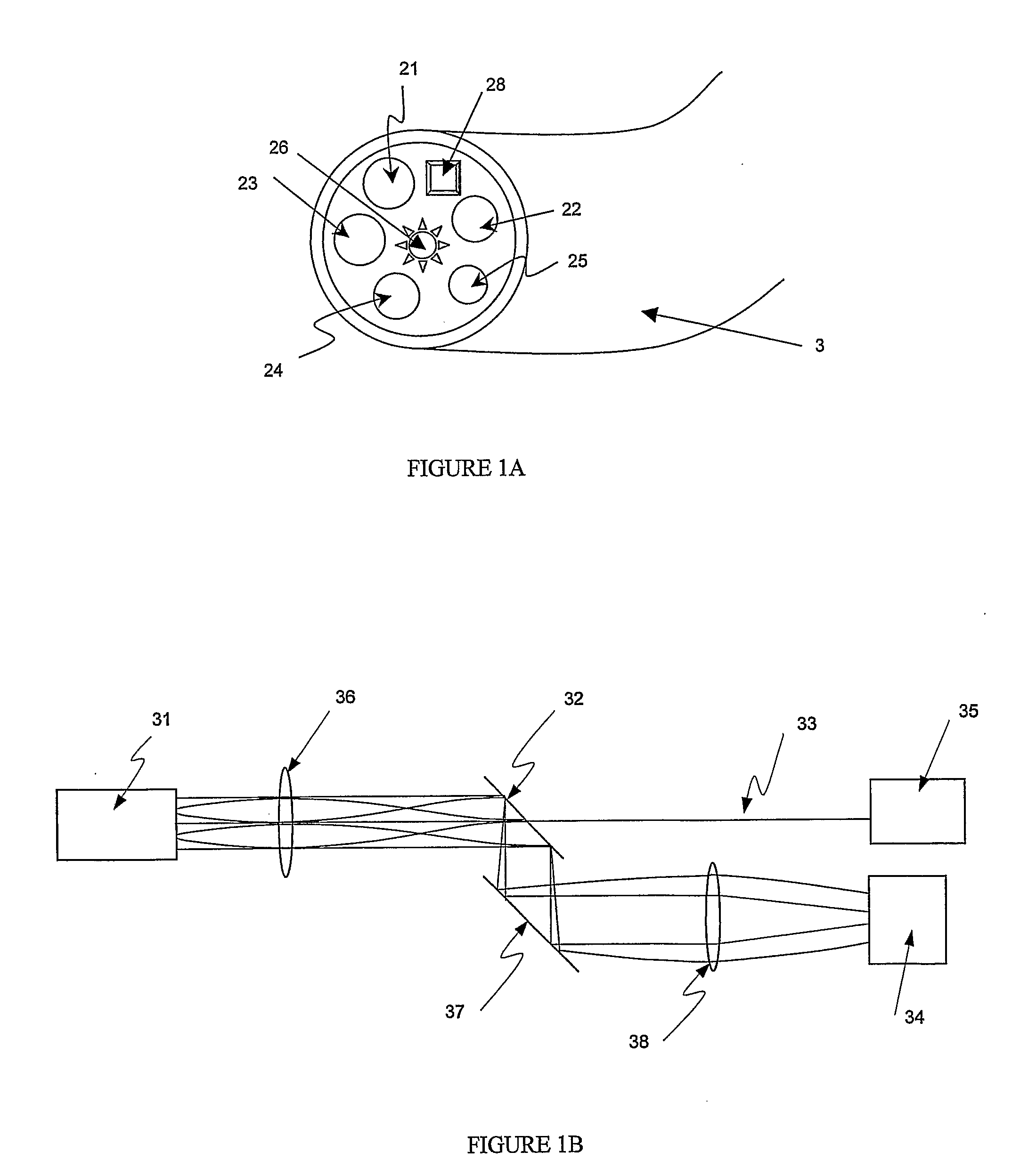
[0042]The approach of the present invention is to obtain quantitative information about the absorption-related and / or scattering-related properties from the diffuse reflectance spectra (DRS) obtained during in vivo endoscopic imaging. Diffuse reflectance relies upon the projection of a broadband light beam into the sample where the light is absorbed, reflected, scattered, and transmitted or back-reflected through the sample material. The back-reflected (back-scattered) light is then collected by the accessory (e.g. an optical fiber) and directed to the detector optics. Only the part of the beam that is s...


PUM


Abstract

The present invention provides a new method and device for disease detection, more particularly cancer detection, from the analysis of diffuse reflectance spectra measured in vivo during endoscopic imaging. The measured diffuse reflectance spectra are analyzed using a specially developed light-transport model and numerical method to derive quantitative parameters related to tissue physiology and morphology. The method also corrects the effects of the specular reflection and the varying distance between endoscope tip and tissue surface on the clinical reflectance measurements. The model allows us to obtain the absorption coefficient (μa) and further to derive the tissue micro-vascular blood volume fraction and the tissue blood oxygen saturation parameters. It also allows us to obtain the scattering coefficients (μs and g) and further to derive the tissue micro-particles volume fraction and size distribution parameters.

Description

BACKGROUND OF THE INVENTION[0001]The present invention relates to the field of optical spectroscopy and more particularly to the method for obtaining information about tissue physiology and morphology using diffuse reflectance spectroscopy. The purpose of the invention is to develop a non-invasive optical method for cancer detection.[0002]Lung cancer is the leading cause of cancer death in North America, and it has the second most common cancer incidence among both men and women. Medical research indicates that cancer can be treated more effectively when is detected early, when lesions are smaller or when tissue is in a precancerous stage. Unfortunately, conventional lung endoscopy (bronchoscopy) based on white light reflectance (WLR) imaging, which is used typically to detect the cancer lesions in the central airways of the lung, can only detect about 25 percent of the lung cancers. Most of these lesions are in the late stage when cancer has progressed and is fatal. This detection ...

Claims


Application Information

Patent Timeline
IPC IPC(8): A61B5/1455A61B6/00
CPCA61B1/043G01N2021/4742A61B5/0066A61B5/0071A61B5/0075A61B5/0084A61B5/0261A61B5/14542A61B5/1459G01J3/02G01J3/0243G01J3/0291G01N21/359G01N21/474G01N21/4795A61B1/2676
Inventor ZENG, HAISHANFAWZY, YASSER SHERIF
Owner PERCEPTRONIX MEDICAL
Learn More