
Spatial light interference microscopy and fourier transform light scattering for cell and tissue characterization

Active Publication Date: 2009-11-26
THE BOARD OF TRUSTEES OF THE UNIV OF ILLINOIS
View PDF6 Cites 248 Cited by
  • Summary
  • Abstract
  • Description
  • Claims
  • Application Information

AI Technical Summary

Benefits of technology

[0021]In accordance with another aspect of the present invention, a flow cytometer is provided that has a flow cell for streaming biological cells within a fluid medium, a source of substantially spatially coherent illumination for illuminating a subset of the biological cells within the flow cell, an objective for collecting light scattered by the illuminated subset of biological cells and for imaging the light in an imaging plane. A dispersing element diffracts light in an imaging plane into at least two diffraction orders, one order comprising a reference beam, with a spatial filtering lens system provided for removing any spatial structure due to the fluid medium from the reference beam. A detector array is used to create an interference signal based on combination of the two diffraction orders, with a processor transforming the interference signal to obtain an angular scattering distribution in a scattering plane associated with the illuminated subset of biological cells. This allows the cells to be characterized on the basis of angular scattering distribution.

Problems solved by technology

However, EM has inherent limitations due to the heavy sample preparation involved, which prohibits investigating cells non-invasively.
However, the current applications of these techniques are, in many cases, limited by specific technological constraints.
First, in all approaches, the fluorescent light detected is very weak, which demands a correspondingly long exposure time, itself limited by photobleaching.
Second, saturation techniques, such as stimulated emission depletion and structured illumination require a high level of excitation power to be delivered to the sample, which raises the issue of photo-damage and ultimately limits the safe exposure time.
Third, approaches based on stochastic photo-switchable dyes operate based on the prior assumption of a sparse distribution of fluorescent molecules, which limits applications to dynamic imaging and long-time investigation.
The great obstacle in this case becomes the fact that, generally, cells of single- and multi-cellular organisms do not absorb or scatter light significantly, i.e., they are essentially transparent, or phase objects.
In PC, a phase shift of π / 2 is introduced between the scattered and unscattered light, which makes the two interfere with greater contrast at the image plane.
While PC is sensitive to minute optical path changes in the cell, down to the nanoscale, the information retrieved is qualitative, i.e., it does not provide the actual phase delay through the sample.
However, it is often the case that the spatial resolution achieved is insufficient.
However, the drawback in the case of particle tracking is that relatively large particles are needed such that they can be tracked individually, which also limits the throughput required for significant statistical average.

Method used


Image

  • Spatial light interference microscopy and fourier transform light scattering for cell and tissue characterization
  • Spatial light interference microscopy and fourier transform light scattering for cell and tissue characterization
  • Spatial light interference microscopy and fourier transform light scattering for cell and tissue characterization

Examples


Embodiment Construction

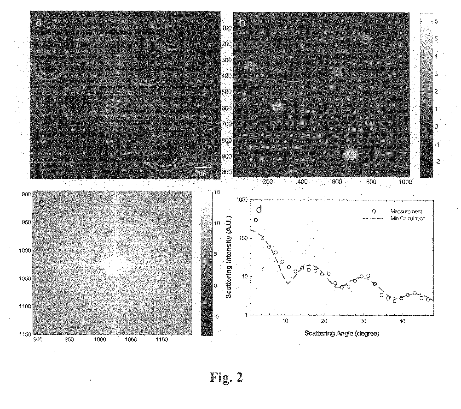
[0065]In accordance with preferred embodiments of the present invention, the high spatial resolution associated with optical microscopy and the intrinsic averaging of light scattering techniques may be combined advantageously in methods referred to collectively as Fourier transform light scattering (FTLS) that may be employed for studying both static and dynamic light scattering.

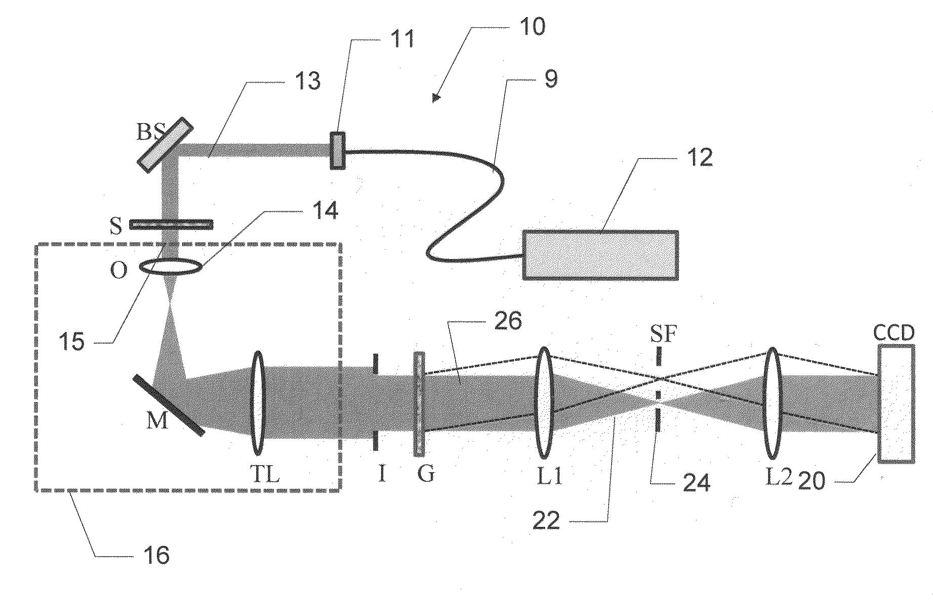
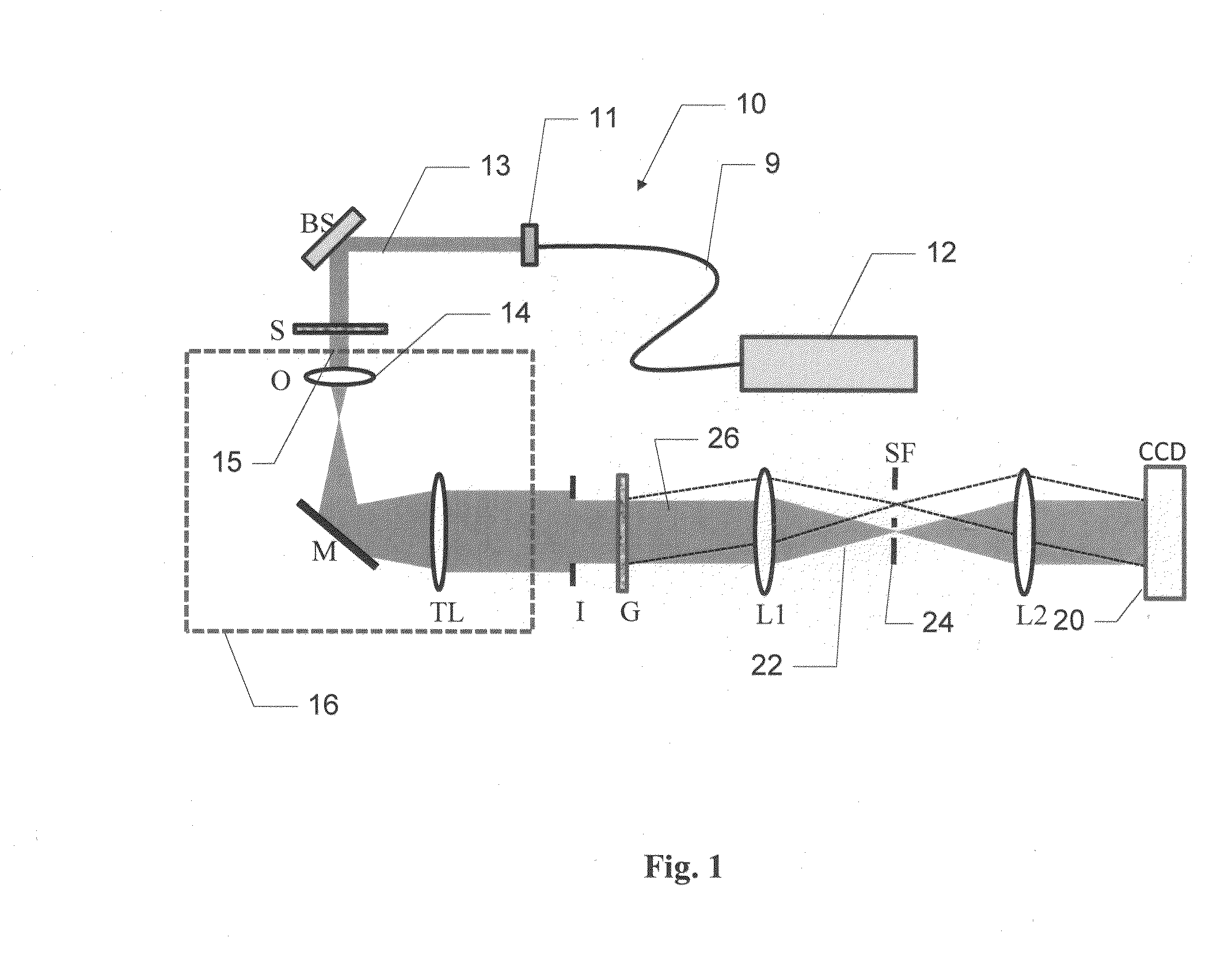
[0066]General features of the present invention are now described with reference to FIG. 1, which depicts a Fourier light scattering apparatus, designated generally by numeral 10.

[0067]As will be described, FTLS provides for retrieving the phase and amplitude associated with a coherent microscope image and numerically propagating this field to the scattering plane. This approach may advantageously provide both the accurate phase retrieval for the ELS measurements and, further, the fast acquisition speed required for DLS studies.

[0068]While the FTLS apparatus 10 is depicted in FIG. 1 in the form of a common-p...


PUM


Abstract

Methods and apparatus for rendering quantitative phase maps across and through transparent samples. A broadband source is employed in conjunction with an objective, Fourier optics, and a programmable two-dimensional phase modulator to obtain amplitude and phase information in an image plane. Methods, referred to as Fourier transform light scattering (FTLS), measure the angular scattering spectrum of the sample. FTLS combines optical microscopy and light scattering for studying inhomogeneous and dynamic media. FTLS relies on quantifying the optical phase and amplitude associated with a coherent image field and propagating it numerically to the scattering plane. Full angular information, limited only by the microscope objective, is obtained from extremely weak scatterers, such as a single micron-sized particle. A flow cytometer may employ FTLS sorting.

Description

[0001]The present application claims the priority of U.S. Provisional Patent Application, Ser. No. 61 / 054,939, filed May 21, 2008, and to U.S. Provisional Patent Application Ser. No. 61 / 204,840, filed Jan. 12, 2009, both of which applications are incorporated by reference herein.[0002]Aspects of the present invention were developed with Government support under NIH Grants HL086870 and HD007333, and under CAREER Award Grant 08-46660 awarded by the National Science Foundation. The Government has certain rights in the invention.TECHNICAL FIELD[0003]The present invention pertains to apparatus and methods for quantitative imaging of the optical phase shift introduced by largely transparent samples, and for analyzing angular scattering over a broad range of angles by a sample that may include single particles and live cells, and, further, to the application of the foregoing apparatus and methods for characterizing cells and tissue.BACKGROUND OF THE INVENTION[0004]For centuries, light micr...

Claims


Application Information

Patent Timeline
IPC IPC(8): G01N21/53G02F1/01
CPCG01N15/0211G01N15/1434G01N15/1459G01N2015/0038Y10S977/881G01N2015/025G01N2015/1454G02B21/0056G02B21/008G01N2015/0222G03H2001/0491G03H2001/2244
Inventor POPESCU, GABRIELDING, HUAFENGWANG, ZHUO
Owner THE BOARD OF TRUSTEES OF THE UNIV OF ILLINOIS
Learn More