

Method to Assess Stability of Proteins
a protein and structural stability technology, applied in the field of methods of evaluating the structural stability of proteins and peptides, can solve problems such as not being chemically or structurally stabl
- Summary
- Abstract
- Description
- Claims
- Application Information
AI Technical Summary
Benefits of technology
Problems solved by technology
Method used
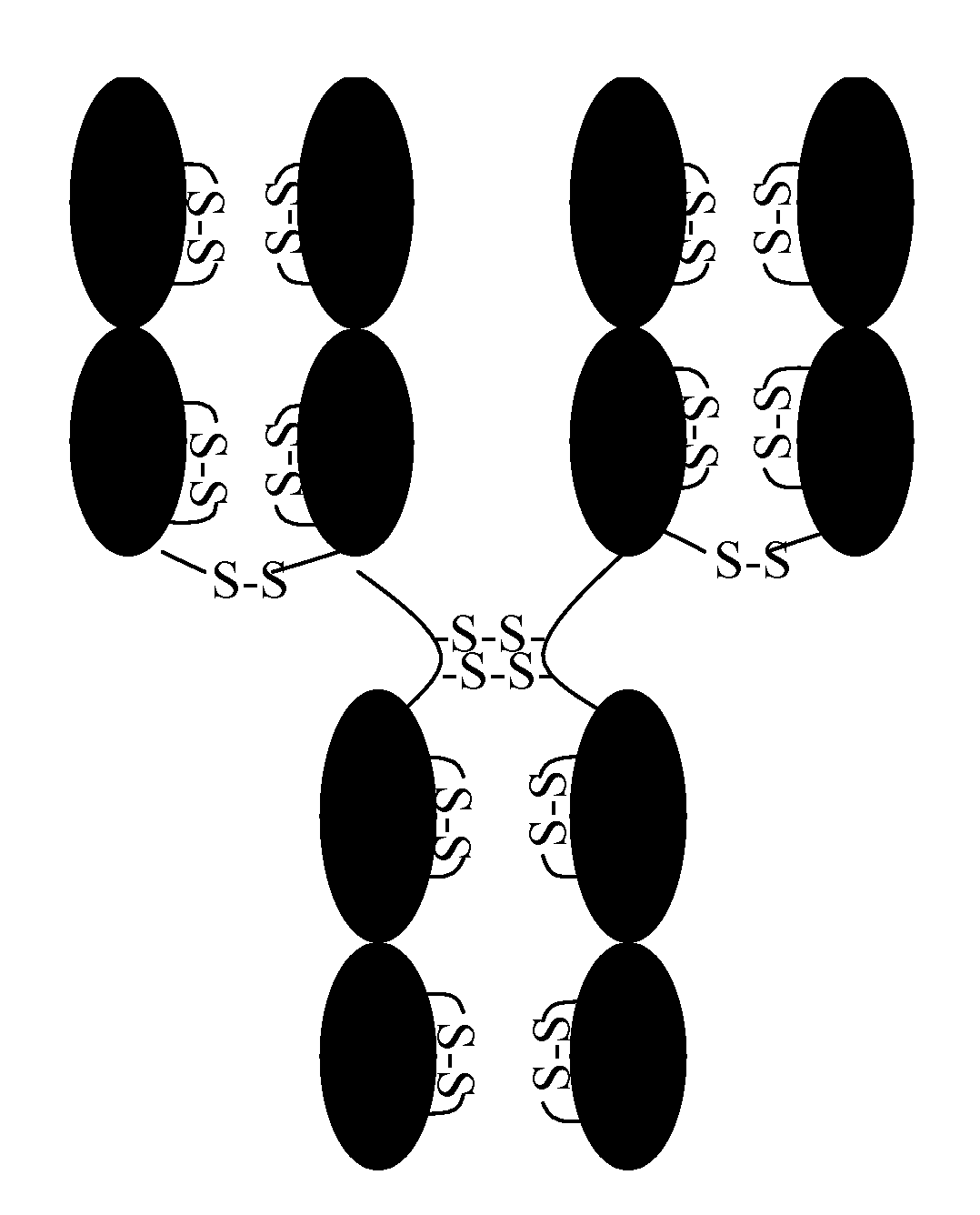
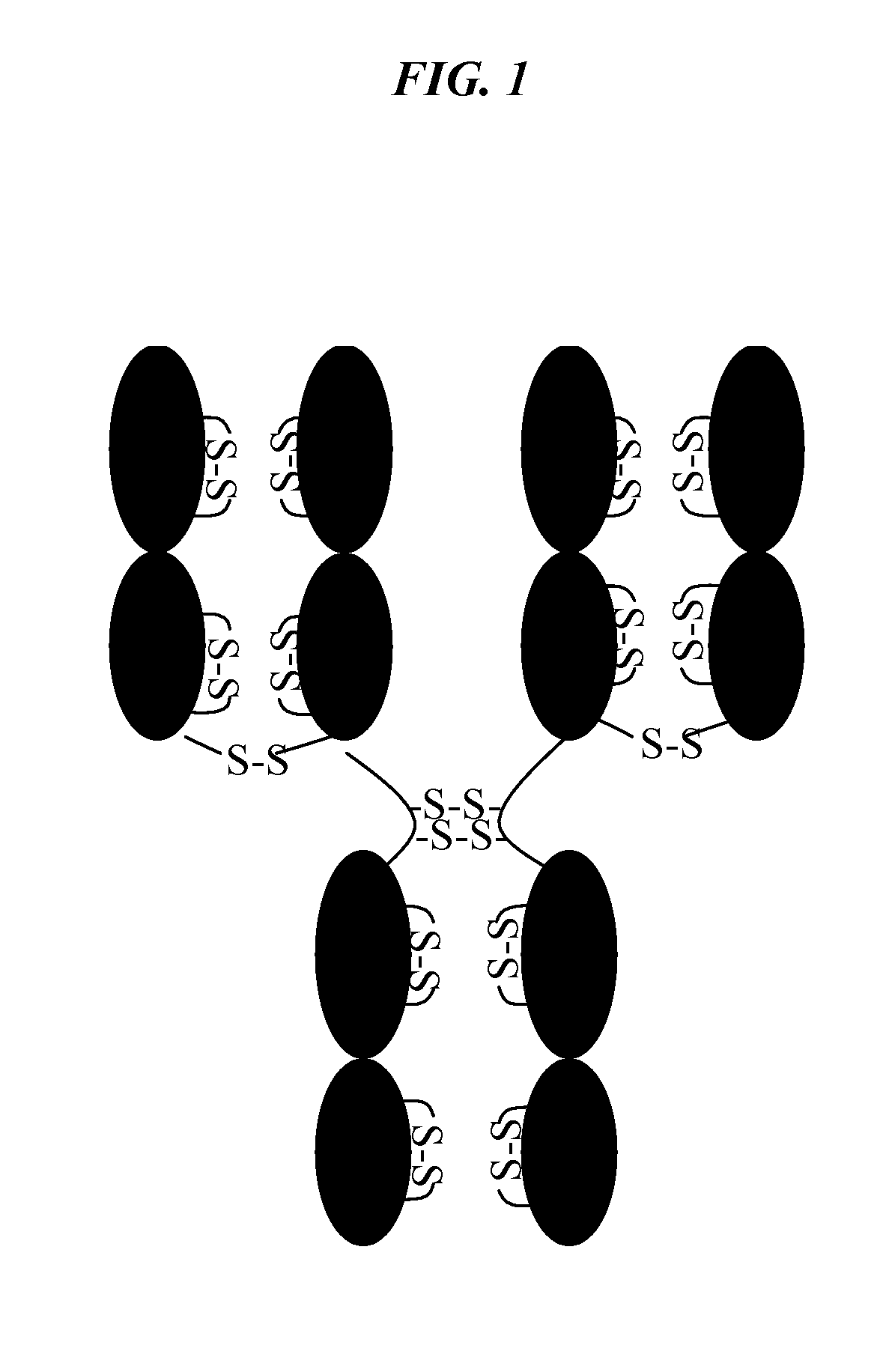
Image

Examples
example 1
Preparation of Standards
[0038]Calibration curves for different concentrations of —SH were prepared using either N-acetyl-L-cysteine or BSA as a standard from 0.02 to 17 μM under denaturing and nondenaturing conditions. The stock solutions for the standards for the calibration curves were prepared in Dulbecco's phosphate-buffered saline (D-PBS), pH 7.3. Buffers were deoxygenated and degassed by sonication under vacuum and then bubbled with argon. NPM was prepared at 10 μM concentration in dimethylformamide (DMF).
[0039]For the reactions, 50 μL of the appropriate BSA (or N-acetyl-L-cysteine) solution were mixed with 250 μL of D-PBS, pH 7.3 with or without 6M Gdn-HCl to the desired final concentration. Three μL of 10 μM NPM were added to this mixture, and the samples were incubated either 5 min, 1 h or 2 h at RT or 25° C. for solutions in D-PBS; and incubated at RT, 37° C., 40° C., 50° C. or 60° C. for those with Gdn-HCl. NPM was added slowly to avoid cloudiness in D-PBS. The reaction w...
example 2
Assay Conditions
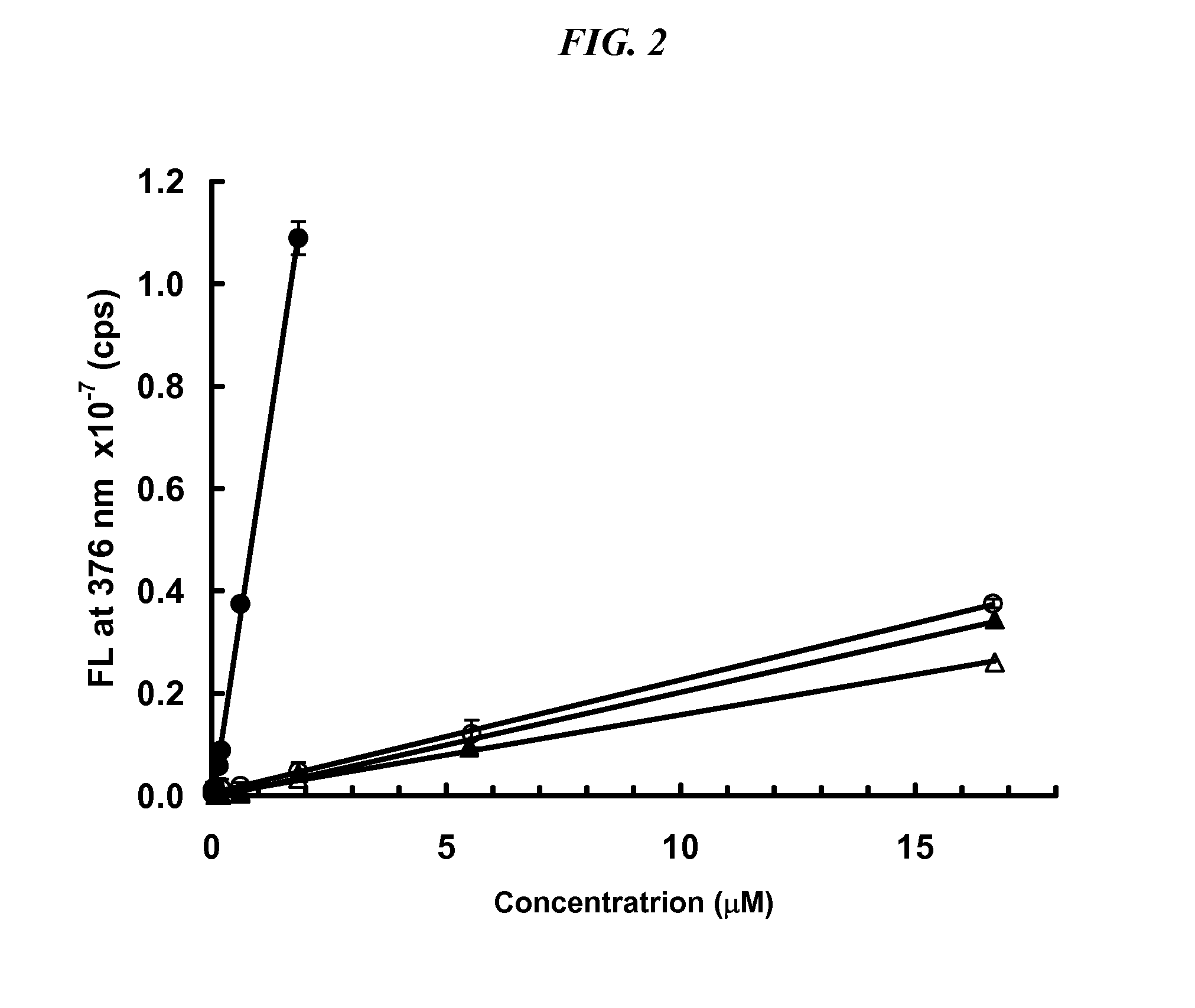
[0044]To determine the optimal conditions for determination of —SH under denaturing conditions, several proteins, BSA and three antibodies were subjected to various amounts of thermal denaturation. Adalimumab is a human anti-TNF antibody, infliximab is a murine-human chimeric anti-TNF antibody, and MAB6 is a human engineered anti-cytokine antibody. The signal obtained for standards and samples analyzed at 60° C., 50° C., 40° C. and 37° C. were compared. The temperature at which the net signal (sample signal minus buffer) was maximal was obtained is 37° C. (FIG. 5). In addition, the effect of reaction time was tested at 5 min, 1 h and 2 h. The optimal reaction time, where signal strength reached a plateau, was 2 h. Experiments at pH 6.0 were also performed. Although signal was obtained, it was significantly reduced as compared to pH 7.3.
[0045]The best results were produced when samples were prepared in a non-reducing buffer (such as Dulbecco's phosphate-buffered salin...
example 3
Stability of Structurally Related Proteins
[0047]Human serum IgG1, and IgG4 antibodies (lambda and kappa light chains) were obtained from Sigma. Monoclonal antibodies; CDG1, a humanized murine Mab which has a human IgG4 heavy chain and kappa LC constant regions (IgG4,κ); Mab13 is a human IgG1 with lambda light chain, Mab59, Mab12 and Mab9.5 are human IgG1 with kappa light chains; and Mab41 and Mab48 are humanized murine Mabs with IgG4 heavy and kappa light chains. All of the Mabs had unique binding specificity and unique hypervariable domains (CDR) domains.
[0048]For analysis of free sulfhydryl, antibodies were prepared at 1 μg / mL (6.7 uM) in D-PBS, pH 7.3 or Tris buffer, pH 7.2 and diluted to 1.1 uM using the appropriate reaction buffer. Solutions at 3 μg / mL were also used, but the data show that the results obtained are comparable to those obtained at 1 μg / ml (using IgG1 lambda and kappa as controls). 50 uL of these solutions of protein were treated following the same procedure used...
PUM
| Property | Measurement | Unit |
|---|---|---|
| temperature | aaaaa | aaaaa |
| volume | aaaaa | aaaaa |
| pH | aaaaa | aaaaa |
Abstract
Description
Claims
Application Information