Assembled Cartilage Repair Graft

a cartilage repair and graft technology, applied in the field of assembled cartilage repair grafts, can solve the problems of reducing affecting the overall quality of life of individuals, and each have limitations ranging from the quality of cartilage repair to the cost and complexity of surgical procedures, and the procedures themselves create defects (i.e. oats). , to achieve the effect of adding extra cost and complications

Inactive Publication Date: 2009-12-17
RTI BIOLOGICS INC
View PDF18 Cites 83 Cited by
  • Summary
  • Abstract
  • Description
  • Claims
  • Application Information

AI Technical Summary

Benefits of technology

[0018]A further advantage of this embodiment, especially when an assembled chondroinductive portion is used, is that the negatively tapered shaft and bore fit provides solid and secure assembly wherein the at least two pieces of chondroinductive demineralized cortical bone are in direct contact with each other and are completely or partially surrounded over at least a part of their surface at the hydration controlled interference fit interface by a piece of osteoconductive cancellous bone.
[0019]An advantage of the assembly methods, and especially of the hydration controlled interference fit or hydration controlled shrink fit, is that the assembled implants are suitable for implantation at an osteochondral site in a human or other mammal without additional internal fasteners or connective elements such as press fit pins, bone pins, sutures, or adhesives. In addition to requiring excess material and additional processing steps, these other fasteners or connective elements add extra cost and complications to the design, manufacture and use of the implants. A preferred embodiment of the present invention provides an assembly that does not comprise separate fasteners or adhesive for holding together the implant.

Problems solved by technology

These types of articular cartilage defects may cause pain, swelling and / or knee locking and thus diminish the individual's overall quality of life.
While these procedures show significant rates of clinical success in the short and medium term, they each have limitations ranging from the quality of the cartilage repair to the cost and complexity of surgical procedures.
Additionally, some of the procedures themselves create defects (i.e. OATS) either as a direct result of the primary procedure or through second site morbidity caused by the recovery of the patient's own tissue.

Method used


Image

  • Assembled Cartilage Repair Graft
  • Assembled Cartilage Repair Graft
  • Assembled Cartilage Repair Graft

Examples


example 1

[0203]This example demonstrates a method of making an embodiment of an assembled implant.

[0204]Cancellous bone was obtained from the condyles of long bones, or optionally from the Talus or heel bone. The bone was cut into blanks with diameters ranging from roughly 6-13 mm and heights from 8-10 mm using a band saw and coring tools. The inner geometry of the cancellous bone was then machined using a dovetail cutter. A 1.5 mm hole was machined in the bottom center of the 8 mm and 10 mm cancellous bone portions. The cancellous bone portions are measured, inspected for quality, packaged, and stored frozen until processing for sterilization. The cancellous bone portions were sterilized, defatted and deantigenized, and soluble protein was removed by subjecting the cancellous tissue to cyclically alternating cycles of pressure and vacuum in the sequential presence of mild sterilizing chemical solutions. Following sterilization, the cancellous bone portions were packaged and stored until ass...

example 2

[0208]This example documents an animal study completed to confirm the chondroinductivity of an implant.

[0209]Prototype implants were implanted into an ectopic site in an athymic nude rat model, resulting in histologic evidence of chondroinduction without any signs of an inflammatory response.

[0210]Specifically, a 4 mm diameter by 2 mm tall disc taken from the demineralized cortical bone portion of Example 1 was implanted in abdominal muscle pouches of athymic nude rats using a modified Urist model, Urist, M. R., “Bone: Formation by Autoinduction,” Science 160:893-894 (1965). The explants were retrieved two weeks later, processed, and evaluated histologically for evidence of new cartilage formation. The control implants made from chemically inactivated demineralized cortical bone material formed only fibrous material within the Haversian canals with minimal evidence of inflammation. More significantly, the implants made from active demineralized cortical bone demonstrated signs of ch...

example 3

[0211]This example documents a cadaver study completed by a practicing surgeon experienced in cartilage repair at an orthopedic clinic to confirm the proper function and methods of use for the present implants.

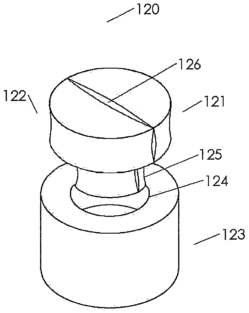
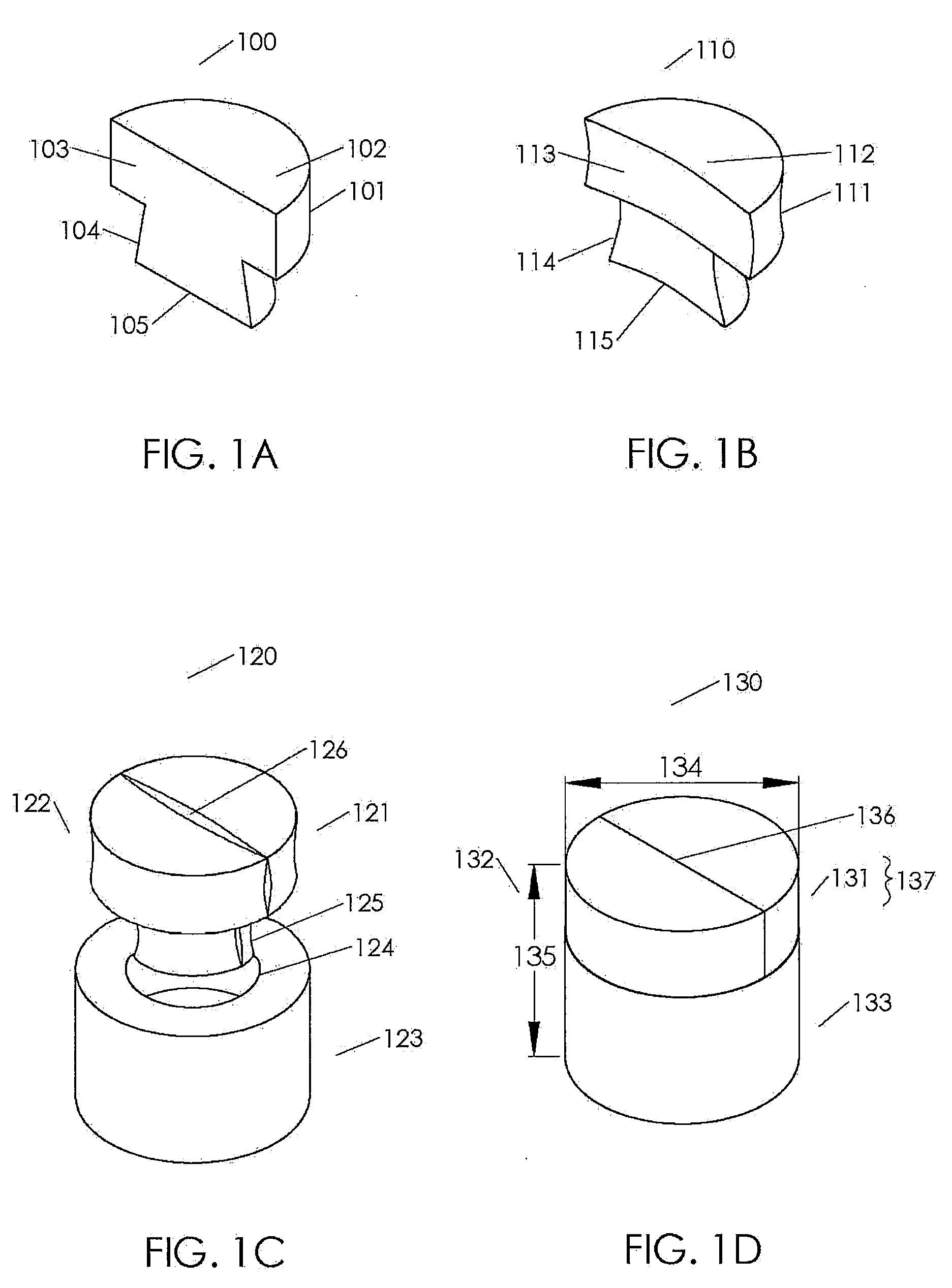
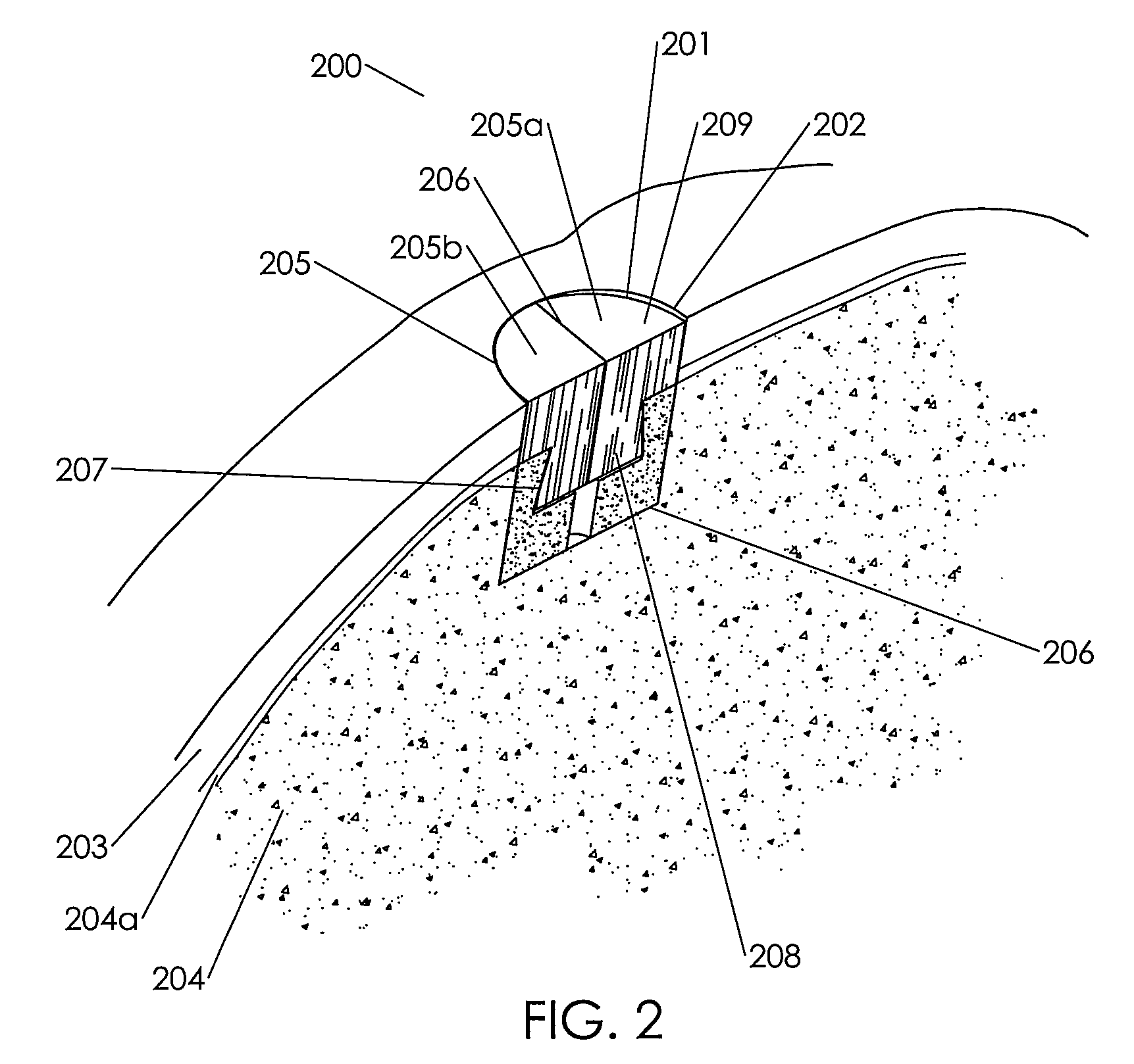
[0212]Several primary-site implants were made to production specifications using an assembled biological implant comprising a two piece chondroinductive demineralized cortical bone cap assembled via a hydration controlled interference fit to a mineralized cancellous bone osteoconductive portion. The assembled implants were implanted into a cadaver knee using an open approach. The implants were left out at room temperature for several minutes and were then placed in room temperature saline for a minimum of ten minutes to ensure consistent final levels of hydration prior to implantation. The surgeon prepared the implantation site by coring out an 8 mm diameter plug from the primary site using an OATS single-use kit. He measured the depth of the defect using the depth gauge and v...


PUM


Abstract

Bifunctional and assembled implants are provided for osteochondral implantation.

Description

BACKGROUND OF THE INVENTION[0001]Focal articular cartilage defects of the knee are not uncommon consequences of injuries from sports, work or activities of daily living. Arthroscopic studies have estimated the occurrence of near full thickness lesions, full thickness lesions or full thickness with boney involvement lesions greater than 1 cm2 to be in the range of 6% of all surgical procedures in the knee. These types of articular cartilage defects may cause pain, swelling and / or knee locking and thus diminish the individual's overall quality of life. The size (area dimensions), depth (partial cartilage tissue to boney involvement), containment, co-morbidities (e.g., ACL tear, meniscal tear, and malalignment) and region of a defect will influence the procedure used by surgeons. Currently, for focal defects smaller than (<) 2.5 cm2, the subject and surgeon have few options: debridement (chondroplasty), marrow stimulation (micro-fracture, abrasionoplasty, or subchondral drilling), o...

Claims


Application Information

Patent Timeline
Patent Type & Authority Applications(United States)
IPC IPC(8): A61F2/02A61F2/28
CPCA61B17/86A61F2002/30929A61F2/30756A61F2002/30057A61F2002/30138A61F2002/302A61F2002/30224A61F2002/30233A61F2002/30299A61F2002/30332A61F2002/30337A61F2002/30485A61F2002/30766A61F2002/30772A61F2002/30878A61F2002/30883A61F2220/0025A61F2220/0033A61F2230/0017A61F2230/0065A61F2230/0069A61F2230/0093A61F2/30771A61F2/28A61F2002/30751A61F2002/30075
Inventor BURSAC, PREDRAGBROWN, LAUREN M.SCHMITT, ERIC J.GROVER, GUY B.
Owner RTI BIOLOGICS INC
Why Eureka
Try Eureka