

Biodegradable and bioabsorbable biomaterials and keratin fibrous articles for medical applications
a biomaterial and keratin fibrous technology, applied in the direction of monocomponent protein artificial filaments, peptide/protein ingredients, bandages, etc., can solve the problems of lack of cell-recognition signals, lack of cell-attachment on the surface of these polymer materials, and hydrophobicity
- Summary
- Abstract
- Description
- Claims
- Application Information
AI Technical Summary
Problems solved by technology
Method used
Image

Examples
example 1
[0034]A membrane was prepared as follows: a 1 wt % PLLA / chloroform / N,N-dimethyl formamide (DMF) solution was prepared by slowly dissolving PLLA pellets (inherent viscosity of 7.0 dl / g, PURAC, Netherlands) into a chloroform solvent at room temperature with stirring. After PLLA was completely dissolved, 10 wt % DMF was added. The solution was then loaded into the 20 ml syringe fitted with a needle, and delivered to an electrode. The solution was pumped and controlled by a syringe pump at a flow rate of 0.3 ml / min. A 10 kV positive high voltage was applied on the electrode. The distance from the tip of the electrode to the grounded collecting plate was 15 cm. A tiny electrospinning jet was formed and stabilized in 30 seconds under these conditions. The collecting plate was movable and controlled by a stepper motor. The collecting plate was continually moved at a rate of 1 mm / sec until a membrane having a relatively uniform thickness of about 100 microns was obtained. Electrospun membra...
example 2
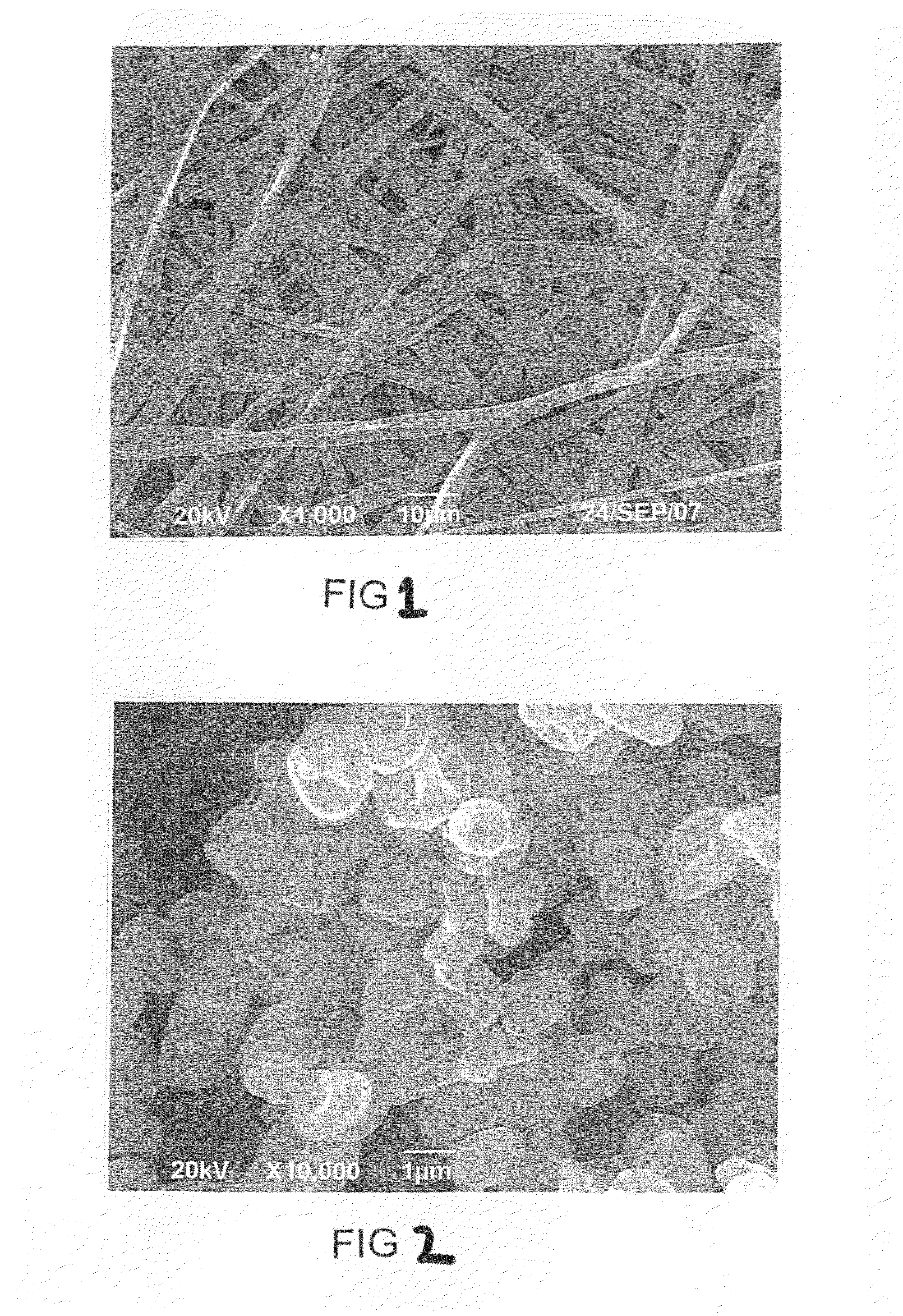
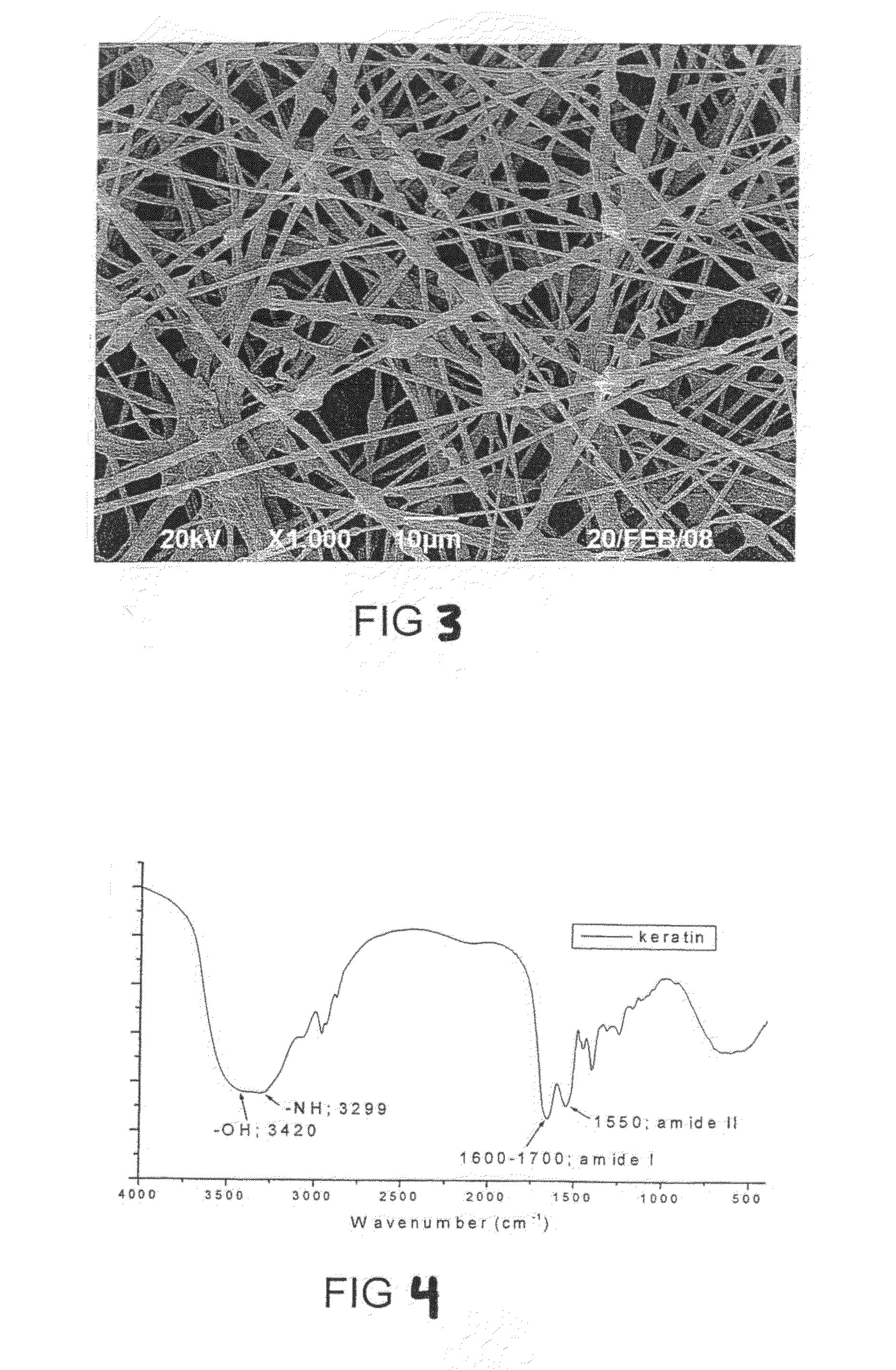
[0036]A biodegradable and bioabsorbable membrane with keratin according to the present invention, fabricated by an electrospinning process, was prepared as follows: 1 wt % keratin powders (FIG. 2) were dispersed in the PLLA / chloroform / DMF solution. The solution was then electrospun at 12 kV. The fibrous membrane was collected at 16 cm (FIG. 3). The membrane was examined by FTIR and SEM.
[0037]Except the parameters mentioned about, the concentration of keratin in the polymer solution also influences the fiber shape. The applied voltage, solution flow rate, distance between capillary and collector are adjusted accordingly.
[0038]FIG. 4 is FTIR spectra of wool keratin. Wavenumbers from 3250 to 3300 cm−1 are the N—H stretch which is in resonance with amide II overtone. Wavenumbers at 1600-1700 cm−1 are mainly the C═O stretching. Wavenumber at 1550 cm−1 is the N—H bending coupled with C—N stretching. FTIR spectra of pure PLLA have no peaks from 1700 to 1500 cm−1. For PLLA and keratin compo...
example 3
[0039]PLLA / keratin (1:1) membranes were immersed phosphate buffer saline (PBS, pH 7.4) at 37° C. for various time periods up to 4 weeks. The degradation medium was changed daily for the first week, one at day 10 and day 14, and then weekly for the rest of the remaining period. Samples were taken out at the end of each sampling time point, i.e., at three hour, 1, 3, 7, 14, and 28 days. The samples removed from the PBS were first rinsed with distilled water and then vacuum dried for 24 h. PLLA / keratin samples before and after degradation were examine by Fourier transform infrared (FTIR) and X-ray photoelectron spectroscope (XPS) The characterizing peaks of PLLA and keratin were used to calculate their ratios after different degradation periods. Along with degradation period, the characterization peaks of keratin decreased correspondingly (FIG. 6). According to the reducing of absorbance in FTIR spectra, the change of keratin in composite was calculated (FIG. 7). In XPS wide scan spect...
PUM
| Property | Measurement | Unit |
|---|---|---|
| Thickness | aaaaa | aaaaa |
| Flow rate | aaaaa | aaaaa |
| Diameter | aaaaa | aaaaa |
Abstract
Description
Claims
Application Information