Compounds Regulating Calreticulin, KDEL Receptor and/or ERP-57 Cell Surface Exposure and Uses Thereof to Evaluate the Efficiency of a Cancer Treatment

a technology of kdel receptor and calreticulin, which is applied in the field of compounds regulating calreticulin, kdel receptor and/or erp-57 cell surface exposure and evaluation of the efficiency of a cancer treatment, can solve the problems of cell death, oversimplification of the dichotomy between immunogenic necrosis and tolerogenic apoptosis, and cancer is the major cause of mortality, so as to reduce or block the immunogenicity of calreticulin

Inactive Publication Date: 2010-01-21
INSTITUT GUSTAVE ROUSSY +1
View PDF1 Cites 3 Cited by
  • Summary
  • Abstract
  • Description
  • Claims
  • Application Information

AI Technical Summary

Benefits of technology

[0240]Furthermore, the location of calreticulin protein at the cell surface may be realised by antibodies anti-calreticulin which detect the endogenous form of calreticulin, a recombinant form and the mimetic form. A method of detection of all forms of calreticulin protein at the cellular surface is also an object of the present invention. This could be done in vitro, ex vivo or in vivo. The methods used to detect this calreticulin protein at the cell surface are well-known from the skilled man of the art. These methods comprise immunochemistry on tissue sections (frozen or paraffined), EIA assays such as ELISA on tumour lysates, confocal immunofluoresence or flow cytometry analyses of cytospins, cell aspirates harvested from tumour beds or autoimmune lesions . . . . One object of the present invention is also to develop a method of quantitative detection of the calreticulin (all forms) at the cellular surface. The immunogenicity of the apoptosis is correlated to the amount of calreticulin present at the cell surface. The more calreticulin at the cellular surface, the more immunogenic apoptosis. This method of detection can serve to predict the immunogenicity of the apoptosis. In addition, the effectiveness of a chemotherapy is correlated to the efficiency of the immune response, therefore the immunogenicity of the apoptosis. The more the immunogenic apoptosis, the more the therapeutic efficiency of a chemotherapy. This method of detection of calreticulin at the cell surface can be used for prediction of immunogenic apoptosis and also for therapeutic efficiency of a chemotherapy. The calreticulin in these methods is used as a predictive marker of both immunogenic apoptosis and therapeutic efficiency of a chemotherapy. This method of quantitative detection can also be advantageous to predict risks of forced apoptosis that becomes too immunogenic. Inhibition of the translocation of the calreticulin at the cellular surface could decrease the immunogenicity of the calreticulin and thus reduce or block (in the best case) the immune response.
[0241]There is a basal amount of calreticulin at the cellular surface and the increased amount of calreticulin allows to predict an immunogenic apoptosis and / or a therapeutic efficiency of a chemotherapy. By comparison between before and after chemotherapy, the amount of calreticulin at the cell surface is generally largely increased and the increase will be a good predictive marker for immunogenic apoptosis, therapeutic efficiency of a chemotherapy, immunogenic viral infection but also for autoimmune diseases or transplantation rejection / GVH disease. The present invention also provides a method of detection of the calreticulin at the cell surface wherein the calreticulin at the cell surface is used as a predictive marker of immunogenic viral infection or autoimmune diseases or transplantation rejection / GVH disease.

Problems solved by technology

Cancer is the major cause of mortality in most industrialized countries.
Chemotherapy leads to the cell death.
Nonetheless, it seems that the dichotomy between immunogenic necrosis versus tolerogenic apoptosis is an oversimplification.
Thus, even after an initially efficient chemotherapy, patients do not develop an efficient antitumourous immune response and then are overcome by chemotherapy-resistant tumourous variants.

Method used


Image

  • Compounds Regulating Calreticulin, KDEL Receptor and/or ERP-57 Cell Surface Exposure and Uses Thereof to Evaluate the Efficiency of a Cancer Treatment
  • Compounds Regulating Calreticulin, KDEL Receptor and/or ERP-57 Cell Surface Exposure and Uses Thereof to Evaluate the Efficiency of a Cancer Treatment
  • Compounds Regulating Calreticulin, KDEL Receptor and/or ERP-57 Cell Surface Exposure and Uses Thereof to Evaluate the Efficiency of a Cancer Treatment

Examples


example 1

CRT Exposure Defines Immunogenic Cell Death

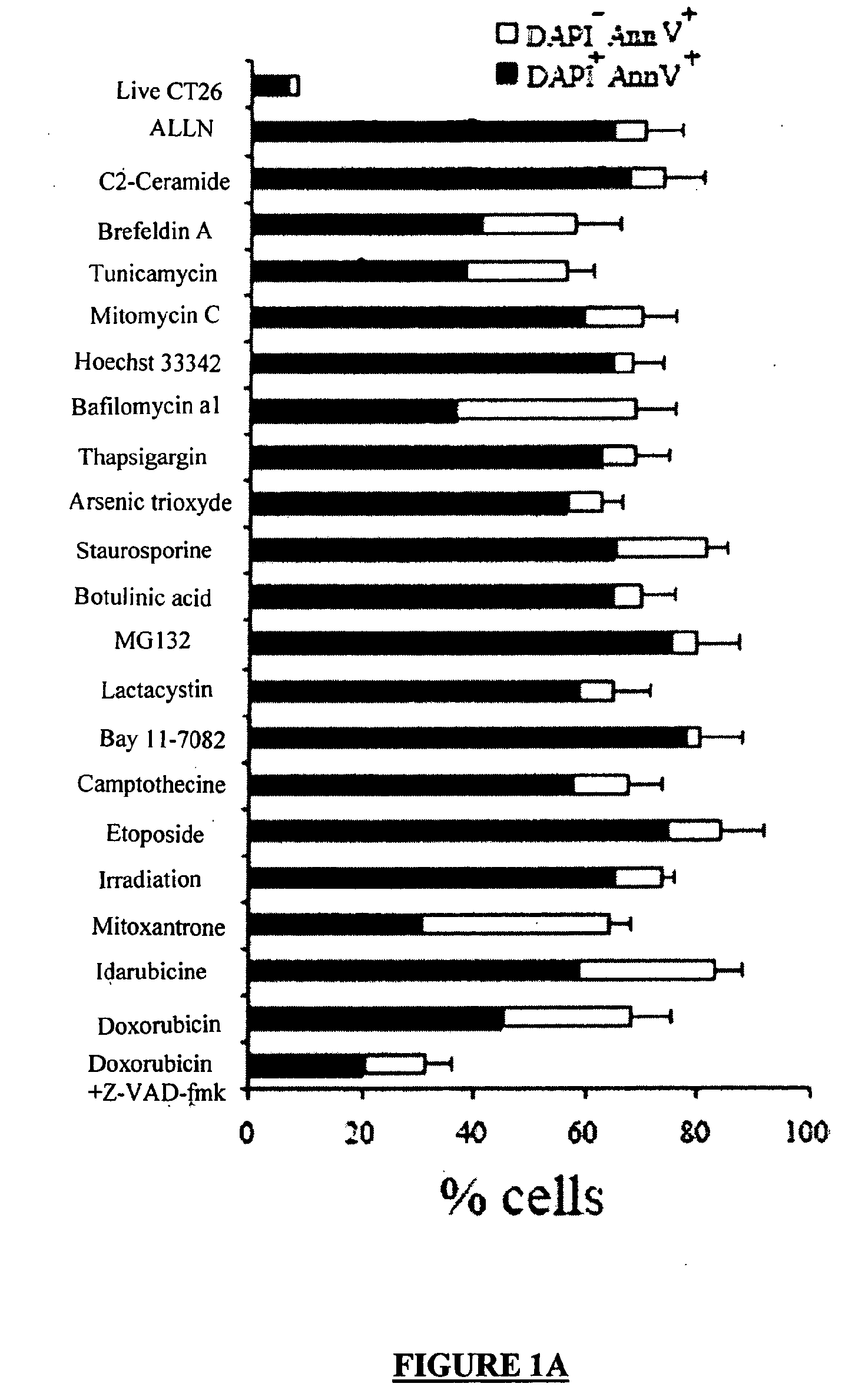
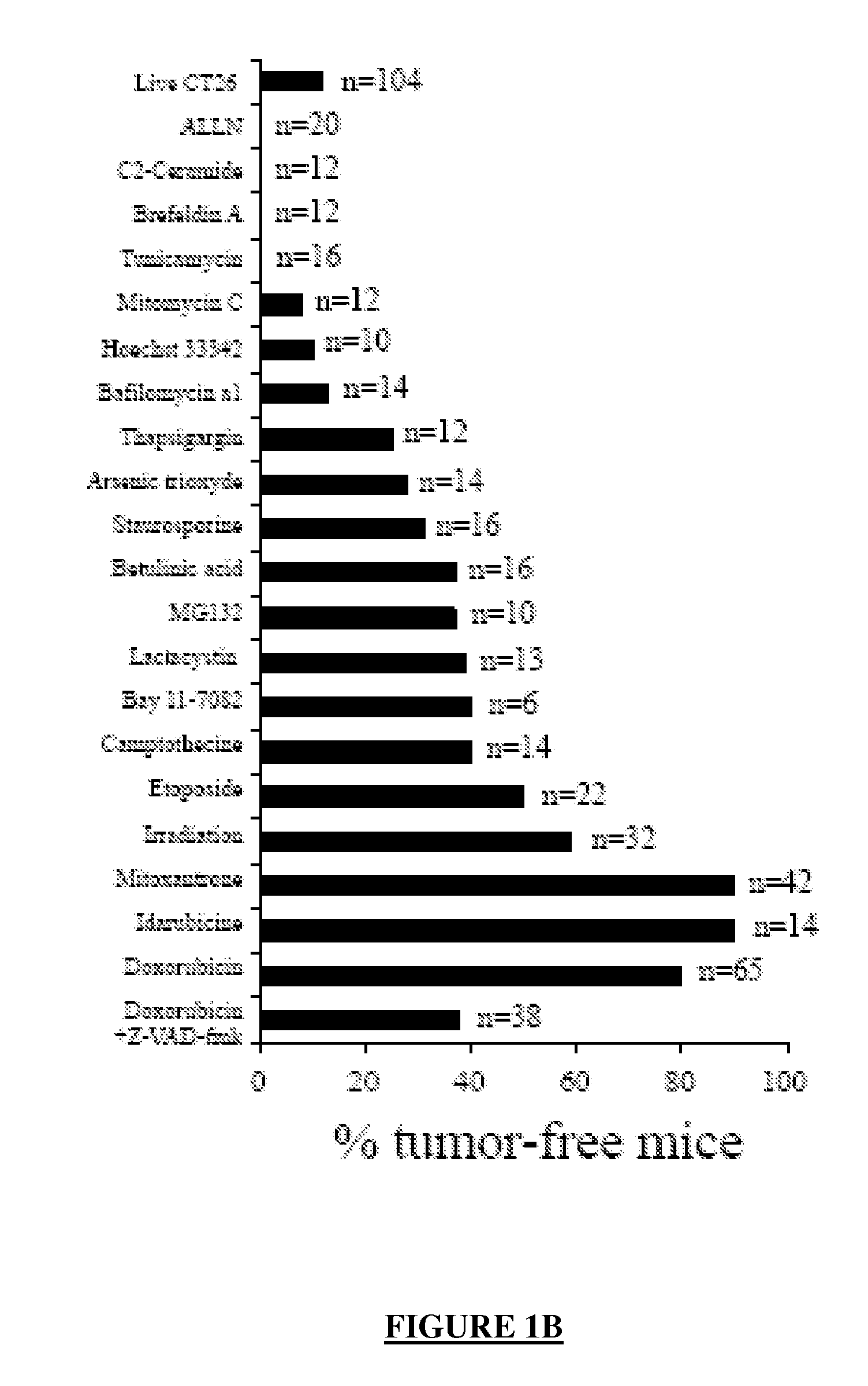
[0334]Dying CT26 tumour cells exposed to a panel of ˜20 distinct apoptosis inducers (all of which induced ˜70±10% apoptosis, as determined by double staining with the vital dye DAPI and the PS-binding dye Annexin V, FIG. 1A) were injected into one flank of immunocompetent BALB / c mice, followed by rechallenge of the animals with live tumour cells injected into the opposite flank 8 days later. Protection against tumour growth then was interpreted as a sign of anti-tumour vaccination (FIG. 1B) because such protection was not observed in athymic (nu / nu) BALB / c mice (Casares, N. et al. J. Exp. Med. 202, 1691-701 (2005).and data not shown). Most apoptosis inducers, including agents that target the endoplasmic reticulum (ER) (thapsigargin, tunicamycin, brefeldin), mitochondria (arsenite, betulinic acid, C2 ceramide) or DNA (Hoechst 33342, camptothecin, etoposide, mitomycin C), failed to induce immunogenic apoptosis, while anthracyclins (doxorubici...

example 2

Requirement of CRT for DC-Mediated Recognition of Dying Tumour Cells

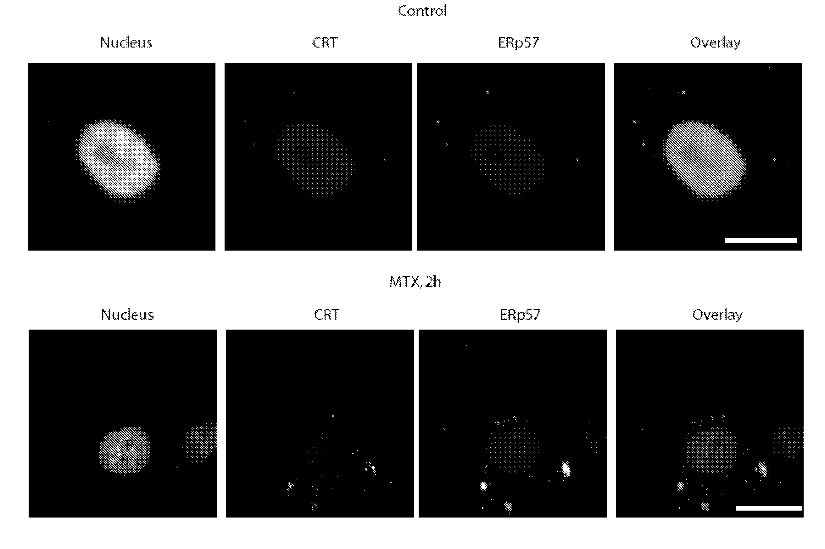
[0335]In view of the established role of CRT as an “eat me” signal (Gardai, S. J. et al. Cell 123, 321-34 (2005); Ogden, C. A. et al. J Exp Med 194, 781-95 (2001)), we decided to further investigate the possible implication of CRT in the phagocytosis of anthracyclin-treated tumour cells by DC, a cell type that is stringently required for mounting an immune response against apoptotic tumour cells (Steinman, R. M., Turley, S., Mellman, I. & Inaba, K. J Exp Med. 191, 411-6 (2000); Casares, N. et al. J. Exp. Med. 202,1691-701 (2005)). Anthracyclin-treated tumour cells acquired the property to be phagocytosed by DC quickly, well before the manifestation of apoptotic changes, within a few hours after treatment with doxorubicin or mitoxantrone (FIG. 3A, FIG. 2S A), correlating with the rapid induction of CRT (FIG. 3B, FIG. 1S A, B) and the acquisition of immunogenicity (FIG. 2S B). The presence of CRT on the surface of tum...

example 3

Requirement of CRT for Immunogencity of Dying Tumour Cells

[0336]The knock-down of CRT compromised the immunogenicity of mitoxantrone-treated CT26 cells, and this defect was restored when rCRT was used to complement the CRT defect induced by the CRT-specific siRNA. This result was obtained in two distinct experimental systems, namely (i) when CT26 tumour cells were injected into the flank of Balb / c mice (or MCA205 cells were injected into C57BI / 6 mice, not shown) to assess the efficacy of anti-tumour vaccination (FIG. 4A) and (ii) when the tumour cells were injected into the foot pad to measure interferon-7 production by T cells from the popliteal lymph node (FIG. 4B). In this latter system, absorption of rCRT to the plasma membrane surface greatly enhanced the immunogenicity of cells that usually fail to induce an immune response such as mitomycin-treated cells (FIG. 4C). Similarly, etoposide-treated cells coated with rCRT elicited a vigorous anti-tumour immune response in vivo, in ...


PUM

PropertyMeasurementUnit
pHaaaaaaaaaa
pHaaaaaaaaaa
pHaaaaaaaaaa

Abstract

The present invention relates to a method for determining the susceptibility of a patient tumour cell to a cancer treatment, which method comprises the detection or measure of CRT, KDEL receptor and/or ERp57 on the surface of a tumour cell.

Description

DOMAIN OF THE INVENTION[0001]The present invention relates to an in vitro or ex vivo method for determining the susceptibility of a tumour cell to a cancer treatment, the method comprising determining whether the cell exposes a functional calreticulin (CRT), KDEL receptor and / or ERp57 on its surface. The invention also relates to particular activators of the exposure of CRT, KDEL receptor and / or ERp57 at the surface of tumour cells, able to induce a reaction from the immune system, and uses thereof, in particular to prepare a pharmaceutical composition to prevent or treat a cancer, or to improve the efficiency of a therapy of cancer in a mammal in need thereof. The present invention further provides kits, methods for detecting CRT, KDEL receptor and / or ERp57 exposure at the surface of a cell, methods for selecting a compound of interest, as well as pharmaceutical compositions and uses thereof.BACKGROUND OF THE INVENTION[0002]Cancer is the major cause of mortality in most industriali...

Claims


Application Information

Patent Timeline
Patent Type & Authority Applications(United States)
IPC IPC(8): C12Q1/37C12Q1/02
CPCG01N33/57492G01N2800/52G01N2500/10
Inventor KROEMER, GUIDOZITVOGEL, LAURENCEPANARETAKIS, THEOCHARIS
Owner INSTITUT GUSTAVE ROUSSY
Why Eureka
Try Eureka