
Detection of a biomarker of aberrant cells of neuroectodermal origin in a body fluid

a biomarker and aberrant cell technology, applied in the field of detection of aberrant cells of neuroectodermal origin in body fluids, can solve the problems of not being able to quantify unable to meet the needs of scavengers, so as to facilitate the making of decisions, reduce the risk of further damage, and reduce the risk of infection.

Inactive Publication Date: 2010-01-28
NEWCASTLE INNOVATION OF IND DEV CENT
View PDF0 Cites 15 Cited by
  • Summary
  • Abstract
  • Description
  • Claims
  • Application Information

AI Technical Summary

Benefits of technology

[0027]Advantageously, at least some forms of assay embodied by the invention may provide a relatively rapid and simple way of providing an indication of the presence or extent of damage to tissues comprising cells of neuroectodermal origin. This can facilitate the making of decisions regarding the administration of suitable treatment to an individual whom presents with stroke, ischaemia or the like, pending further medical evaluation of the individual. Moreover, the reliance on ultrasound scans, computed axial tomography (CAT) scans, positron emission tomography (PET), magnetic resonance imaging (MRI) and nuclear magnetic resonance (NMR) scanning methods to identify the presence and / or extent of brain and other neuronal damage may also be reduced thereby providing significant health cost savings. In addition, assays as described herein in one or more forms may provide a rapid, cost effective way of monitoring damaged or injured such tissue, or for example, progression of neurological or other diseases and conditions which result in damage and the like to cells of neuroectodermal origin.

Problems solved by technology

The lack of oxygen impairs several highly energy-dependent transport and scavenger systems in the brain.
Excess glutamate in the synaptic cleft causes additional neurons to depolarise, triggering an excitotoxic state which can damage or kill neurons.
The disadvantage of serum biomarkers enolase and S100 is that they not suitable for quantifying the risk of further damage after ischaemic events in the human brain.
Currently, there are no biomarkers available predictive of hypoxia induced cell damage.
However, they are non-specific and both techniques currently cannot be used to selectively image neurons exposed to hypoxic conditions.

Method used


Image

  • Detection of a biomarker of aberrant cells of neuroectodermal origin in a body fluid
  • Detection of a biomarker of aberrant cells of neuroectodermal origin in a body fluid
  • Detection of a biomarker of aberrant cells of neuroectodermal origin in a body fluid

Examples


example 1

Detection of GLAST1b in Tissue

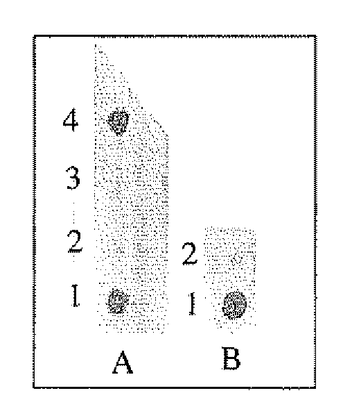
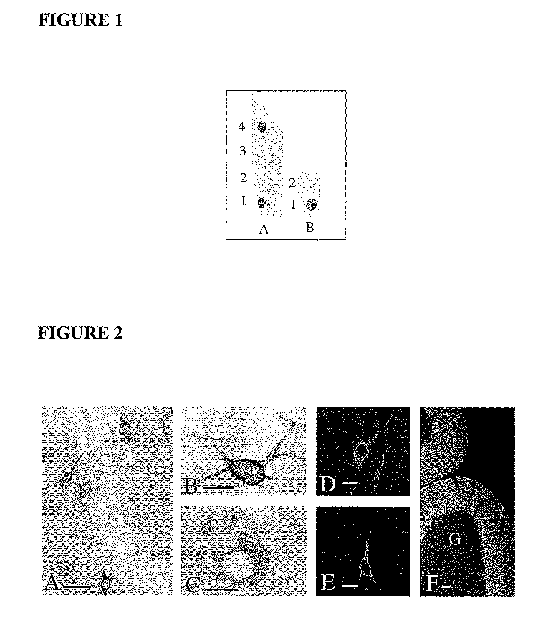
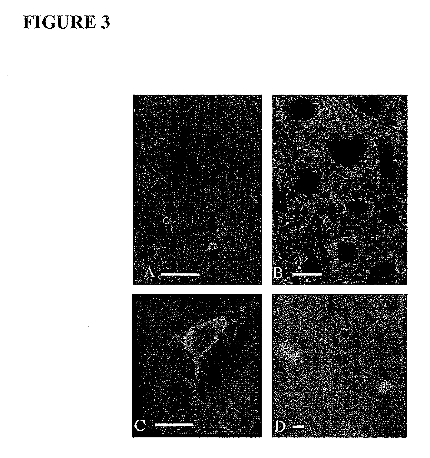
[0068]Antibodies were raised against a unique amino acid synthetic peptide corresponding to the amino acids encoded by the splice site between exons 8 and 10 of GLAST to enable selective detection of GLAST1b (the antibodies are available from Prof. David Pow, The University of Newcastle, Newcastle, NSW, Australia). The aim of the present study was to determine if GLAST1b was present in the CNS and if so, its cellular compartmentalization.

1. Methods

[0069]Animal experiments complied with the guidelines of the National Health and Medical Research Council (NHMRC, Australia). Antisera were generated in rabbit [14], using the unique 11 amino acid peptide H2N-QIITIRDRLRT (SEQ ID No. 1) of GLAST1b (referred to hereafter as peptide 1), which spans the splice region between exons 8 and 10, (see Macnab L T and Pow D V, (2007) [24], the contents of which is incorporated herein in its entirety by cross-reference). The peptide was coupled to porcine thyroglobulin, (S...

example 2

Expression of GLAST1b in Pig Brain

[0084]The distribution of GLAST1b in the hypoxic neonatal pig brain was examined. In this model, the damage is variable between animals as assessed by independent blind scoring conducted by histological analysis on cresyl violet stained sections. Some animals typically experience only damage to white matter whilst others experience damage to either restricted regions of grey matter or in the most severe cases, to large areas of grey and white matter.

2.1 Methods

[0085]Animal experiments complied with the guidelines of the National Health & Medical Research Council (NHMRC) (Australia).

2.1.1 Animal Preparation

[0086]One day old pigs were anaesthetised using propofol (10 mg / kg / h) and alfentanil (50 μg / kg / h) iv. The pigs were intubated and ventilated using a neonatal ventilator, with oxygen and air to maintain arterial CO2 at 35-45 mmHg and oxygen saturation 92-96%. A radiant warmer was used to maintain rectal temperature at 39.0±0.5° C. Following stabilis...

example 3

D-glutamate is Accumulated by GLAST1b

[0112]A study was undertaken to evaluate accumulation of D-glutamate by GLAST1b. Briefly, hypercanic hypoxia was induced in one day old pigs essentially as described in Example 2.1.1. Control pigs were subjected to anaesthesia but no hypoxia and also allowed to recover for 72 hours as described above. The pigs were euthanased by an overdose of sodium pentobarbital, and the brains rapidly removed and placed into ice cold oxygenated artificial cerebrospinal fluid (CSF) (Ames media). 250 μm-thick slices were to room temperature before warming to 36° C., for the performance of transport studies. The temperatures used were slightly higher that those typically used for electrophysiology, and thus closer to physiological normality as transporter activity is greatly reduced if the temperature is significantly lowered. The neuroprotective effects of hypothermia that are evident at lower temperatures are also avoided since they are contraindicated in these...


PUM


Abstract

Assays and kits for detecting aberrant cells of neuroectodermal origin in a body fluid of an individual, comprising testing for expression of GLAST1b as a biomarker of the cells are disclosed. Intact GLAST1b and / or fragments thereof may be detected in the fluid. Alternatively, another analyte indicative of the expression of GLAST1b by the cells may be detected. The assay is particularly suitable for detecting expression of aberrant neuronal populations such as resulting from brain hypoxia. The fluid can be cerebrospinal fluid (CSF).

Description

CROSS-REFERENCE TO RELATED APPLICATIONS[0001]This application claims the benefit under 35 U.S.C. §119 of Australian provisional application No. 2008901400, filed Mar. 22, 2008, and U.S. provisional application Ser. No. 61 / 120,695, filed Dec. 8, 2008, the entire contents of which are incorporated herein by reference.FIELD OF THE INVENTION[0002]The invention provides methods for the detection of GLAST1b in a body fluid of an individual as a biomarker of aberrant cells of neuroectodermal origin. The methods have application, although not exclusively, in evaluating the extent of damaged, degenerating or dying neurons and / or glial cells as a result of injury, trauma or neurological diseases or conditions.BACKGROUND OF THE INVENTION[0003]Brain hypoxia is a patho-physiological condition characterised by a decrease of oxygen supply to the brain. It is caused by reduced blood supply or blood in which there is low oxygen concentration. The lack of oxygen impairs several highly energy-dependen...

Claims


Application Information

Patent Timeline
IPC IPC(8): G01N33/53
CPCG01N33/6896G01N2800/28G01N2333/705
Inventor POW, DAVID V.
Owner NEWCASTLE INNOVATION OF IND DEV CENT
Learn More