
Secreted Luciferase Fluorescent Protein Conjugate Nucleic Acid Construct and Uses Thereof

a technology of conjugate nucleic acid and secreted luciferase, which is applied in the field of biological system real-time analysis system, biological system material and method, can solve the problem of inability to quantitate the efficiency of secretion

Inactive Publication Date: 2010-02-11
THE GENERAL HOSPITAL CORP
View PDF1 Cites 5 Cited by
  • Summary
  • Abstract
  • Description
  • Claims
  • Application Information

AI Technical Summary

Benefits of technology

[0009]The present invention is based, on part, on the discovery that a secreted form of luciferase conjugated to a fluorescent protein is capable of acting as a dual reporter for the assessment of biological processes in cells. The analysis of the secreted form of the luciferase-fluorescent protein conjugate can be monitored without the need to lyse the cells. This permits the convenient detection, using conventional calorimetric methods and / or fluorescent assays, of the luciferase-fluorescent protein conjugate produced by the cell at different time points. Therefore, one embodiment is a method for real-time monitoring of biological systems using the luciferase-fluorescent protein conjugate protein.
[0015]In one embodiment, the methods of the invention can be used for monitoring the protein secretion or protein secretory pathway. In such an embodiment, the methods of the invention described herein are advantageous over previous secretory pathway assays, in that the methods of the present invention enable real time analysis of the secretory pathway by combining the visualization of the Gaussia luciferase-fluorescent protein conjugate with quantitation of the level of secretion of Gaussia luciferase-fluorescent protein conjugate. The methods in such an embodiment enable monitoring of the primarily entry of the secreted protein (in this case Gaussia luciferase-fluorescent protein conjugate) into the endoplasmic reticulum (ER) and co-localization with ER, as assessed by a punctuate localization pattern which co-staining with ER markers. Thus, the methods of the invention have the advantage of analyzing the processing of a secreted protein (in this case Gaussia luciferase-fluorescent protein conjugate) at distinct positions in the secretory pathway, namely the ER entry points, combined with the ability to monitor quantitatively the level of secretion by bioluminescence. Thus, the methods of this invention are the most sensitive assay to analyze the secretory pathway in living cells.

Problems solved by technology

There are several advantages to this system in that the movement of the protein can be synchronized by temperature shifts and visualized by fluorescent microscopy, with the disadvantage that the efficiency of secretion can not be quantitated.
However, to date such a system that efficiently accomplishes the goals of dual quantification with visualization of protein secretion in real time has not been described.

Method used


Image

  • Secreted Luciferase Fluorescent Protein Conjugate Nucleic Acid Construct and Uses Thereof
  • Secreted Luciferase Fluorescent Protein Conjugate Nucleic Acid Construct and Uses Thereof
  • Secreted Luciferase Fluorescent Protein Conjugate Nucleic Acid Construct and Uses Thereof

Examples


example 1

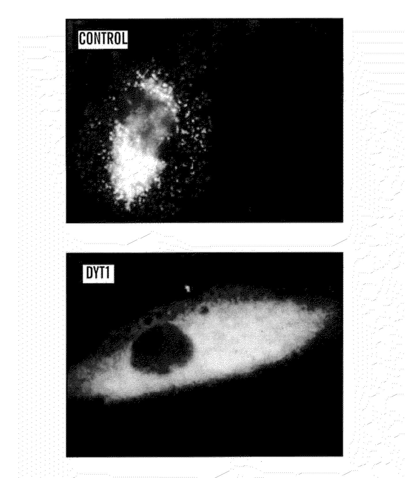
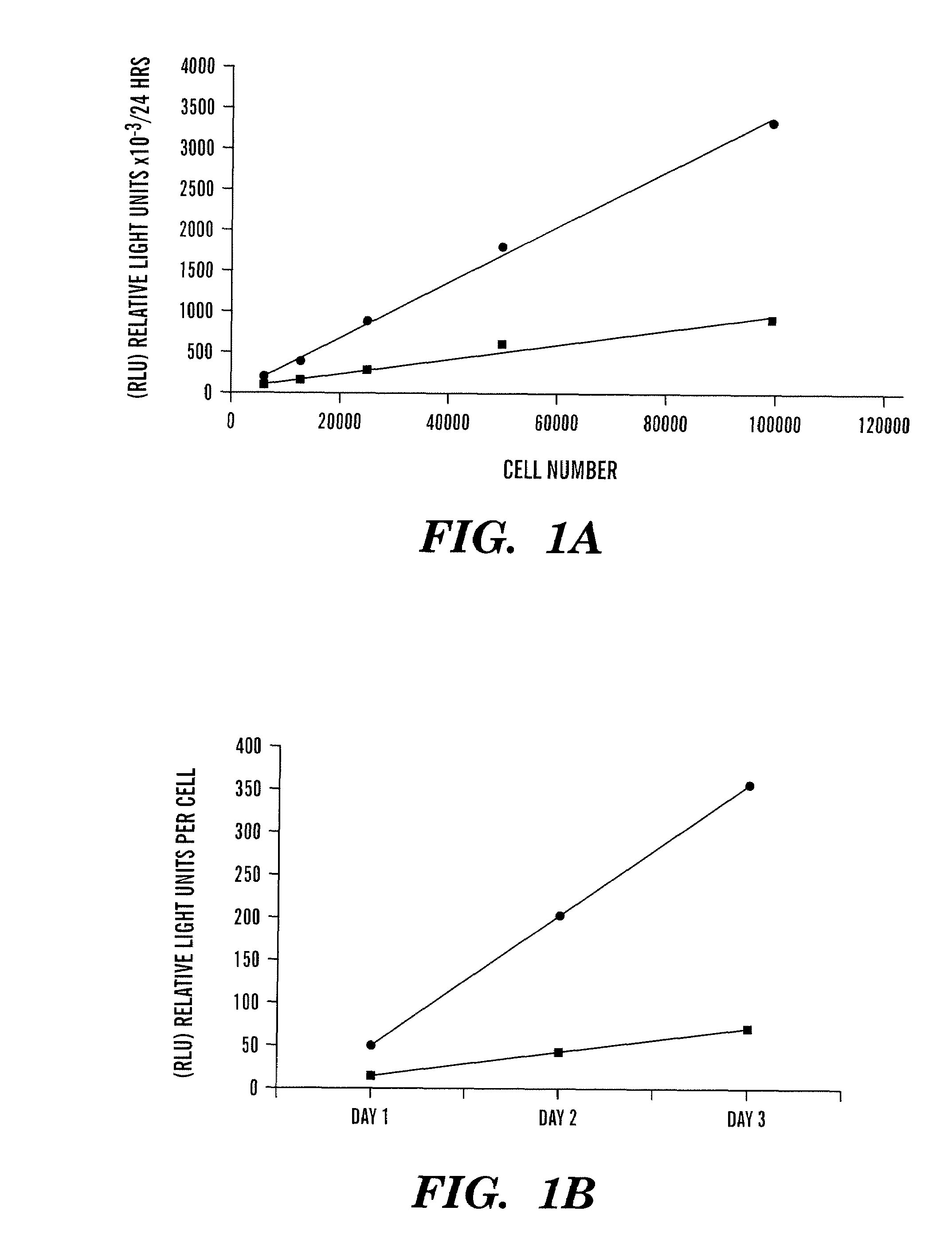
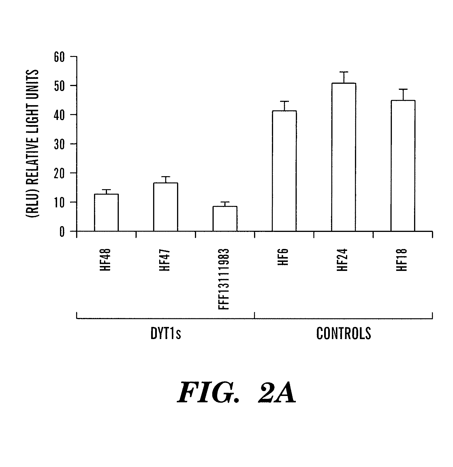
[0151]Monitor the transit of proteins through secretory pathway. Gluc-YFP fusion protein is used as reporter protein to monitor processing through the secretory pathway. As an illustrative example, the processing of Gluc-YFP was assessed in fibroblasts from normal patients and from patients affected with DYT1, an early onset torsion dystonia that is a dominantly inherited movement disorder characterized by sustained, involuntary muscle contractions and abnormal posturing (Fahn, 1988). A specific mutation underlies most cases of DYT1 dystonia—a GAG deletion in the coding region of the DYT1 gene encoding torsinA which results in a loss of a glutamic acid residue (ΔE302 / 303) in the carboxy terminal region of the protein (Ozelius et al., 1997; Kabakci et al., 2004).

[0152]Levels of Gluc activity were measured in cells and media, and the intracellular location was visualized using a Gluc-yellow fluorescent protein (Gluc-YFP) fusion protein following infection of cells with a lentivirus ve...

example 2

[0162]Monitor Promoter Activity. The Gluc assay can also be used to monitor promoter activity with high sensitivity and extended linear range spanning over 7 orders of magnitude. When the Gaussia luciferase cDNA which is codon optimized for mammalian gene expression (hGluc) is expressed under a promoter e.g. CMV, the activity of this promoter can be easily measured overtime by taking an aliquot of the supernatant, adding coelenterazine and measuring the photon counts using a luminometer (FIG. 7). Since the Gaussia luciferase is naturally secreted, the assay is performed on small samples of the conditioned media, with no need to lyse the cells, which makes it much faster and more convenient than assays with other luciferases such as firefly which is used in the SUPERLIGHT™ luciferase reporter gene assay (Bioassays, CA) where cell lysis is required. Since the cells are not disrupted, conditioned media can be sampled repeatedly for time course studies and cells can be used for further ...

example 3

[0163]Monitoring signaling pathway and transcriptional activation. The Gluc assay can also be used to monitor signaling pathways and transcriptional activation. The following promoters responsive to different transcription factors were cloned upstream of hGluc cDNA: the wild-type p53-activated fragment 1 (WAF1, 2.4 kb); early growth response factor (Egr-1); and five tandem repeats of the transcriptional factor nuclear factor-κB (5NFκB). Upon transfection with each of these constructs, the activity of each promoter could be monitored by taking an aliquot of the cell medium, adding coelenterazine and measuring photon counts using a luminometer (FIG. 8). Further, the NFκB response to ionizing-radiation and to radiomimetic drugs, such as bleomycin, and chemotherapeutic drugs, such as doxorubicin and etoposide could also be monitored overtime in real-time using hGluc under control of the 5NFκB (FIG. 9).


PUM

PropertyMeasurementUnit
temperatureaaaaaaaaaa
volumeaaaaaaaaaa
pHaaaaaaaaaa

Abstract

The present invention relates generally to methods to monitor the transport of proteins through the secretory pathway, and methods to monitor ER stress. In particular, the present invention relates to methods to monitor, in real-time, the processing of protein through the secretory pathway, which can be monitored both at a subcellular level by florescence visualization and quantitatively by detecting the secreted luciferase reporter protein. The present invention also relates to methods to assess biological processes in cells, in particular the secretory pathway and ER stress, as well as methods to identify agents which augment or inhibit the secretory pathway and / or ER stress. The present invention also relates to compositions and nucleic constructs encoding a secreted luciferase-fluorescent protein conjugate for methods to monitor protein trafficking in the cell by simultaneous detection of fluorescence and luciferase secretion.

Description

CROSS REFERENCED APPLICATION[0001]This application is a 371 National Phase Entry Application of co-pending International Application PCT / US2007 / 021810 filed Oct. 12, 2007, which designated the U.S., and claims the benefit under 35 U.S.C 119(e) of U.S. Provisional Patent Application Ser. No. 60 / 851,110 filed Oct. 12, 2006, the contents of which are incorporated herein by reference in their entirety.GOVERNMENT SUPPORT[0002]This invention was made with Government support under NS28384 and 5P01 NS037409 awarded by the National Institutes for Health (NIH) and National Institute of Neurological Disorders and Stroke (NINDS). The Government of the United States has certain rights in the invention.FIELD OF THE INVENTION[0003]The invention relates to materials and methods for the identification and assessment of biological systems. More particularly, the invention relates to a system for real-time analysis of biological systems using a secreted luciferase and a fluorescent protein conjugate a...

Claims


Application Information

Patent Timeline
Patent Type & Authority Applications(United States)
IPC IPC(8): C12Q1/66C07H21/00
CPCC07K2319/60C12Q1/66C12N2799/027
Inventor TANNOUS, BAKHOS A.BREAKEFIELD, XANDRAHEWETT, JEFFREY W.
Owner THE GENERAL HOSPITAL CORP
Learn More