Spectroscopically enhanced imaging

a spectroscopic enhancement and imaging technology, applied in the direction of spectroscopy diagnostics, instruments, catheters, etc., can solve the problems of rarely identifying normal tissue as cancerous, unnecessary biopsies, etc., and achieves less expensive, high noise level, and sufficient dispersion

Inactive Publication Date: 2010-03-18
NEWTON LAB
View PDF26 Cites 22 Cited by
  • Summary
  • Abstract
  • Description
  • Claims
  • Application Information

AI Technical Summary

Benefits of technology

[0012]The delivery fibers can be used to simply indicate the area of the tissue that is being analyzed. Alternatively, the delivery fibers can be used to couple narrow-band laser light into the tissue at those points on the tissue where the distal tips of the delivery fibers are imaged. The collection fibers are imaged at different spots on the tissue, separate from those areas where the narrow-band laser light enters the tissue. The scattering through the tissue can thus be measured. The local hemoglobin concentration can be measured by comparing the scattering in the tissue at several wavelengths, specifically where hemoglobin absorption is significant and at wavelengths where it is not significant. Imaging spectrometers can separate the light exiting one collection fiber from another and have sufficient dispersion to separate laser sources from each other. In the preferred embodiment of this detection system three delivery fibers, three collection fibers and six laser wavelengths are used to obtain 18 different combinations of wavelength and scattering distance in a single exposure. This allows a much more precise measurement of both the scattering spectrum in the tissue and the hemoglobin concentration in the tissue. Superior measurements will yield more precise predictions of the likely presence or absence of cancer.
[0013]Imaging spectrometers and thermo-electrically-cooled, two-dimensional CCD's are sensitive but relatively slow because of the time required to digitize the signal in each pixel. Faster CMOS imaging arrays are available but can have higher noise levels. When a high resolution spectrum is not required or when the illumination source is a laser, the detectors can be made with optical filters and high speed photomultipliers. These detection systems can return quantified results in less than a second which may be important if measurements need to be taken quickly in succession, such as for comparing measurements in one tissue area to measurements in a neighboring area. A preferred embodiment of this type of detection system utilizes three delivery fibers, a plurality of light sources such as, six laser light sources, three collection fibers and a rotating three-color filter wheel. The same 18 combinations of scattering distances and colors described in imaging spectrometer system above can be obtained in a smaller, less expensive package and with a reduced collection time.

Problems solved by technology

Fewer unnecessary biopsies would be taken, however, if the method also had high specificity, meaning that it would rarely identify normal tissue as cancerous.

Method used


Image

  • Spectroscopically enhanced imaging
  • Spectroscopically enhanced imaging
  • Spectroscopically enhanced imaging

Examples


Embodiment Construction

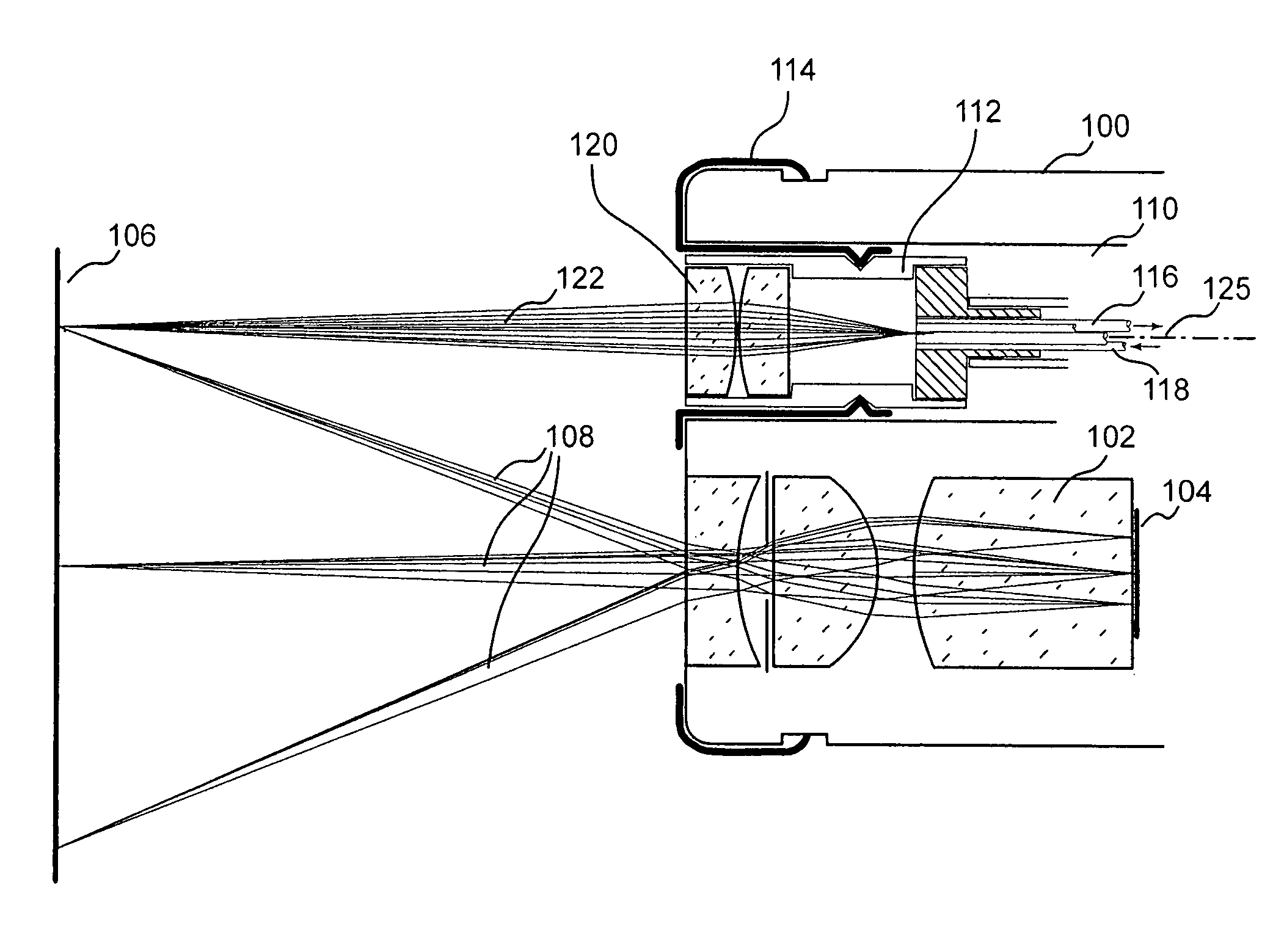
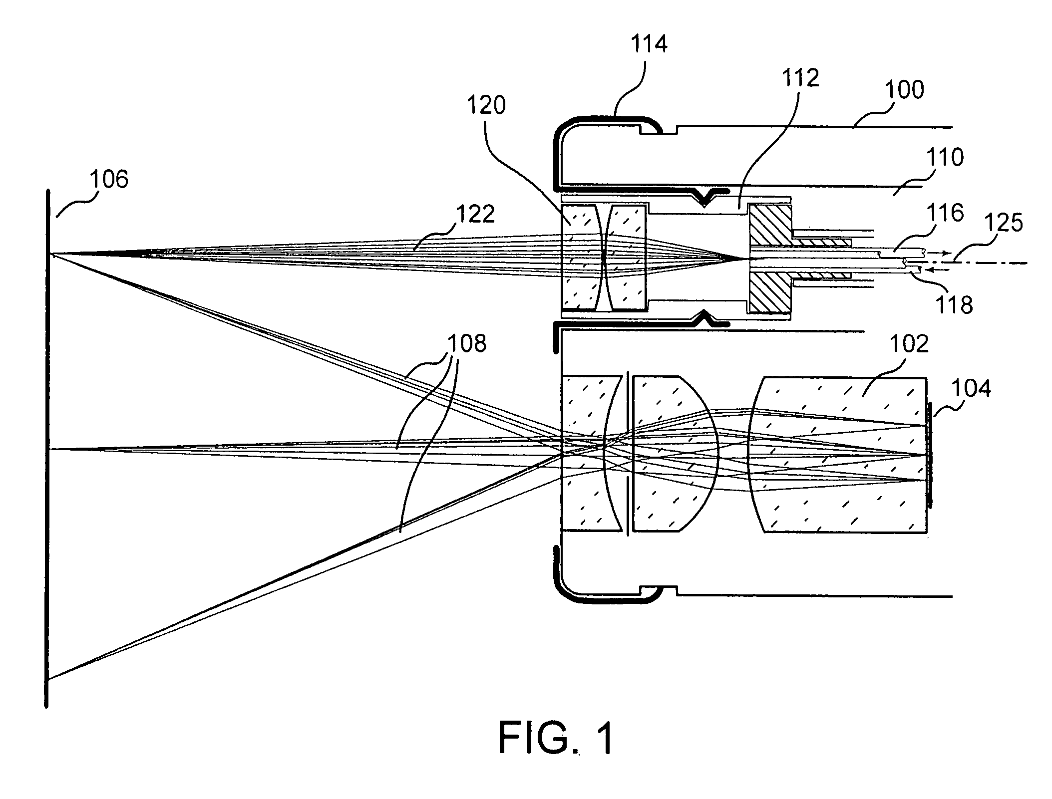
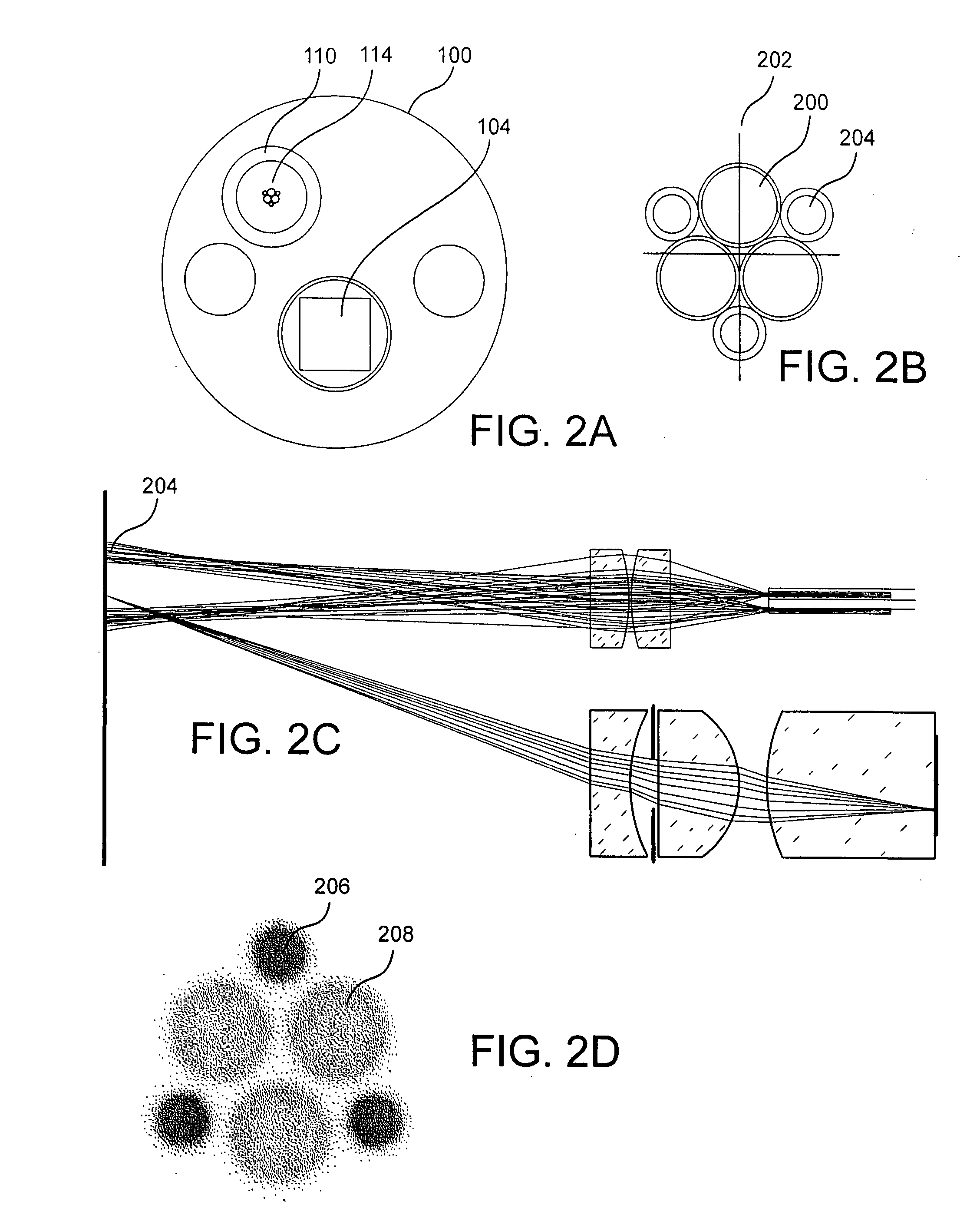
[0038]Autofluorescence endoscope systems to date demonstrate high sensitivity for the detection of cancerous or precancerous lesions. These areas are indicated by a reduction in the level of tissue autofluorescence. Visual detection of such regions is straightforward but often results in false positive readings since there are benign conditions which can cause the same effect. A method which results in a high number of false positive readings is described as one with low specificity. To improve the specificity of autofluorescence endoscopy additional information beyond a visual assessment of the reduction in fluorescence intensity can be taken. Spectral information, resulting from the dispersion or filtering of the intrinsic fluorescence and / or white light reflected from the tissue has been shown to be effective at diagnosing cancerous tissue. Similarly, information available from measurements of light scattering in the tissue can be used to classify tissue types and measure the con...


PUM


Abstract

The present invention provides systems and methods for the spectroscopic determination of the physical characteristics of the tissue under observation by an autofluorescence or other endoscope without the requirement of contacting the tissue directly. The optical probe contained in the endoscope itself is passive and may be either built into the endoscope or positioned in a biopsy channel of same. The spectroscopic information, combined with other information provided by the endoscope such as total fluorescence, improves the sensitivity and specificity of the identification of precancerous or cancerous lesions.

Description

CROSS REFERENCE TO RELATED APPLICATIONS[0001]This application claims the priority of U.S. Provisional Application No. 60 / 861,871 filed on Nov. 30, 2006 and entitled SPECTROSCOPICALLY ENHANCED IMAGING; and U.S. Provisional Application No. 60 / 874,650 filed on Dec. 13, 2006 and entitled SPECTROSCOPICALLY ENHANCED IMAGING, which are hereby incorporated by reference herein.STATEMENT REGARDING FEDERALLY SPONSORED RESEARCH OR DEVELOPMENT[0002]N / ABACKGROUND OF THE INVENTION[0003]Autofluorescence imaging endoscopes can detect precancerous and cancerous lesions in the lung, colon and other body areas. Normal tissue, when illuminated with ultraviolet or violet light, will emit relatively weak fluorescence in the visible spectrum. This autofluorescence can be imaged by endoscopes which are not sensitive to, or which filter out, the much stronger excitation light. Precancerous and cancerous tissue, for a number of reasons such as increased hemoglobin concentration, exhibit reduced fluorescence w...

Claims


Application Information

Patent Timeline
Patent Type & Authority Applications(United States)
IPC IPC(8): A61B1/06
CPCA61B1/00167A61B1/00188A61B1/018A61B1/043A61B1/05A61B1/0638A61B1/00096A61B5/0075A61B5/0084G01N21/474G01N21/645G01N21/6486A61B1/07
Inventor FULGHUM, STEPHENVON ROSENBERG, CHARLESFELD, MICHAEL
Owner NEWTON LAB
Why Eureka
Try Eureka