Pig model for breast cancer, mitochondria related protein folding disorders and/or epidermolysis bullosa simplex

a technology of epidermolysis and mitochondria, applied in the field of genetically modified pigs, can solve the problems of affecting the ability of the organelle to produce energy, affecting the survival rate of the pig, and the majority of breast cancer incidence remains unexplained

Inactive Publication Date: 2010-05-06
AARHUS UNIV
View PDF1 Cites 3 Cited by
  • Summary
  • Abstract
  • Description
  • Claims
  • Application Information

AI Technical Summary

Benefits of technology

[0051]A tenth aspect relates to a method for screening the efficacy of a pharmaceutical composition, said method comprising the steps of
[0052]A eleventh aspect relates to a method for screening the efficacy of a pharmaceutical composition, said method comprising the steps of
[0053]A eleventh aspect relates to a method for treatment of a human being suffering from breast cancer, said method comprising the initial steps of
[0054]Mitochondria Related Protein Folding Disorder...

Problems solved by technology

However, their value as animal models for many human diseases is quite limited due to differences in mice compared to humans.
Although many epidemiological risk factors, and biological co-factors and promoters have been identified, the majority of breast cancer incidence remains unexplained, and the primary cause is still unknown.
Oxidative phosphorylation generates most of the cell's ATP, and any impairment of the organelle's ability to produce energy can have serious consequences.
Moreover, deficient mitochondrial metabolism may generate reactive oxygen species, which is extremely deleterious for the cell.
The mutated enzyme results in impairment of the reaction that leads to condensation of carbamyl phosphate and ornithine to form citrulline.
This impairment leads to reduced ammonia incorporation, which, in turn, causes symptomatic Hyperammonemia.
However, the relationship between the protein misfolding, which often occurs outside the mitochondria, and the mitochondrial dysfunction remains unclear.
Signs and symptoms can include irritability, depression, small involuntary movements, poor coo...

Method used


Image

  • Pig model for breast cancer, mitochondria related protein folding disorders and/or epidermolysis bullosa simplex
  • Pig model for breast cancer, mitochondria related protein folding disorders and/or epidermolysis bullosa simplex
  • Pig model for breast cancer, mitochondria related protein folding disorders and/or epidermolysis bullosa simplex

Examples


example 1

[0328]Differences in developmental competence between sow (2.5 years, 170 Kg in weight) derived oocytes and gilt (5.5˜6 months, 75 Kg in weight) derived oocytes were investigated through ZF and ZI PA after 44 h in vitro maturation. Four combined groups were investigated in 3 identical replicates: (1) ZF oocytes from sows (2) ZI oocytes from sows (3) ZF oocytes from gilts (4) ZI oocytes from gilts. For ZF activation, a single DC pulse of 0.85 KV / cm for 80 μs was applied, while a single 1.25 KV / cm pulse was used to activate ZI oocytes. Following 7 days culture as described above, the percentage of blastocysts per activated embryo was determined.

[0329]The in vitro developmental competence of parthenogenetically activated oocytes derived from either sows or gilts was investigated. As shown in Table 1, the blastocyst rates of parthenogenetically activated oocytes from sows were significantly higher than those from gilts, either after ZF or ZI PA.

TABLE 1Blastocyst development of Day 7 par...

example 2

[0331]The feasibility of modified porcine HMC was investigated in 6 identical replicates, with IVF and in parallel ZF PA as controls. The more competent sow oocytes (according to Example 1) were used in Example 2. Seven days after reconstruction and / or activation, the number of blastocysts per reconstructed embryo and total cell numbers of randomly selected blastocysts were determined.

[0332]More than 90% of oocyte fragments derived from morphologically intact oocytes could be recovered for HMC after the trisection. In average, 37 embryos could be reconstructed out of 100 matured oocytes. The developmental competence of all sources of porcine embryos is shown in Table 2. On Day 7, the development of reconstructed embryos to the blastocyst stage was 17±4% with mean cell number of 46±5, while the blastocyst rates for IVF, and ZF PA were 30±6% and 47±4% (n=243, 170, 97) respectively.

TABLE 2In vitro development of embryos produced by HMC, IVF and ZF PANo. ofblastocystMean cellEmbryoembry...

example 3

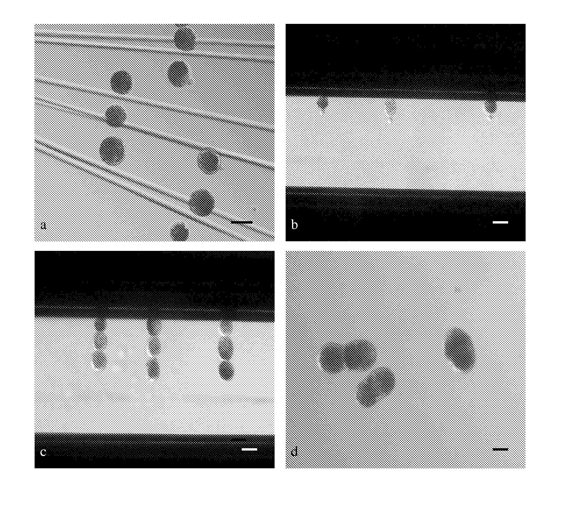
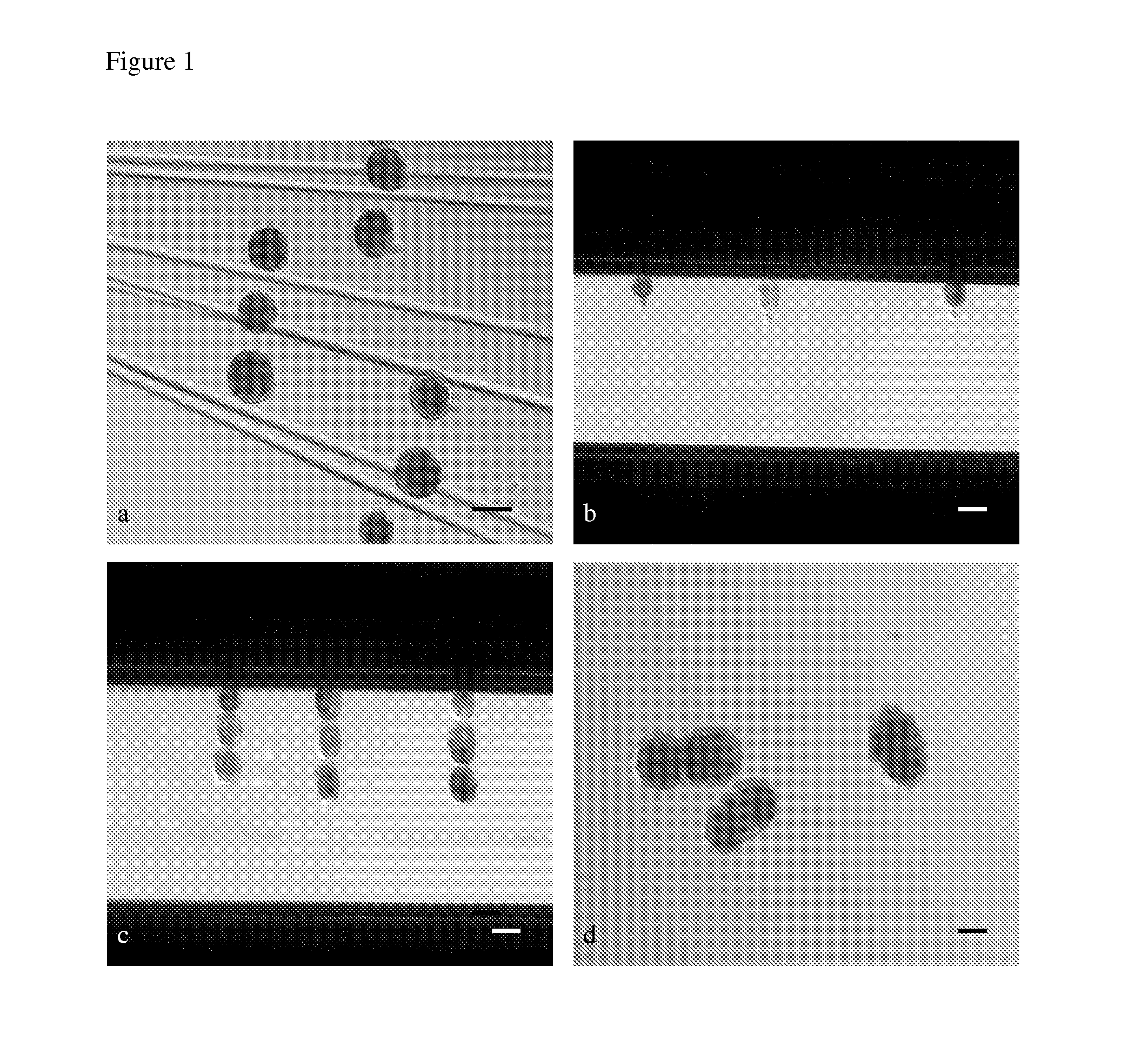
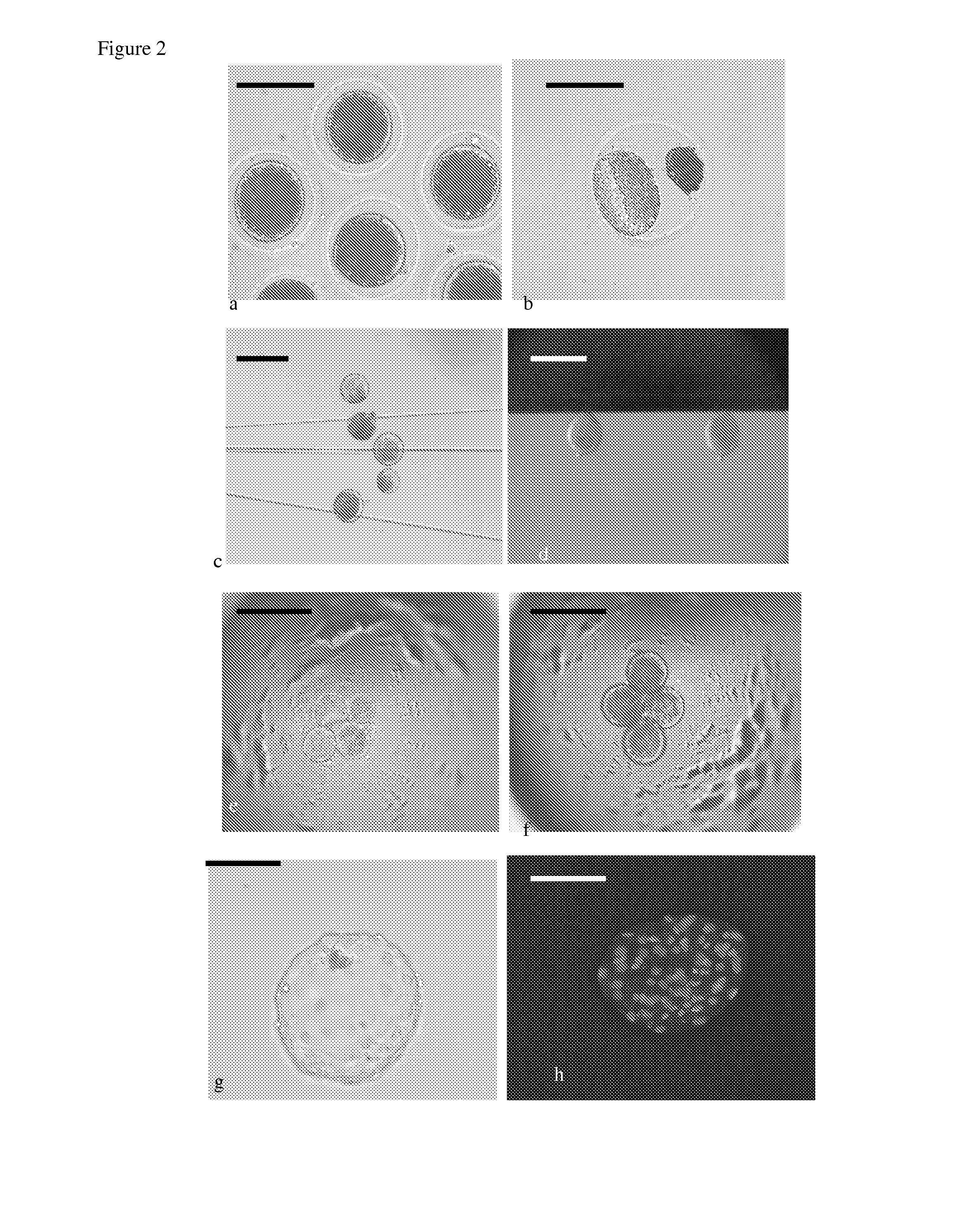
[0335]Vitrification of hand-made cloned porcine blastocysts produced from delipated in vitro matured oocytes.

[0336]Recently a noninvasive procedure was published for delipation of porcine embryos with centrifugation but without subsequent micromanipulation (Esaki et al. 2004 Biol Reprod. 71, 432-6).

[0337]Cryopreservation of embryos / blastocysts was carried out by vitrification using Cryotop (Kitazato Supply Co, Fujinomiya Japan) as described previously (Kuwayama et al. 2005a; 2005b). At the time of vitrification, embryos / blastocysts were transferred into equilibration solution (ES) consisting of 7.5% (V / V) ethylene glycol (EG) and 7.5% dimethylsulfoxide (DMSO) in TCM199 supplemented with 20% synthetic serum substitute (SSS) at 39° C. for 5 to 15 min. After an initial shrinkage, embryos regained their original volume. 4-6 embryos / blastocysts were transferred into 20 ul drop of vitrification solution (VS) consisting of 15% (V / V) EG and 15% (DMSO) and 0.5M sucrose dissolved in TCM199 su...


PUM

PropertyMeasurementUnit
Fractionaaaaaaaaaa
Fractionaaaaaaaaaa

Abstract

The present invention relates to a genetically modified pig as a model for studying breast cancer, mitochondria related protein folding disorders and/or epidermolysis bullosa simplex. The modified pig model displays one or more phenotypes associated with any of said disorders. Disclosed is also a modified pig comprising a modified endogeneous BRCA1 and/or BRCA 2 gene, and/or a modified ornithine transcarbamylase gene, and/or a modified Keratin 14 gene and/or a transcriptional or translational product or part thereof. The invention further relates to methods for producing the modified pig; and methods for evaluating the effect of a therapeutical treatment of breast cancer, mitochondria related protein folding disorders and/or epidermolysis bullosa simplex; methods for screening the efficacy of a pharmaceutical composition; and a method for treatment of a human being suffering from breast cancer, mitochondria related protein folding disorders and/or epidermolysis bullosa simplex are disclosed.

Description

FIELD OF INVENTION[0001]The present invention relates to a genetically modified pig as a model for studying breast cancer, mitochondria related protein folding disorders and / or epidermolysis bullosa simplex, wherein the pig model expresses at least one phenotype associated with said disease. The invention further relates to methods by which the genetically modified pig is produced. In addition, methods for evaluating the response of a therapeutical treatment of breast cancer, mitochondria related protein folding disorders and / or epidermolysis bullosa simplex, for screening the efficacy of a pharmaceutical composition, and a method for treatment of human being suffering from breast cancer, mitochondria related protein folding disorders and / or epidermolysis bullosa simplex are disclosed.BACKGROUND OF INVENTION[0002]Transgenic, non-human animals can be used to understand the action of a single gene or genes in the context of the whole animal and the interrelated phenomena of gene activ...

Claims


Application Information

Patent Timeline
IPC IPC(8): G01N33/00A01K67/00C12N15/00
CPCA01K67/0271A01K67/0275A01K2217/052A01K2227/108A01K2267/0331C12N15/8509C12N15/8778C12N2800/30C12N2800/90
Inventor KRAGH, PETER MICHAELBOLUND, LARS AXELSORENSEN, CHARLOTTE BRANDTCORYDON, THOMAS JUHLGREGERSEN, NIELSBROSS, PETER
Owner AARHUS UNIV
Why Eureka
Try Eureka