Intraoperative Tissue Mapping and Dissection Systems, Devices, Methods, and Kits

a tissue mapping and tissue technology, applied in the field of surgical navigation and control, can solve the problems of consuming significant surgical time, affecting the accuracy of surgical results,

Inactive Publication Date: 2010-07-01
TRILLIUM PRECISION SURGICAL
View PDF17 Cites 240 Cited by
  • Summary
  • Abstract
  • Description
  • Claims
  • Application Information

AI Technical Summary

Benefits of technology

[0020]An intraoperative device for mapping an area of tissue during a surgical procedure is also provided. This device comprises at least one excitation element for interacting with a tissue of interest with a stimulus to generate a detectable signal; a remote interrogation element; and a marking element for creating a detectable mark associated with a location on a tissue. The detectable signal can be, for example, a measure of the electrical impedance measured between the excitation element and the remote interrogation element. Additionally, the excitation element can be moveable. Furthermore, in some aspects, the detectable signal can be a measure of the electrical impedance measured between the remote interrogation element and a plurality of moveable excitation electrodes. The marking element or elements can be adapted and configured to create the detectable mark associated with the location of a tissue of interest. Furthermore, the excitation element stimulus for stimulating the tissue of interest can be any of a variety of suitable stimulus, including, for example, electrical stimulation, magnetic stimulation, mechanical stimulation, acoustic stimulation, optical stimulation, thermal stimulation, electromagnetic stimulation, mechanical vibration, ultrasound, stimulus arrays, and imaging. The detectable signal detected can be an electrical signal, a mechanical signal, an electromagnetic signal, a magnetic signal, a thermal signal, ultrasound, detection arrays, imaging methods, or any other suitable detectable signal. Additionally, the detectable mark could, in some cases, be a surface cautery of the tissue of interest, and / or a dye selected from India ink, Prussian blue, crystal violet, or any other suitable dye. In some instances, fluorescent dye or radioactive material may be desirable for use as a detectable mark. In other cases, the detectable mark might be a particle, a quantum dot, a carbon nanotube, a paramagnetic particle, a ferromagnetic particle, a metallic particle, a radioactive particle, or a colored particle. Paraffin wax, wax, sucrose solution, or any other suitable material that forms a gel upon deposition are also suitable for use as a detectable mark. Furthermore, optical marks, or marks made by an electrochemical reaction can be used without departing from the scope of the invention. The marking element can be further adapted and configured to create multiple detectable marks on the tissue of interest. Each of the multiple detectable marks can further identify multiple different types of tissues. Furthermore, different types of marking elements can be used to mark each of the discrete tissue types such that the sensory signal enables the user to distinguish between the different tissue types.
[0031]Still another method is provided for selectively removing tissue at a surgical site. This method comprises: positioning a reference element within or adjacent to a tissue of interest; probing the area surrounding the tissue of interest using a probe element wherein a dissection element is operably connected to the probe element; detecting the location of the reference element with the probe element; and dissecting the tissue the reference element thereby protecting the tissue nearest the reference element.

Problems solved by technology

Veins, arteries and nerves can be difficult to distinguish from one another in situ.
This approach fails when proper dissection planes are not identified.
Searching for the location of specific nerves and vessels can consume significant surgical time and carries the risk of injury to delicate structures.
Current practice relies upon anatomical landmarks, but anatomical variations, disease, trauma and scar tissue can slow the process of identifying anatomical features and increase the risk of injury.
Unintended damage to these structures can result in significant complications.
Trauma to sensor nerves can result in numbness and loss of function.
Damage to motor nerves can result in loss of function.
Identification of the facial nerve matrix is critically important and the nerve branching within the parotid can be quite complex.
However, with this approach the risk of nerve trauma is elevated further because unambiguous identification of nerve segments is difficult.
Other examples of surgeries that have significant risk of collateral damage, include, for example, pelvic surgery, spine surgery, and radical prostatectomy which carries a significant risk of impotence and the potential for incontinence due to trauma to the nerves adjacent or surrounding the prostate and trauma to the urethral sphincters.
Locating the ureter during pelvic surgery can be time consuming and carries the risk of injury, particularly when there is scarring or tumors in the adjacent or surrounding tissue.
As will be appreciated by those skilled in the art, injury to the ureter during pelvic surgery can result in impaired function, infection, and other complications.
Surgical procedures can result in potentially avoidable complications.
Intraoperative ultrasound has also been employed to identify structures, but does not discern between specified vessels and hasn't been employed for closed loop control of ablation.

Method used


Image

  • Intraoperative Tissue Mapping and Dissection Systems, Devices, Methods, and Kits
  • Intraoperative Tissue Mapping and Dissection Systems, Devices, Methods, and Kits
  • Intraoperative Tissue Mapping and Dissection Systems, Devices, Methods, and Kits

Examples


example 1

Sample Tissue

[0115]An example proximity system 900 is illustrated in FIG. 14. In this example, the proximity system uses a modified Parkell Sensimatic Electrosurge 500-SE 904 dissection element which was modified so that the activation pedal 910 was in series with a reed relay 909. A Pepper & Fuchs NBN4 inductive proximity sensor element 907 was then affixed to a wire electrode 905 that was connected to the active output of 904 and covered with an insulator 906. Fresh raw chicken breast 1, was used as a test tissue sample and was rested on a grounding electrode 911 and a steel plate reference element 902. The sensing element 907 was expected to be triggered by the reference element 902 at the proximity limit 903. Output of the sensing element 907 was connected to an operational amplifier 908 configured as an inverter and to the +12 V voltage by the 220 k ohm resistor 914. The output of 908 was connected to the relay 909. When the sensor element 907 was not triggered the output of th...

example 2

Prostatic Urethra

[0116]Another proximity system could be adapted from the proximity system in FIG. 14, using a Foley catheter with, for example, aluminum film coating. The modified catheter would then be inserted into the urethra and would serve as the reference element. This system would be used to preserve the prostatic urethra during a modified radical prostatectomy procedure where the prostate is resected but the urethra is left intact. As noted elsewhere this would have the advantage of minimizing trauma to the urethra and associated structures and is expected to result in shorter recovery times and less urinary incontinence and other complications.

example 3

Radical Prostatectomy

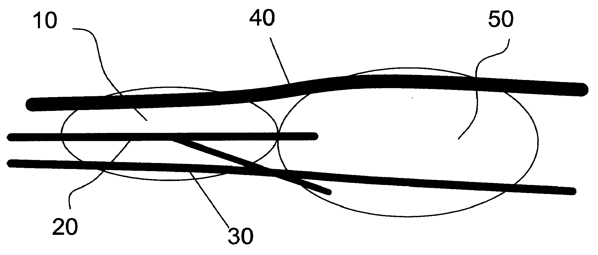
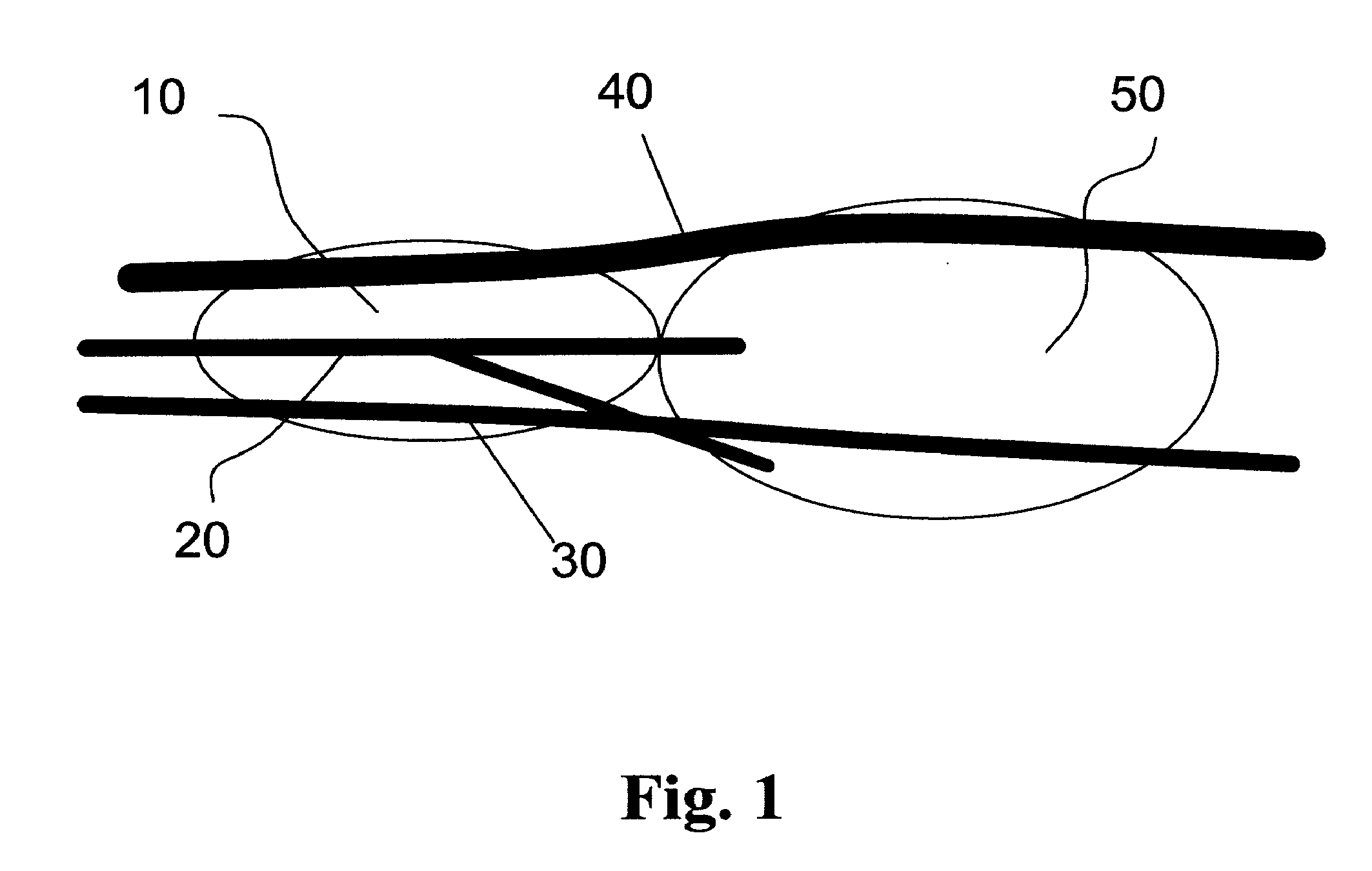
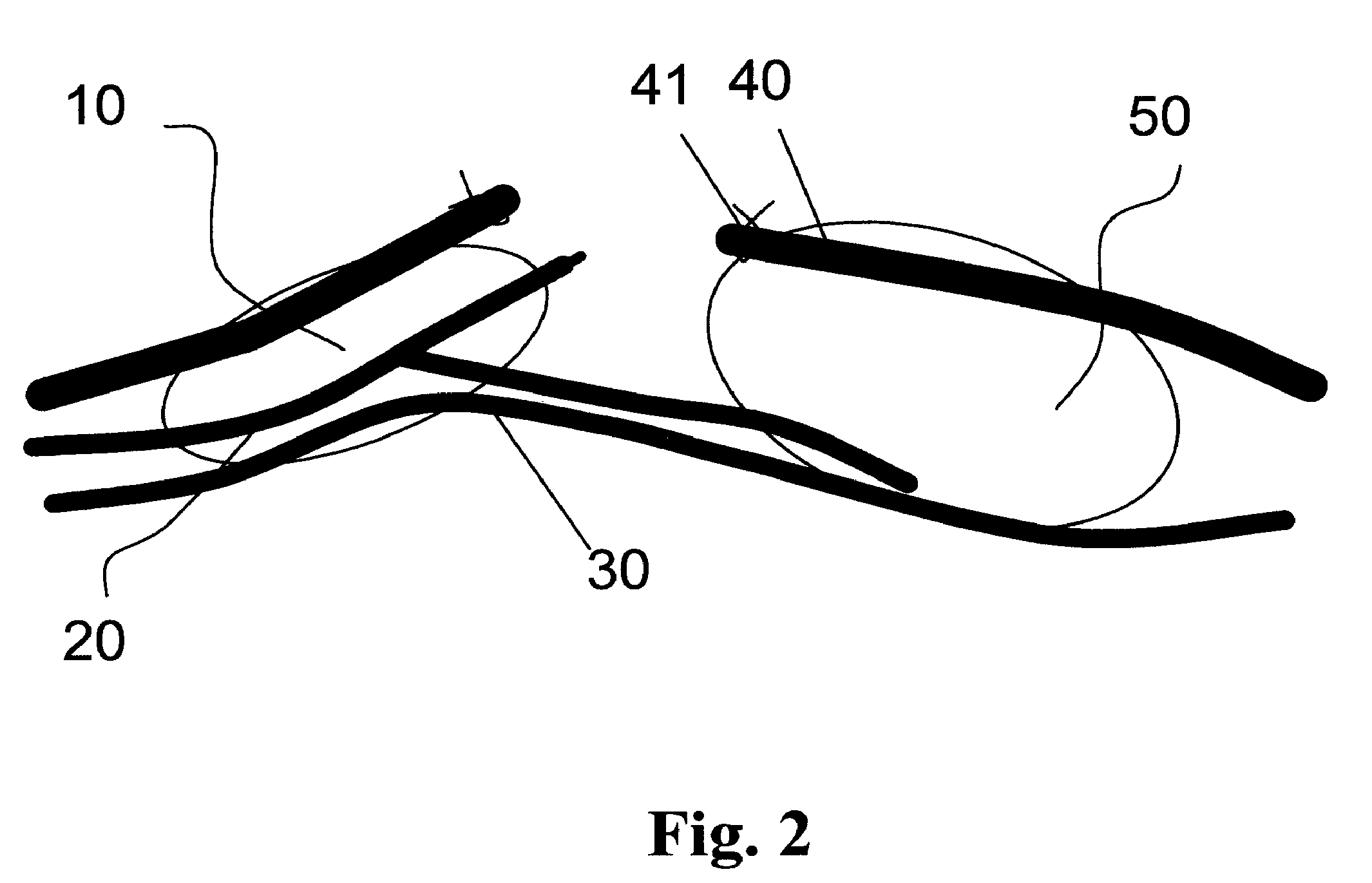
[0117]Another proximity system could be used to target a dissection of an interface between the neurovascular bundles 30 and the prostate 10FIG. 3, during radical prostatectomy. In this example, six neodymium magnets (NdFeB) reference elements coated with Teflon, cylindrically shaped, 500 um in diameter and 1 mm long, would be inserted before radical prostatectomy procedure transanally using intraoperative ultrasound imaging and a biopsy trochar with a tip modified to hold and deploy each reference element. Each reference element would be positioned at the prostate-neurovascular bundle interface 32, three on the left and three on the right. A lap aroscopic tool, such as a tissue retrieval system, would then be adapted to carry, for example, a three axis magnetic sensor such as a Honeywell HMC1023 Three-Axis Magnetic Sensor surface mount in a package with white orientation markings, sensor element. Thereafter, a robotic surgical system, such as the DaVinci roboti...


PUM


Abstract

Intraoperative devices are described that assist the surgeon in identifying the location and characteristics of tissues and structures. Devices are also described that have the added capability of marking the location of the identified tissues and structures. This invention also includes devices that can selectively ablate adjacent tissues while avoiding damage and trauma to the identified tissues and structures by combining ablation with sensing, where sensing of either tissue properties, markings made by another device or surgeon, or a reference probe can be used. Devices are also described that protect tissue in the proximity of reference markings or probes by closed loop inhibition of the ablation process. The devices, systems, methods and kits described are adapted and configured to facilitate locating a target structure or target tissue within a body of a mammal, including nerves, peripheral nerves, blood vessels, and tubes such as the ureter. The devices, systems and methods may discriminate between different tissues by exploiting the electrical, mechanical, and physiological properties of the body.

Description

CROSS-REFERENCE[0001]This application claims the benefit of U.S. Provisional Application No. 60 / 875,355, filed Dec. 18, 2006, by Rolfe Carter Anderson entitled Surgical Assistance Systems, and U.S. Provisional Application 60 / 992,985, filed Dec. 6, 2007 by Rolfe C. Anderson entitled Closed Loop Dissection Using Reference Probes, which applications are incorporated herein by reference.BACKGROUND OF THE INVENTION[0002]This invention relates generally to surgical navigation and control. In particular, the invention provides intraoperative devices that assist the surgeon in identifying the location and characteristics of tissues and structures. Devices are also described that have the added capability of marking the location of the identified tissues and structures. This invention also includes devices that can selectively ablate tissues that are adjacent or surrounding a target identified tissue while avoiding damage and trauma to the identified tissues and structures by combining ablat...

Claims


Application Information

Patent Timeline
Patent Type & Authority Applications(United States)
IPC IPC(8): A61B5/05
CPCA61B17/22004A61B2090/3987A61B17/3403A61B18/02A61B18/042A61B18/14A61B2017/00022A61B2017/00026A61B2017/00053A61B2017/00411A61N1/05A61N1/36014A61B90/36A61B2090/3975A61B90/98A61B90/39A61B2090/3933A61B2090/3937A61B2090/395A61B2090/3954A61B17/32
Inventor ANDERSON, ROLFE C.
Owner TRILLIUM PRECISION SURGICAL
Why Eureka
Try Eureka