

Method for non-autologous cartilage regeneration
a cartilage regeneration and non-autologous technology, applied in the field of tissue engineering, can solve the problems of poor mechanical properties of fibrocartilage and degeneration, and achieve the effects of reducing the risk of tissue overgrowth in the joint, poor mechanical properties, and degeneration over tim
- Summary
- Abstract
- Description
- Claims
- Application Information
AI Technical Summary
Benefits of technology
Problems solved by technology
Method used
Image

Examples
example 1
Material and Methods
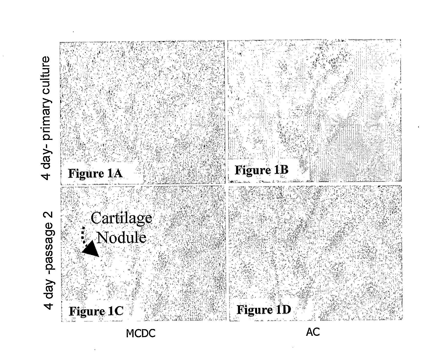
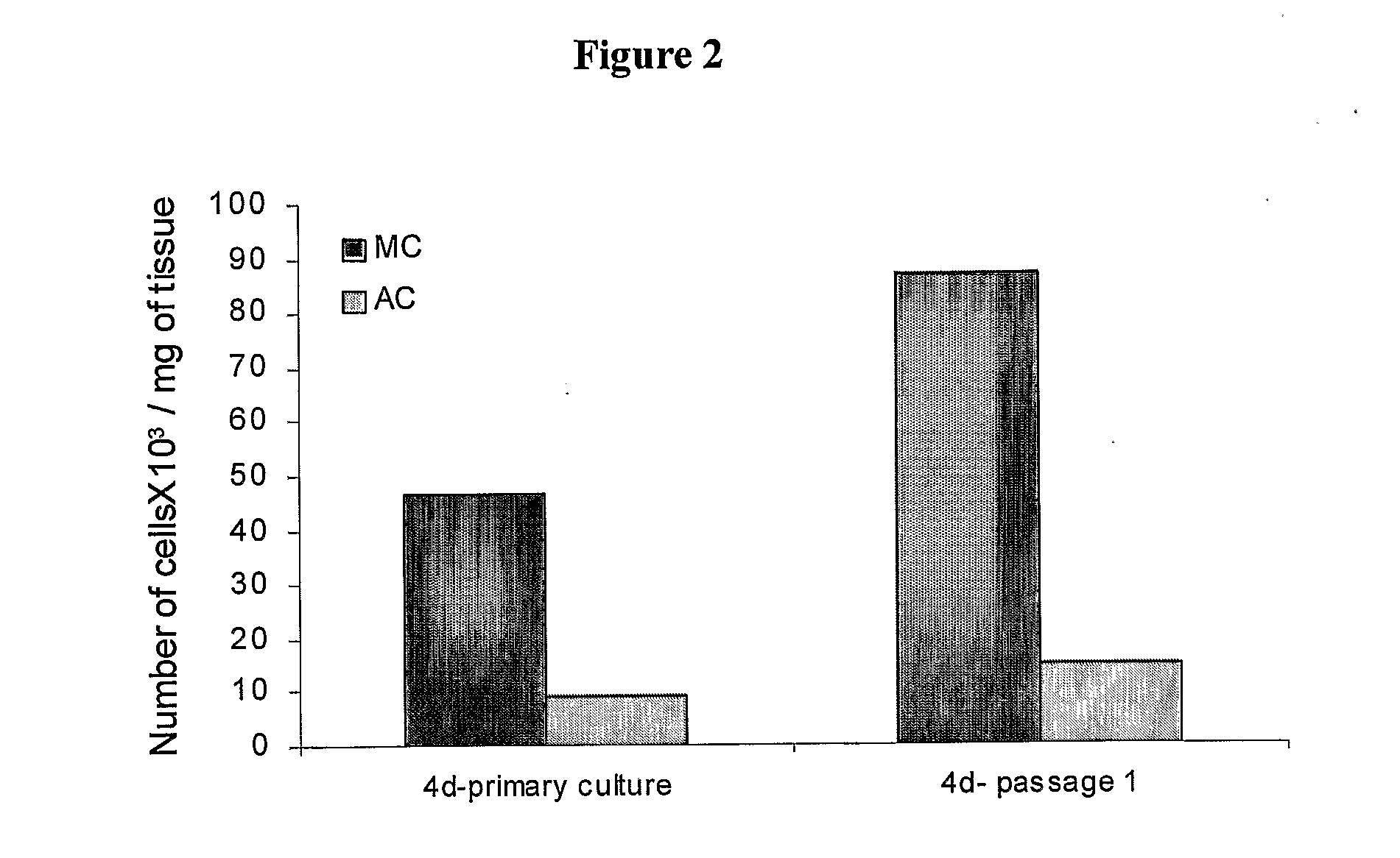
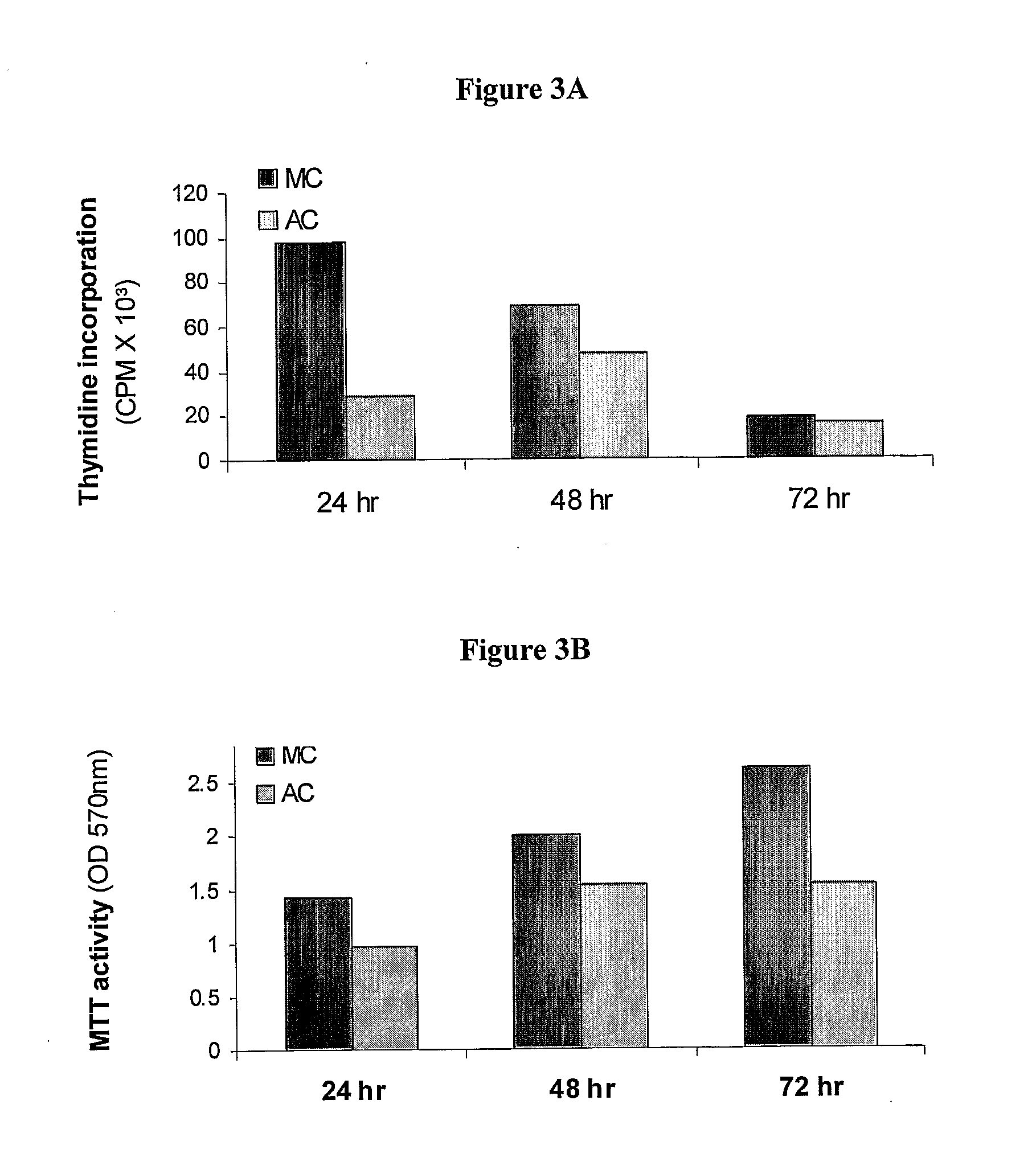
[0105]Neonatal piglet (24 hr old, male or female, ˜1.6 kg weight) was deeply anesthetized with 5 ml Iustil (200 mg / ml sodium pentobarbiton, injected into the heart). Mandibular condyles were aseptically dissected, freed of any soft tissue and subjected to successive collagenase digestion (37° C.; 0.1% type II collagenase, cat No. C-6885, Sigma Co., St. Louis, Mo., USA). Following the 1st digestion (25 min), which mainly separates adjacent soft tissues, the next 4 successive digestions (45 min each) yielded homogenous population of chondrocytes. These mandibular condyle derived chondrocytes are denoted MC. For the sake of comparison, chondrocytes were also separated from the femoral distal condyles and are referred to herein AC.
[0106]Dulbecco's Modified Eagle's Medium (DMEM) (cat No. 010551, Biological Industries, Bet Ha'Emek, Israel) supplemented with 1 mM sodium pyruvate, 10% fetal calf serum (FCS), and 1% penicillin / streptomycin (cat No. 03...
example 2
Cartilage Film (Membrane)
[0140]Porcine mandibular condyle-derived chondrocytes from the second passage, were plated on cover glasses at a concentration of 5×10⁵ cells / ml in 35 mm culture wells and cultured under same conditions as described above for MCDC. Cells were re-fed every 48 hr. At 12 days post-plating, an intact cartilage film developed. The cartilage film was rigid enough to be transferred and re-plated in a new culture dish. Throughout the first 24 hr, cartilage film was cultured with only 1 ml of medium to let it adhere to the plate. Further culturing continued under ordinary conditions. Development of the cartilage culture originating from the original cartilage film was followed morphologically and biochemically.
[0141]Cartilaginous Film Formed by MC-Derived Chondrocytes
[0142]Mandibular condyle-derived chondrocytes rapidly differentiate in culture into cartilage forming cells, which secrete increasing amounts of type II collagen and aggrecan. Four days post cells platin...
example 3
Xenotransplantation of MCDC Cells
[0148]Adjuvant induced rheumatoid arthritis (AIA) is an accepted experimental model for rheumatoid-induced articular damages. AIA rats undergo an acute disease phase lasting about 30 days and characterized by severe edema in the joints in general and in the knee joint in particular. When edema disappears, most rats still drag their hind legs due to the severe damage of the articular cartilage.
[0149]Seven AIA female Lewis rats were left to recover for 30 days post the acute AIA phase. Four sets of two AIA rats each were included in each treatment. Two million MCDC cells derived from rat (1), mouse (2) or porcine (3) were injected into the affected knee joints in 0.5 ml of PBS. Each cellular source was injected to both hind knees of two rats. One AIA rat was left untreated as a positive control (C+). One of its knees was injected with PBS alone. Morphological results were compared to those of an intact-naive rat as a negative control (C−).
[0150]Rats we...
PUM
| Property | Measurement | Unit |
|---|---|---|
| Time | aaaaa | aaaaa |
| Time | aaaaa | aaaaa |
| Time | aaaaa | aaaaa |
Abstract
Description
Claims
Application Information