Navigation System for Cardiac Therapies

a navigation system and cardiac therapy technology, applied in the field of image guided surgery, can solve the problems of not optimizing the pacing lead procedures currently performed today for use in heart failure treatment, not facilitating the actual guiding of the medical device to a targeted tissue area for medical treatment, and not optimizing the placement, so as to reduce the exposure to fluoroscopy, improve the outcome, and identify precisely

Inactive Publication Date: 2010-08-19
MEDTRONIC NAVIGATION INC
View PDF100 Cites 124 Cited by
  • Summary
  • Abstract
  • Description
  • Claims
  • Application Information

AI Technical Summary

Benefits of technology

[0015]A navigation system is provided including a catheter carrying single or multiple localization sensors, a sensor interface, a user interface, a controller, and a visual display. Aspects of the present invention allow for the location of a catheter advanced within an internal space within the human body, for example within the cardiovascular structures, to be identified in two, three or four dimensions in real time. Further aspects of the present invention allow for accurate mapping of a tissue or organ, such as the heart or other soft tissue, and / or precise identification of a desired location for delivering a medical lead or other medical device or therapy while reducing the exposure to fluoroscopy normally required during conventional catheterization procedures. These types of therapies include, but are not limited to, drug delivery therapy, cell delivery therapy, ablation, stenting, or sensing of various physiological parameters with the appropriate type of sensor. In cardiac applications, methods included in the present invention compensate for the effects of respiration and the beating heart that can normally complicate mapping or diagnostic data. Aspects of the present invention may be tailored to improve the outcomes of numerous cardiac therapies as well as non-cardiac therapies, such as neurological, oncological, or other medical therapies, including lung, liver, prostate and other soft tissue therapies, requiring the use of a catheter or other instrument at a precise location.
[0016]The steerable catheter provided by the present invention features at least one or more, location sensors located near the distal end of an elongated catheter body. The location sensors may be spaced axially from each other and may be electromagnetic detectors. An electromagnetic source is positioned externally to the patient for inducing a magnetic field, which causes voltage to be developed on the location sensors. The location sensors may be each electrically coupled to twisted pair conductors, which extend through the catheter body to the proximal catheter end. Twisted pair conductors provide electromagnetic shielding of the conductors, which prevents voltage induction along the conductors when exposed to the magnetic flux produced by the electromagnetic source. Alternatively, the sensors and the source may be reversed where the catheter emits a magnetic field that is sensed by external sensors.
[0017]By sensing and processing the voltage signals from each location sensor, the location of the catheter tip with respect to the external sources and the location of each sensor with respect to one another may be determined. The present invention allows a two, three, or four-dimensional reconstruction of several centimeters of the distal portion of the catheter body in real time. Visualization of the shape and position of a distal portion of the catheter makes the advancement of the catheter to a desired position more intuitive to the user. The system may also provide a curve fitting algorithm that is selectable based upon the type of catheter used, and based upon the flexibility of the catheter, based upon a path finding algorithm, and based upon image data. This enables estimated curved trajectories of the catheter to be displayed to assist the user.
[0019]The catheter body is formed of a biocompatible polymer having stiffness properties that allow torsional or linear force applied to a handle at the proximal end to be transferred to the distal end in such a way that the catheter may be advanced in a desired direction. The catheter body includes multiple lumens for carrying conductors to sensors located at or near the distal end of the catheter and a guide wire extending from a proximal handle to the distal catheter tip. The guide wire aids in steering the catheter through a venous pathway, or other body lumens, and can be manipulated at its proximal end to cause bending or curving of the distal catheter tip.

Problems solved by technology

However, use of such fluoroscopic imaging throughout a procedure exposes both the patient and the operating room staff to radiation, as well as exposes the patient to contrast agents.
Such mapping catheters are useful in identifying an area of tissue that is either conducting normally or abnormally, however, some mapping catheters may not aid in actually guiding a medical device to a targeted tissue area for medical treatment.
Also, pacing lead procedures currently performed today for use in heart failure treatment are not optimized.
In this regard, the lead placement is not optimized due to the lack of having real-time anatomic information, navigation and localization information, hemo-dynamic data, and electrophysiological data.
Thus, pacing leads are currently simply “stuffed” into the heart without any optimization being performed due to lack of information that can be used for this optimization.
Advancement of a guide catheter or an over-the-wire lead through a vessel pathway and through cardiac structures requires considerable skill and can be a time-consuming task.
This type of procedure also exposes the patient to an undesirable amount of radiation exposure and contrast agent.
With regard to navigating an instrument through a moving body structure, difficulties arise in attempting to track such an instrument using known tracking technology as the instrument passes adjacent or through a moving body structure, since the virtual representation of the instrument may be offset from the corresponding anatomy when superimposed onto image data.
Other difficulties with cardiac procedures include annual check-ups to measure early indications for organ rejection in heart transplant patients.
This type of invasive procedure creates significant trauma, such as skull holes, dura opening, fiber destruction, and other cerebral structural damage or leakage.
However, this type of vascular technique has various drawbacks.
For example, this type of disposable IVUS catheter is extremely expensive.
Moreover, the ultrasound transducer embedded within the catheter provides only visualization on one side of the catheter, typically orthogonal to the catheter body, and therefore does not provide any forward views or other views about the catheter.

Method used


Image

  • Navigation System for Cardiac Therapies
  • Navigation System for Cardiac Therapies
  • Navigation System for Cardiac Therapies

Examples


Embodiment Construction

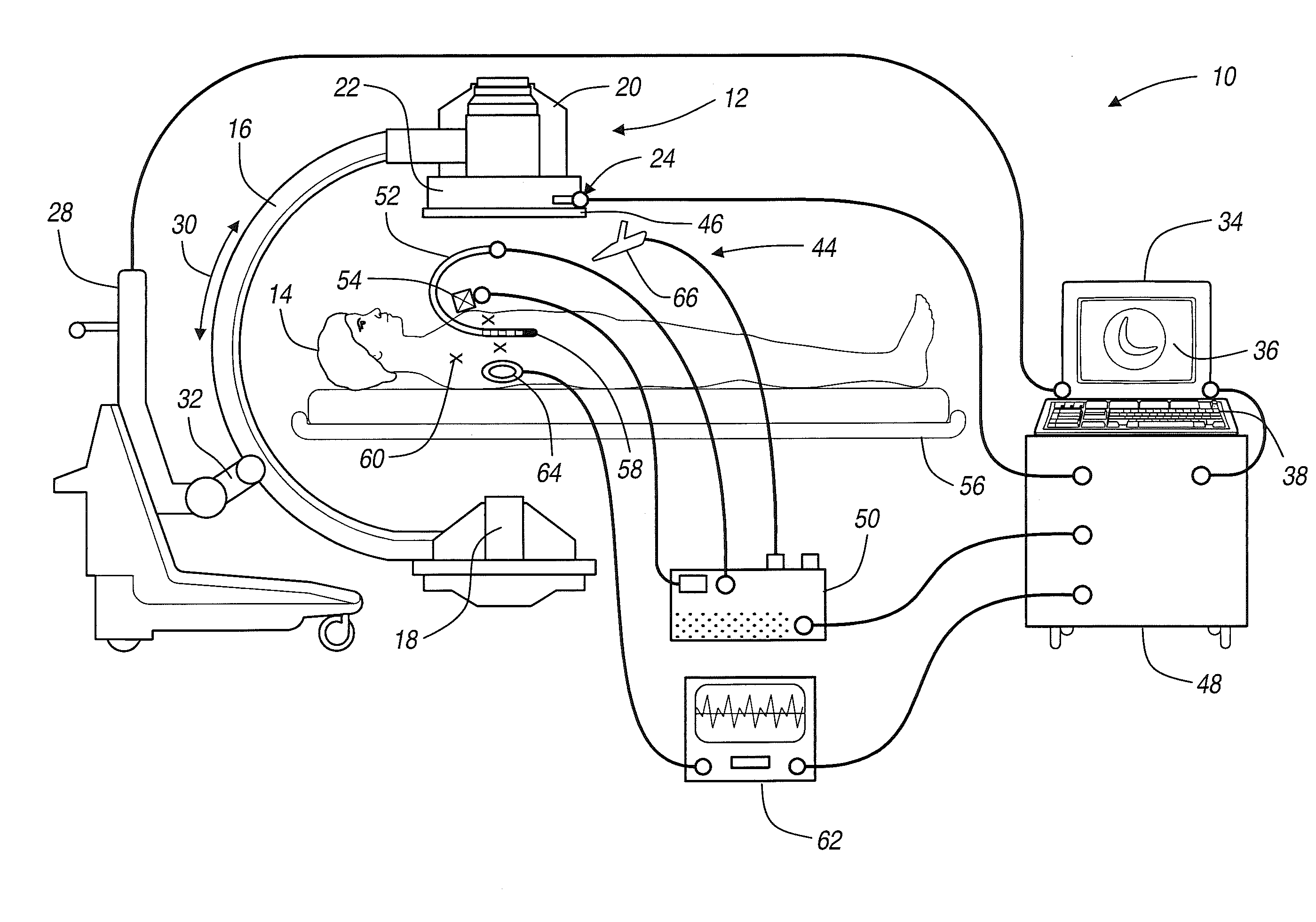
[0055]The following description of the preferred embodiment(s) is merely exemplary in nature and is in no way intended to limit the invention, its application, or uses. As indicated above, the present invention is directed at providing improved, non-line-of-site image-guided navigation of an instrument, such as a catheter, balloon catheter, implant, lead, stent, needle, guide wire, insert and / or capsule, that may be used for physiological monitoring, delivering a medical therapy, or guiding the delivery of a medical device in an internal body space, such as the heart or any other region of the body.

[0056]FIG. 1 is a diagram illustrating an overview of an image-guided catheter navigation system 10 for use in non-line-of-site navigating of a catheter during cardiac therapy or any other soft tissue therapy. It should further be noted that the navigation system 10 may be used to navigate any other type of instrument or delivery system, including guide wires, needles, drug delivery syste...


PUM


Abstract

An image guided navigation system for navigating a region of a patient includes an imaging device, a tracking device, a controller, and a display. The imaging device generates images of the region of a patient. The tracking device tracks the location of the instrument in a region of the patient. The controller superimposes an icon representative of the instrument onto the images generated from the imaging device based upon the location of the instrument. The display displays the image with the superimposed instrument. The images and a registration process may be synchronized to a physiological event. The controller may also provide and automatically identify an optimized site to navigate the instrument to.

Description

CROSS-REFERENCE TO RELATED APPLICATIONS[0001]This application is a continuation of U.S. patent application Ser. No. 10 / 619,216 filed on Jul. 14, 2003, which is a continuation-in-part of U.S. patent application Ser. No. 10 / 299,969 filed on Nov. 19, 2002, now U.S. Pat. No. 7,599,703, issued Oct. 6, 2009, which is hereby incorporated by reference.FIELD OF THE INVENTION[0002]The present invention relates generally to image guided surgery, and more specifically, to systems and methods for using one or more medical images to assist in navigating an instrument through internal body structures, in particular for navigating a catheter in a moving body structure, such as the heart, during a surgical procedure.BACKGROUND OF THE INVENTION[0003]Image guided medical and surgical procedures utilize patient images obtained prior to or during a medical procedure to guide a physician performing the procedure. Recent advances in imaging technology, especially in imaging technologies that produce highl...

Claims


Application Information

Patent Timeline
Patent Type & Authority Applications(United States)
IPC IPC(8): A61B6/00A61B1/005A61B1/012A61B5/0215A61B5/06A61B6/12A61B17/00A61B17/34A61B19/00
CPCA61B1/00071A61B6/507A61B1/012A61B5/0215A61B5/06A61B5/413A61B5/7285A61B6/12A61B6/463A61B6/541A61B10/06A61B17/3478A61B19/5244A61B19/56A61B2017/00243A61B2017/00247A61B2018/00392A61B2019/504A61B2019/505A61B2019/507A61B2019/5238A61B2019/5251A61B2019/5272A61B2019/5276A61B2019/5291A61B2019/5295A61B2019/562A61B2019/564A61B2019/566A61B1/0058A61B8/4254A61B2034/104A61B2034/2072A61B2090/367A61B2034/254A61B2034/256A61B34/20A61B34/25A61B2090/376A61B2034/105A61B2034/107A61B2034/2051A61B2090/378A61B2090/365A61B2034/252A61B5/743A61B5/062A61B5/068
Inventor VERARD, LAURENTHUNTER, MARK W.BZOSTEK, ANDREW
Owner MEDTRONIC NAVIGATION INC
Why Eureka
Try Eureka