Quantitative spectroscopic imaging

a spectroscopic imaging and quantitative technology, applied in the field of quantitative spectroscopic imaging, can solve the problems of biopsy and undersampling, and achieve the effect of accurate diagnosis and clinical applicability

Inactive Publication Date: 2010-09-30
MASSACHUSETTS INST OF TECH
View PDF42 Cites 8 Cited by
  • Summary
  • Abstract
  • Description
  • Claims
  • Application Information

AI Technical Summary

Benefits of technology

[0006]Contact probe techniques are promising, but like biopsy, suffer from undersampling. To overcome this, wide area light collection and imaging in fluorescence and reflectance tissue diagnosis, is used to provide quantitative analysis. A preferred embodiment uses a model-based, quantitative approach to wide field imaging that is referred to herein as quantitative spectroscopic imaging (QSI). Data are collected by means of a non-contact “virtual” probe, imaged at the tissue surface. This virtual probe is then raster scanned to interrogate a wide tissue area (for example, in a range of 1-4 cm2), using one or more spots (1 mm2) at a time. The quantitative measurements of tissue properties enable the spectra for each pixel to be analyzed using probe methodology. Hence, the QSI images are directly interpretable in terms of histological features, thus providing an accurate diagnosis.
[0007]A preferred embodiment of the present invention involves the design, construction, calibration, and the clinical application of this QSI system. Measurements using physical tissue models (“phantoms”) demonstrate the accuracy of QSI. Ex vivo spectral images of a resected colon adenoma demonstrate its ability to diagnose and image malignant lesions. In addition, in vivo spectral images from a hyperkeratotic lesion on the ventral surface of the tongue further demonstrates clinical applicability.

Problems solved by technology

Contact probe techniques are promising, but like biopsy, suffer from undersampling.

Method used


Image

  • Quantitative spectroscopic imaging
  • Quantitative spectroscopic imaging
  • Quantitative spectroscopic imaging

Examples


Embodiment Construction

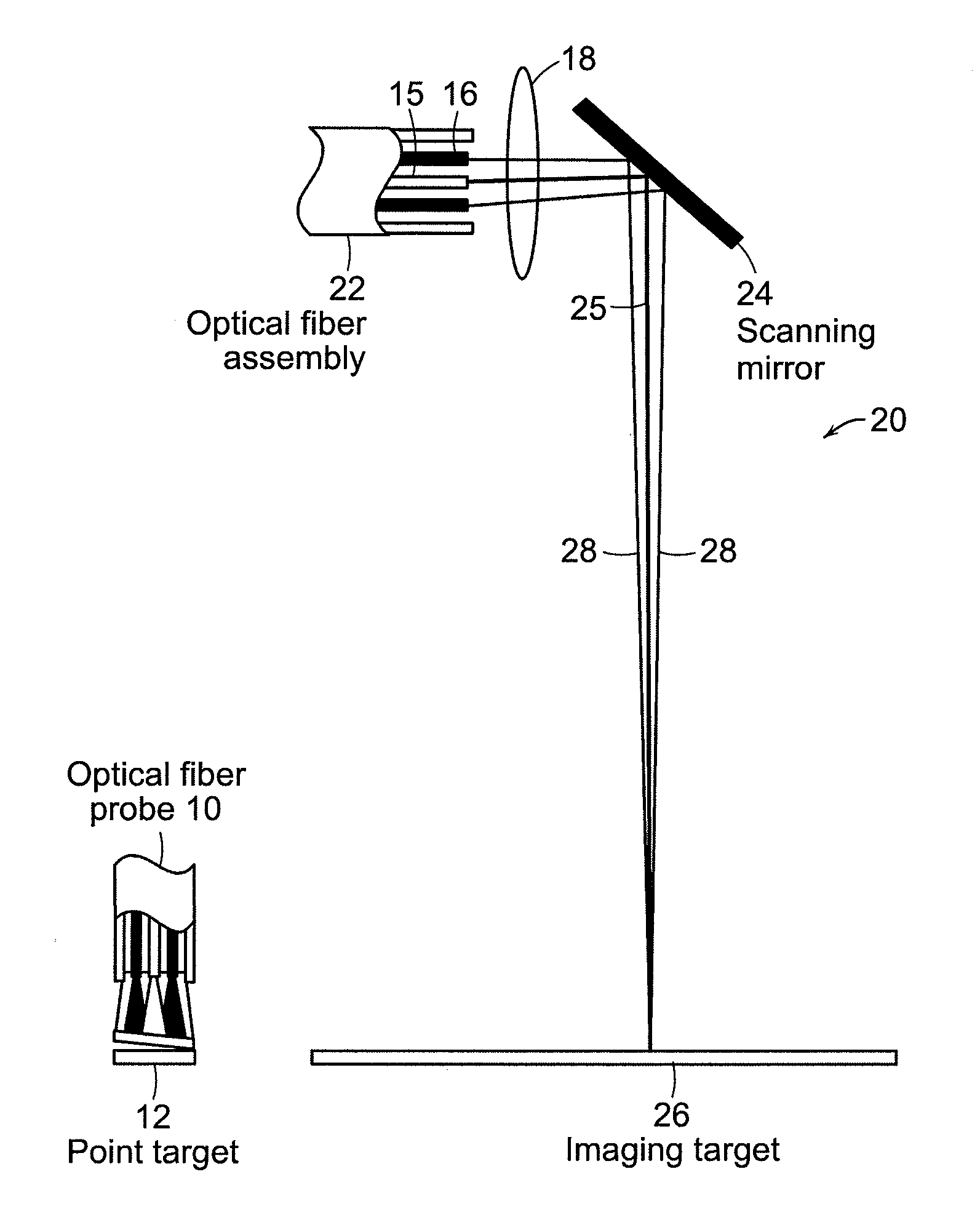
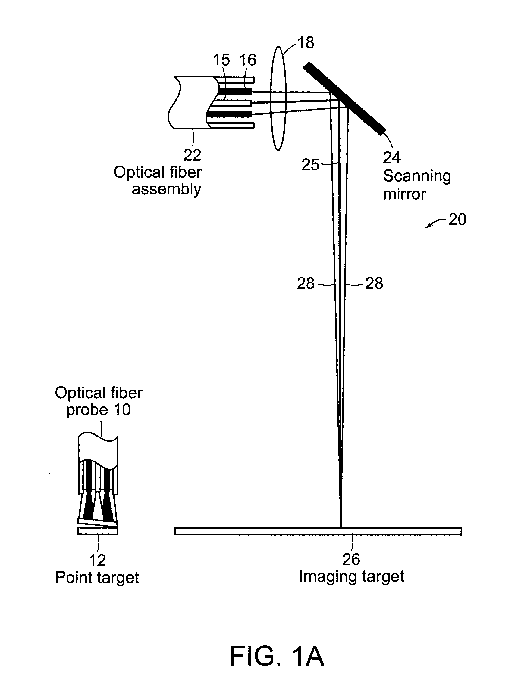
[0019]The present invention relates to the use of a scanning light region to quantitatively measure objects at a distance. Instead of the quantitative methodology of contact probes, such as those described by: J. W. Tunnell et al., “Instrumentation for multi-modal spectroscopic diagnosis of epithelial dysplasia,” Technol. Cancer Res. Treat. 2, 505-514 (2003), the entire contents of which is incorporated herein by reference, the present invention provides a wide area imaging instrument. In the contact probe 10 geometry, such as seen in FIG. 1A where the optical fiber probe 10 consists of a single light delivery fiber surrounded by six collection fibers. All fibers are fused together at the tip to form an optical shield approximately 1 mm long. This arrangement of fibers and quartz shield shaped at a 17 degree angle at the tip provides a reproducible geometry of overlapping excitation and collection cones, creating a fixed distance between fiber tips and tissue and a sampling spot 12 ...


PUM


Abstract

The present invention relates to a fully quantitative spectroscopy imaging instrument for wide area detection of early cancer (dysplasia). This instrument provides quantitative maps of tissue biochemistry and morphology, making it a powerful surveillance tool for objective early cancer detection. The design, construction, calibration, and diagnostics applications of this system is described with the use of physical tissue models. Measurements were conducted on a resected colon adenoma, and the system can be used for vivo imaging in the oral cavity.

Description

CROSS REFERENCE TO RELATED APPLICATION[0001]This application claims priority to U.S. Application 61 / 194,457 filed on Sep. 26, 2008 the entire contents of which are incorporated herein by reference.BACKGROUND OF THE INVENTION[0002]Early detection is essential for managing cancer, since treatment is much more successful when lesions are diagnosed at an early, noninvasive stage. Current cancer diagnosis often employs visual inspection of a wide area of tissue followed by biopsy of suspicious sites. This practice is problematic for two reasons: (1) early cancers are not always detectable by visual inspection, so, unavoidably, unnecessary biopsies are taken for precautionary reasons and invisible lesions are missed; and (2) biopsy suffers from undersampling, the results are subjective, and the resulting pathology can be subject to low inter-observer agreement. Furthermore, biopsy results often are not available immediately, resulting in delayed treatment and patient anxiety. Much attenti...

Claims


Application Information

Patent Timeline
Patent Type & Authority Applications(United States)
IPC IPC(8): A61B6/00A61B1/06
CPCA61B1/043A61B5/0071A61B5/4255A61B5/0084A61B5/0088A61B5/0075
Inventor YU, CHUNG-CHIEHLAU, CONDONDASARI, RAMACHANDRAFELD, MICHAEL
Owner MASSACHUSETTS INST OF TECH
Why Eureka
Try Eureka