

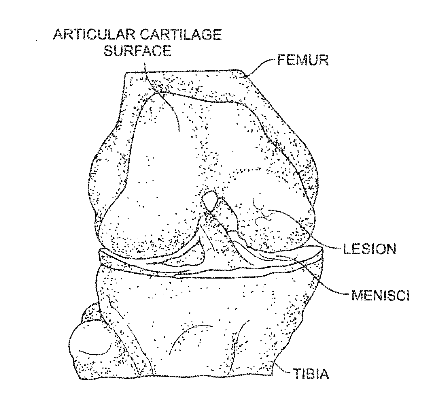
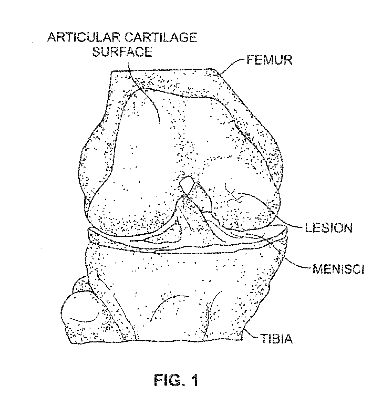
Cartilage particle tissue mixtures optionally combined with a cancellous construct
a cartilage particle and tissue mixture technology, applied in the field of preparation and constructs, can solve the problems of articular cartilage lesions generally not healing, joint pain or severe restriction, and limited articular cartilage regeneration
- Summary
- Abstract
- Description
- Claims
- Application Information
AI Technical Summary
Problems solved by technology
Method used
Image

Examples
example 1
Measurement of Demineralized Construct Porosity
[0167]The percentage of porosity and average surface pore diameter of a cancellous construct demineralized cap member according to the present invention can be determined utilizing a microscope / infrared camera and associated computer analysis. A microscope / infrared camera was used to produce the images of FIGS. 3A and 3B, which provide a visual assessment of the porosity of a demineralized cap of a construct according to the present invention. Such images were analyzed using suitable microscopy and image analysis software; for example, Image Pro Plus® (Media Cybernetics, Inc., Bethesda, Md.). The number and diameter of pores and the relative porosity of a demineralized member of a construct can be characterized using techniques known to those skilled in the art.
[0168]It is noted that, for allograft constructs, the number and diameter of pores and the relative porosity of the demineralized members will vary from one tissue donor to anoth...
example 2
Tissue Extraction and Particularization
[0169]A process of cartilage particle extraction may be applied to any of a number of different soft tissue types (for example, meniscus tissue). In an embodiment, cartilage is recovered from deceased human donors, and the tissue is treated with a soft tissue process.
[0170]Fresh articular cartilage is removed from a donor using a scalpel, taking care to remove the cartilage so that the full thickness of the cartilage is intact (excluding any bone). Removed cartilage is then packaged in double Kapak® bags for storage until ready to conduct chemical cleaning of the allograft tissue. In one example, the cartilage can be stored in the refrigerator for 24-72 hours or in the freezer (e.g., at a temperature of −70° C.) for longer-term storage.
[0171]Chemical cleaning of cartilage tissue is then conducted according to methods known to those skilled in the art. Subsequent to chemical cleaning, the cartilage is lyophilized so as to reduce the water conten...
example 3
Extraction of Proteins from Human Cartilage Using Extraction and Subsequent Dialysis
[0176]In another example, growth factors may be physically and / or chemically isolated from cartilage particles, and dialyzed using a suitable agent. The growth factors are thereby isolated for subsequent analysis and / or quantification. In an embodiment, 0.3 g of cartilage particles were weighed out for each donor. The cartilage particles were transferred to tubes containing 5 ml of extraction solution (4M guanidine HCl in Tris HCL). The cartilage particles were incubated at 4° C. on an orbital shaker at 60 RPM for 24 hours, followed by dialysis (8 k MWCO membrane dialysis tube) in 0.05M TrisHCL or PBS for 15 hrs. at 4° C. The dialysis solution was then replaced and the dialysis continued for another 8 hrs. at 4° C. The post-dialysis extracts were stored at −70° C. until an Enzyme-linked Immunosorbent Assay (ELISA) was run.
PUM
| Property | Measurement | Unit |
|---|---|---|
| size | aaaaa | aaaaa |
| size | aaaaa | aaaaa |
| size | aaaaa | aaaaa |
Abstract
Description
Claims
Application Information