
Cardiomyocyte-like cell clusters derived from hbs cells

Inactive Publication Date: 2010-11-04
CELLARTIS AB (SE)
View PDF24 Cites 5 Cited by
  • Summary
  • Abstract
  • Description
  • Claims
  • Application Information

AI Technical Summary

Benefits of technology

[0026]Accordingly, the present invention provides a relative simple means of obtaining a suitable cardiomyocyte-like cell product for med

Problems solved by technology

Human cardiomyocytes can be isolated from heart biopsies but the procedure is complicated and it is difficult to obtain viable cell preparations.
In addition, the access to human heart tissue is very limited and it is thus not possible to isolate large number of cells.
In addition, due to the lack of donor material as well as the somewhat problematic procedure of cell isolation, human primary cardiomyocytes are not currently available for in vitro testing during pre-clinical drug discovery.
However, relatively little is currently known about how to control and manipulate hBS cell differentiation to produce exclusive populations of specific cell types.

Method used


Image

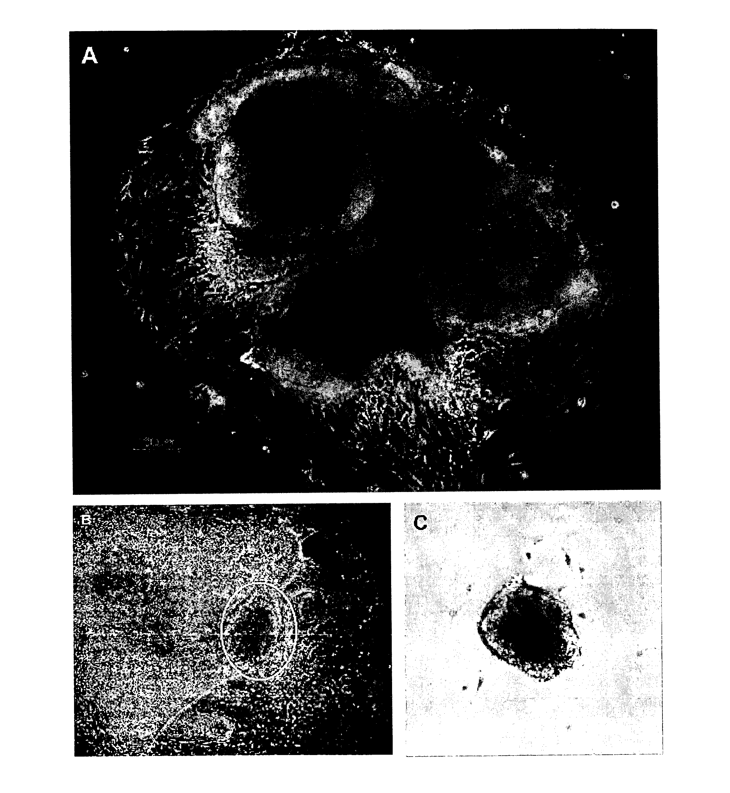
  • Cardiomyocyte-like cell clusters derived from hbs cells
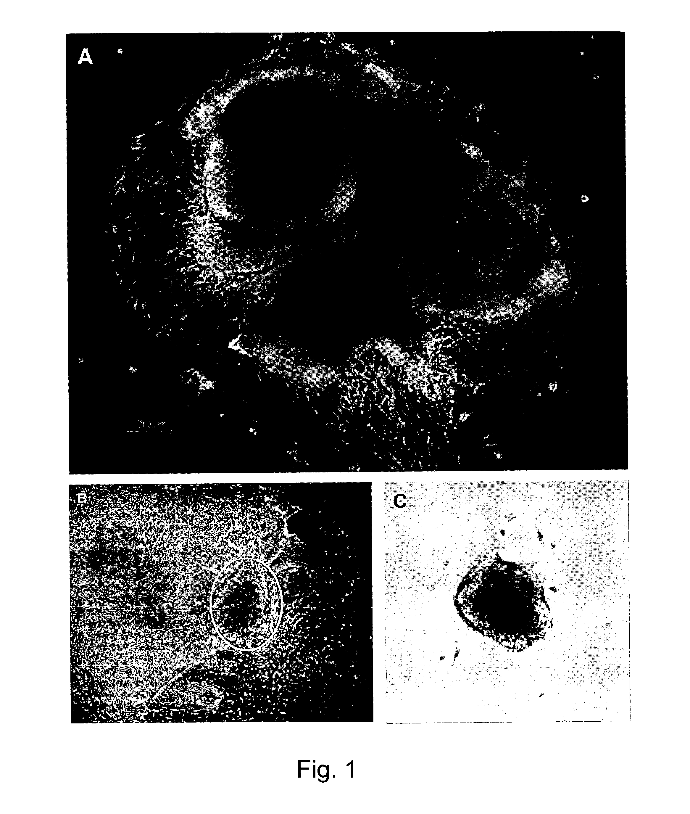
  • Cardiomyocyte-like cell clusters derived from hbs cells
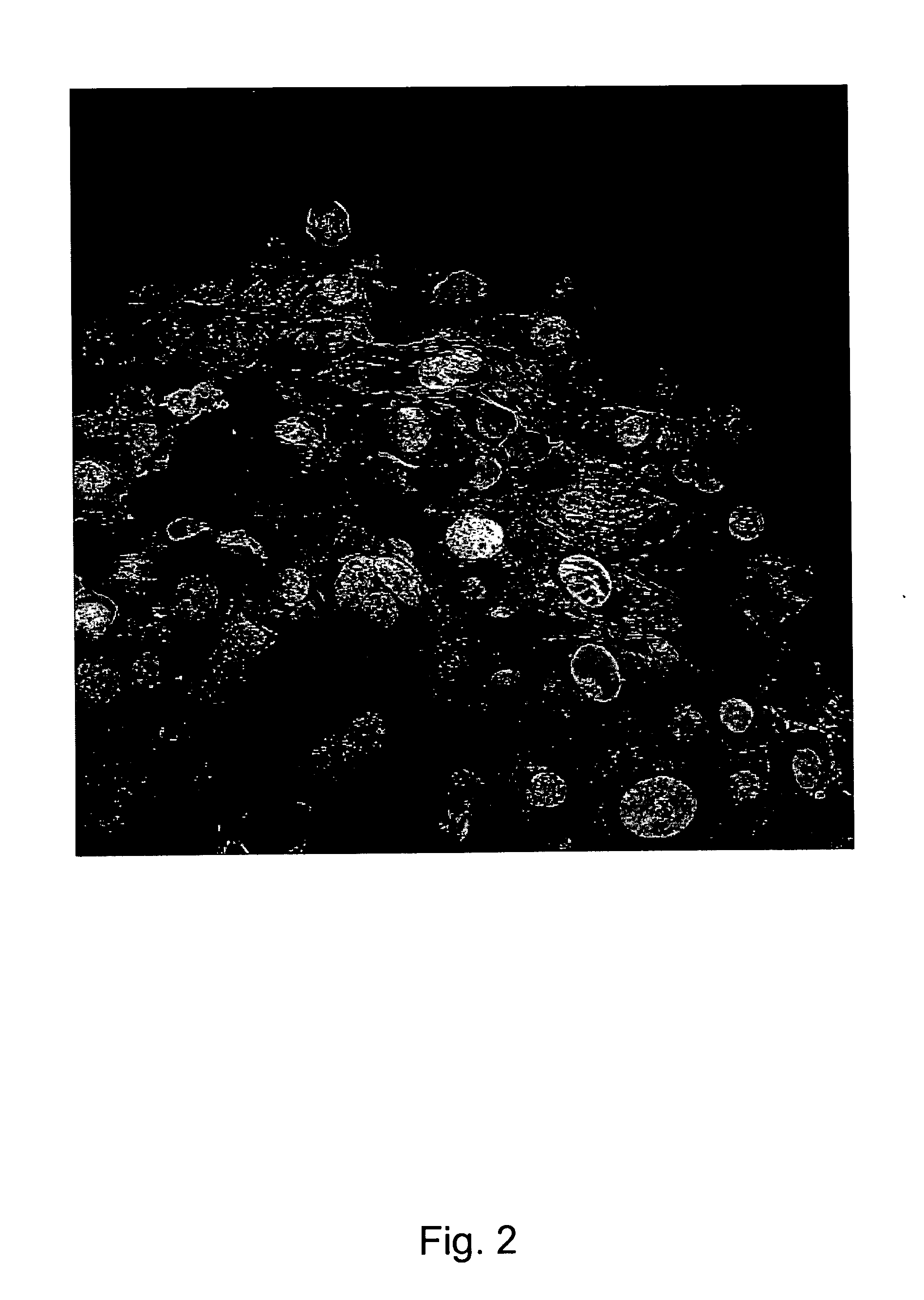
  • Cardiomyocyte-like cell clusters derived from hbs cells

Examples


example 1

Differentiation of hBS Cells to Cardiomyocyte-Like Cell Clusters

[0253]Spontaneously contracting cells were derived from undifferentiated hBS cells cultured on MEF cells (Heins et al 2004 Stem Cells). The cell lines used for this experiment could be the hBS cell line SA002, SA121, SA001, SA002.5, SA461 (Cellartis AB, Goteborg, Sweden, http: / / www.cellartis.com) and they can be propagated as described Heins et al. 2004. These cell lines are listed in the NIH stem cell registry and the UK Stem Cell bank. The hBS cells were detached from the feeder layer by incubation with collagenase IV (200 U / ml), for 10-15 minutes at 37° C. The cell suspension was transferred to a 15 ml tube, and after the cells had sedimented, the supernatant was removed. The colonies were resuspended in Knock Out DMEM supplemented with 20% FBS, 1% penicillin-streptomycin, 1% Glutamax, 0.5 mmol / l β-mercaptoethanol and 1% non-essential amino acids (all from Invitrogen, Carlsbad, Calif.) and dissociated mechanically in...

example 2

Micro Array Analysis of Cardiomyocyte-Like Cell Clusters (CMLC) Derived from hBS Cells

[0256]Cell Culture and Differentiation

[0257]The hBS cell line SA002 (Cellartis AB, Goteborg, Sweden, http: / / www.cellartis.com) was propagated as described Heins et al. 2004. This cell line is listed in the NIH stem cell registry and the UK Stem Cell bank. Differentiation of hBS cells was performed as described in Example 1 above. Using light microscopy clusters of spontaneously contracting cardiomyocyte-like cell clusters (CMLC) are frequently observed when hBS cells are differentiated through this protocol. For microarray experiments, three separate samples (biological replicates) of CMLC were collected. For comparison purposes, two independent samples from undifferentiated hBS cells and one sample from a mixed population of differentiated cells (i.e., no contracting CMLC present) were also collected for the analysis. The mixed population was collected as the remaining population of differentiated...

example 3

Real-Time QPCR of CMLC Derived from hBS Cells

[0300]Cell Culture and RNA Extraction

[0301]Spontaneously contracting CMLC were derived from undifferentiated hBS cells (SA002) (see Example 1). The hBS were detached from the MEF feeder layer by incubation with collagenase IV (200 U / ml), for 10-15 minutes at 37° C. The suspension was transferred to a 15 ml tube, and after the cells had sedimented, the supernatant was removed. The colonies were re-suspended in culture medium and dissociated mechanically into small aggregates of undifferentiated cells using a pipette. This cell suspension was distributed (200 μl / well) into a 96 well plate (Costar, Corning, untreated, v-bottom) and centrifuged for 5 min at 400×g. The plate was then incubated at 37° C. for 6-8 days. The 3D structures that formed were then transferred to gelatine coated dishes containing culture medium. After a couple of days spontaneously contracting clusters of cells (CMLC) were observed. Some of these clusters were harveste...


PUM


Abstract

A cluster is provided comprising cardiomyocyte-like cells, wherein the cluster has i) contracting cells, ii) cells that are electrically connected, and expresses iii) cardiac markers including Nkx.2.5, troponin and myosin, iv) markers for functional adrenergic receptors, v) markers for functional muscarinic receptors, vi) markers for functional ion-channels including hERG, Na+, Ca2+ and K+ channels, vii) one or more endodermal markers selected from the group consisting of AFP, TF, APOA2, AHSG, SERPINA1, APOA1, APOC3, TTR1 APOB, and RBP4. A method for preparing the clusters and methods utilizing the clusters in drug discovery and toxicity screenings are described.

Description

FIELD OF THE INVENTION[0001]The present invention relates to novel cardiomyocyte-like cell clusters (CMLC) derived from hBS cells and to the potential use of such cardiomyocyte-like cell clusters in e.g., pharmaceutical drug discovery and development, toxicity testing, cell therapy and medical treatment. The clusters contain cells expressing endodermal, mesodermal as well as cardiac markers. The invention also relates to a method for preparing the cluster, to composition comprising one or more CMLC for use in therapy and toxicity testing. The compositions are stable after storage of the composition at a temperature of at least −80° C. for at least 2 years, i.e. the characteristics and viability of the clusters are not substantially changed during this storage period.BACKGROUND OF THE INVENTION[0002]Mature cardiomyocytes are considered terminally differentiated cells and as such they have no, or very low, proliferative capacity. Human cardiomyocytes can be isolated from heart biopsie...

Claims


Application Information

Patent Timeline
IPC IPC(8): A61K35/12C12N5/073C12Q1/02A61P9/00C12N5/077
CPCC12N5/0657C12N2501/115C12N2501/16C12N2506/02C12N2501/70C12N2501/999C12N2503/02C12N2501/235A61P9/00
Inventor SARTIPY, PETERKESSON, KAROLINAAMEEN, CAROLINESYNNERGREN, JANEDAHLENBORG, KERSTINSTEEL, DANIELLA
Owner CELLARTIS AB (SE)
Learn More