Gene expression signatures in enriched tumor cell samples

a gene expression and tumor cell technology, applied in the field of gene expression signatures in enriched tumor cell samples, can solve the problems of antibody entail a substantial loss of metastatic tumor cells, cancer is not sufficiently distinguished from normal cells, and ctcs are rar

Active Publication Date: 2010-11-25
THE RES FOUND OF STATE UNIV OF NEW YORK
View PDF22 Cites 27 Cited by
  • Summary
  • Abstract
  • Description
  • Claims
  • Application Information

AI Technical Summary

Benefits of technology

[0030]The methods that embody the invention provide tumor cell signatures that, in turn, enable methods of identifying, in an undiagnosed subject, a variety of tumor cell types, and distinguishing among them. The methods allow the artisan to distinguish between normal cells and benign, malignant, primary, metastatic, or metastasized cells. The methods also can distinguish between normal cells and tumor cells of epithelial lineage, including, without limitation, colorectal cancer, ovarian cancer, and breast cancer cells. The methods also allow the artisan to distinguish between benign vs. malignant cells, primary tumor vs. metastatic cells, and different tumor cells of epithelial lineage such as colorectal vs. breast cancer cells for example.
[0031]In one methodological embodiment of the invention, the method comprises a gene expression signature for metastatic tumor cells in ascites fluid obtained from ovarian cancer patients, which gene signature is effective in distinguishing samples of metastatic tumor cells in ascites from late stage primary ovarian tumor cells, and is also effective in distinguishing benign ovarian tumor cells from early stage malignant ovarian tumor cells.

Problems solved by technology

However, CTCs are currently detected by epithelial lineage markers or tumor-associated markers that are neither sufficient in distinguishing cancer from normal cells nor adequate in identifying specific types of tumors.
For example, current CTC enrichment methods using an anti-Epithelial Cell Adhesion Molecule (EpCAM) antibody may entail a substantial loss of metastatic tumor cells due to downregulation of the EpCAM gene in these cells (Pantel et al., 1999; Sabile et al., 1999; Thurm et al., 2003; Choesmel et al., 2004).
However, CTCs are rare.
However, it is not known to date which member(s) of the KRT family are specific for the cancer stem or progenitor cell type among CTCs.
Although some of these differentiated epithelial markers have been used to isolate and identify CTCs and DTCs (Pantel et al., 2008), the true identity of the tumor progenitor cell population remains undefined for the majority of human cancers due to the lack of markers that are specific for that type of CTC (van't Veer and Bernards, 2008; Sawyers, 2008).
There remains an unresolved quantification problem, however: there are no ‘ideal’ endogenous control genes; that is, genes that do not deviate between tumor and normal cells in blood from different individuals (de Kok et al., 2005).
However, false negative results can occur, since these antigens are not present in all the cells of a tumor (Paterlini-Brechot and Benali, 2007).
Therefore, the detection of exfoliated abnormal cells by routine cytopathology is often limited.
Compounding this heterogeneity problem, the frequency of neoplastic cells present in each clinical specimen is variable, which biases and complicates the quantification of differential gene expression in randomized mixed populations.
These cells do not contain high quality RNA and thus present technical problems for molecular analyses (Karczewski et al., 1994).
The detection of metastatic cells is particularly challenging.
This method typically has several limitations: (1) the subsequent sample processing is complicated, (2) cell viability cannot readily be established, and (3) selection of the cells to be dissected is based mainly on morphological criteria, which has a high frequency of giving rise to false-positive results.b) Physical characteristics of tumor cells, such as shape, size, density or electrical charge, can also be used (Vona et al., 2000).
However, these methods may not effectively enrich viable tumor cells from normal cells.
That is, 500-1,000 fold cell enrichment is often found to be relatively modest enrichment which generates substantial background noise adversely affecting further molecular analysis.
In addition, enrichment methods based on physical separation techniques are often cumbersome, lengthy, and involve steps (e.g. more than 2-3 rounds of centrifugation) that can result in cellular damage.c) Antibody-based techniques are a more recent development.
However, cells that exist in clusters or clumps are discarded during the FACS process, and in some instances, for example, ovarian cancer, most of the cells are present as aggregates, making FACS CTC or CEC detection highly ineffective.
Despite its great power for enrichment, there are also inherent limitations associated with all of the antibody-based cell separation methods.
The most serious one is that cancer cells usually express putative tumor-specific antigens to variable degrees (Sabile et al., 1999); hence it is easy to lose a large and potentially non-random subset of tumor cells during the collection.
Overall, such antibody-based cell separation methods have a higher than desired false-negative rate.
Therefore, it is still a challenging task to detect the presence of thousands of cancer cells in one ml of blood (Gulati and Acaba, 1993).
Invadopodia are not found on differentiated normal blood cells or on primary tumor cells, and they do not function effectively on dead or dying cells.
However, it is not possible to resolve these cell types in a mixed population due to a lack of markers specific for these cell types, be they cancer cells or endothelial cell progenitors (van't Veer and Bernards, 2008; Sawyers, 2008).
Enrichment makes any particular cell type highly accessible to genetic analysis, but leaves one uncertain about the cell to which the analysis should apply.

Method used


Image

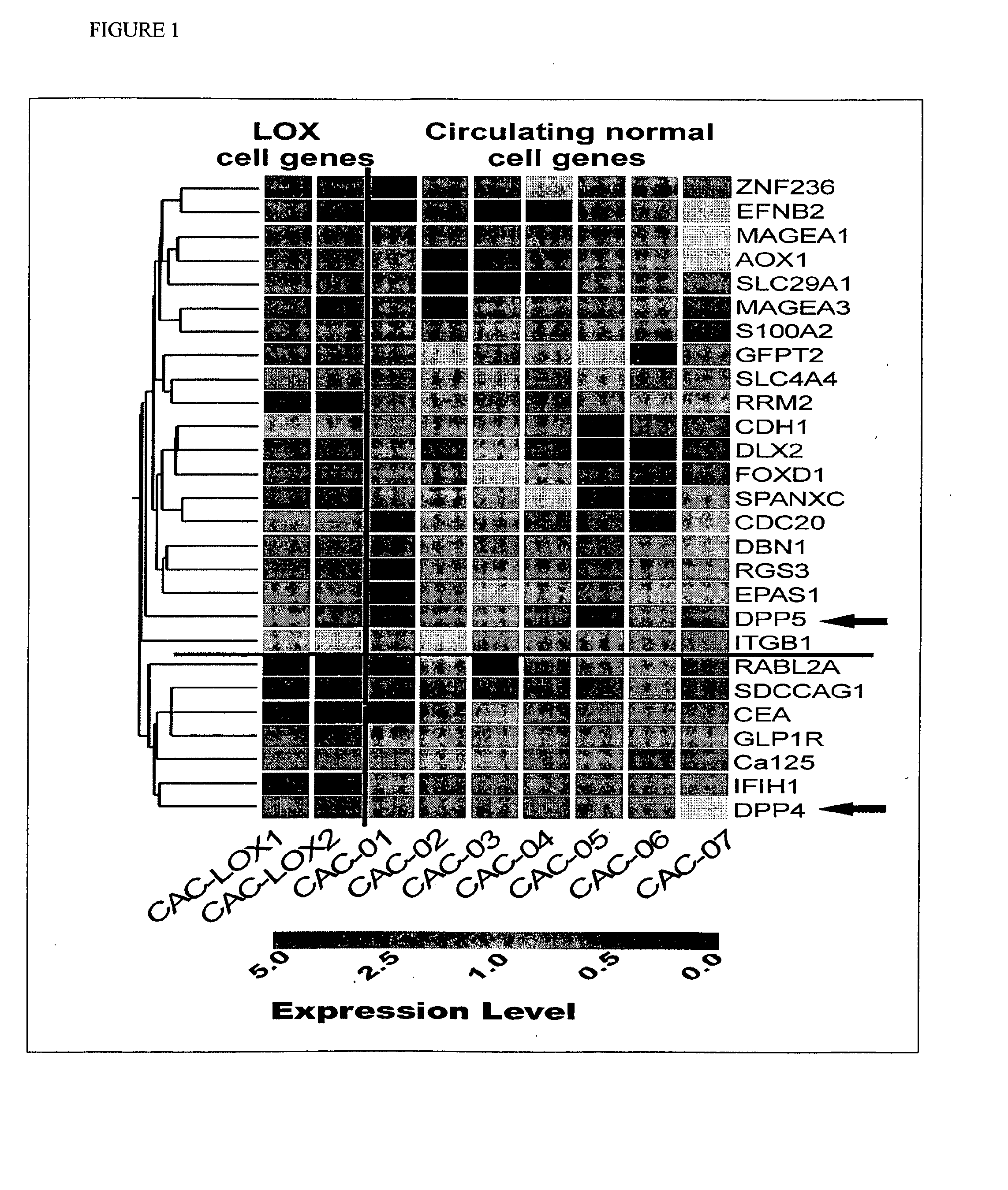
  • Gene expression signatures in enriched tumor cell samples
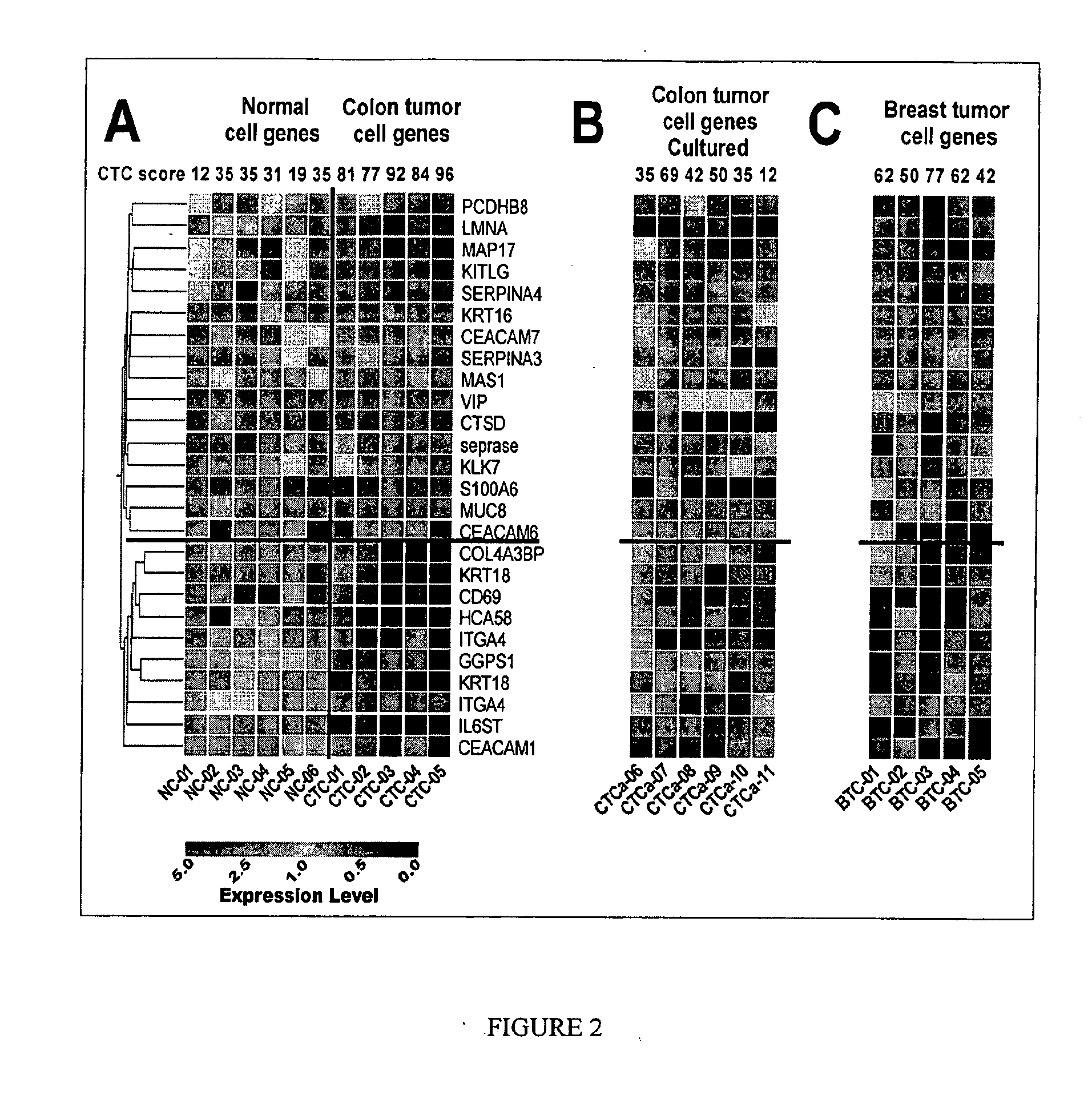
  • Gene expression signatures in enriched tumor cell samples
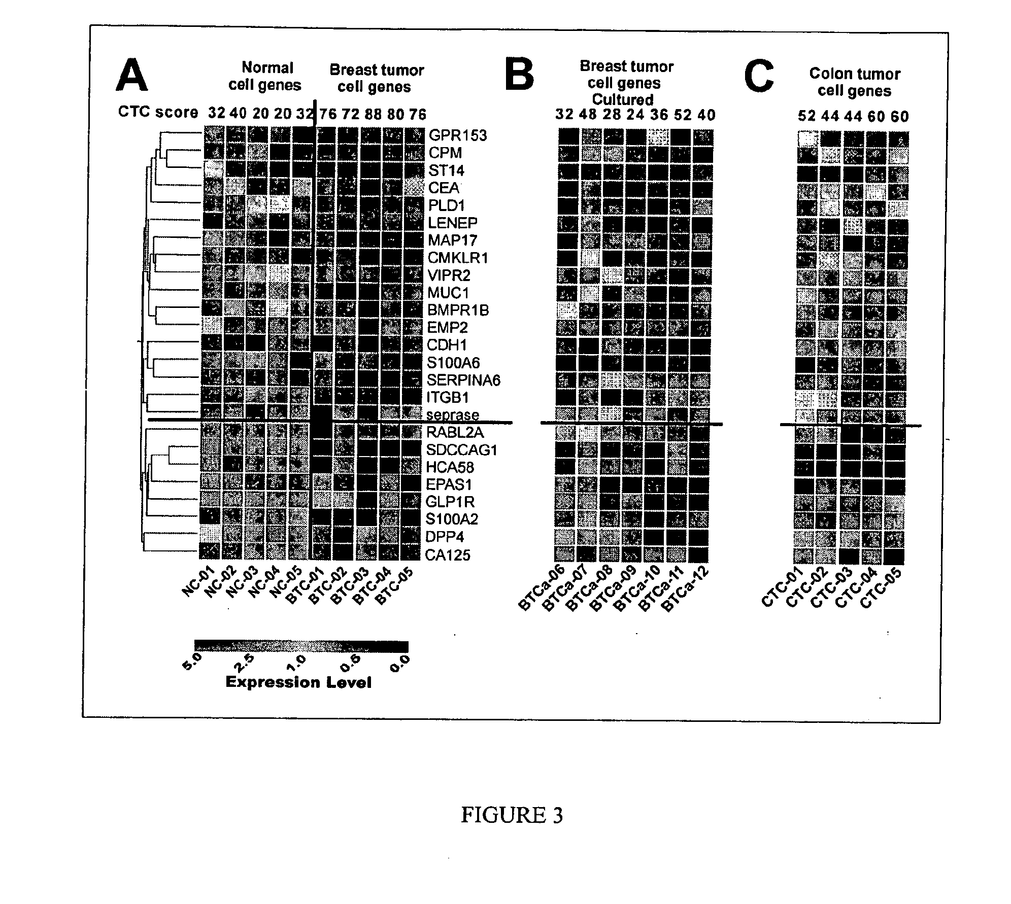
  • Gene expression signatures in enriched tumor cell samples

Examples


example 1

Gene Expression Signatures of Metastatic Tumor Cells in Blood of Patients with Malignant Solid Tumors

[0232]Patients and Healthy Blood Donors. Prior to treatment, cancer patients, including patients with colorectal and breast cancers, as well as healthy individuals were admitted to the study after written, informed consent was obtained. Fresh blood samples and relevant pathological data were obtained from the Stony Brook University Medical Center and the Veteran Administration Medical Center at Northport, N.Y., respectively. The clinical follow-up data included tumor type and stage, as assessed by the treating physician.

[0233]Blood Sample Preparation. Between 4 to 16 ml (median=8 ml) of peripheral blood were collected in Vacutainer™ tubes (Becton Dickinson, Franklin Lakes, N.J.; green top, lithium heparin as anticoagulant). Blood samples were delivered to the laboratory at room temperature within one to four hours from collection, and processed immediately. The CAM-initiated rare cel...

example 2

Gene Expression Signature of Metastatic Tumor Cells in Patients with Ovarian Cancer

[0260]Here, we describe the enrichment of metastatic tumor cells in ascites (MTCA), which contained greater than 25% of cells positive for cytokeratin and CA125, from ovarian cancer patients using a novel collagen adhesion matrix (CAM) method that isolated cells invading collagen. Using the RNA extracted from MTCA fractions and comparing it with the RNA extracted from primary tumor cells in the ovary (PTC), we identified a MTCA signature, in which 21 upregulated genes were common between primary and metastatic tumor cells and 16 genes were upregulated only in metastatic tumor cells. We then selected five genes from the former and four genes from the latter to test in a similar set of samples using real-time RT-PCR. The MICA signature was used to distinguish samples of metastatic tumor cells in ascites from late stage primary tumor cells, and cells from benign and early stage ovarian tumors. Finding no...

example 3

[0272]To validate the 17-candidate colorectal cancer CTC marker genes (DTR, PSCA, MUC3A, THRA, CD79B, NOTCH1, MUCDHL, NEUROG2, MDS028, KRT18, FOLH1, CD44, FCER2, SCF, CD7, SOX1 and TEKT3), the CTC score of a sample was used in the testing set of cellular samples that consists of 9 normal, 9 colorectal cancer and 20 breast cancer (FIG. 16B-C). The CTC score of a sample was determined as the percentage of the 17-candidate colorectal cancer CTC marker genes that exhibited upregulated expression. CTC scores for the 9 normal samples were 0-12%, whereas the CTC scores of the 9 colorectal cancer samples were 94-100% and the 20 breast cancer samples were 24-100%. There was a significant difference of the mean±standard error CTC score between normal and cancer patients with colorectal and breast tumor types (FIG. 16C): 2.67±1.45 in normal samples as compared with 98.67±0.88 in colorectal cancer samples and 69.20±5.53 in breast cancer samples (p-value<0.0001). CTC scores varied significantly ...


PUM

PropertyMeasurementUnit
Ficoll Paque density gradient centrifugationaaaaaaaaaa
diameteraaaaaaaaaa
epithelial cell adhesionaaaaaaaaaa

Abstract

The invention is embodied in methods for finding gene expression signatures of circulating melanoma cells, ovarian, breast, colorectal cancer cells, and circulating endothelial progenitor cells, which signatures are effective in distinguishing the circulating cancer cell from normal circulating cells and can also distinguish between different types of circulating cancer cells.

Description

[0001]This invention was made with government support under Grant No. R42CA108247 and R41CA103462 awarded by the National Institutes of Health. The Government has certain rights in this invention. This application claims priority to U.S. Provisional Patent Application 60 / 994,592, which is incorporated herein by reference in its entirety for all purposes.FIELD OF THE INVENTION[0002]Embodiments of the present invention relate to methods of finding gene expression signatures in rare cells in blood and other body fluids. In particular, these embodiments relate to methods of identifying and distinguishing endothelial progenitor cells in the blood from circulating tumor cells including metastatic cells and, further, distinguishing among circulating tumor cells that originate from diverse tumors of epithelial lineage.BACKGROUND OF THE INVENTION[0003]Malignant tumors and metastatic lesions of epithelial tissues are the most common form of cancer and are responsible for the majority of cance...

Claims


Application Information

Patent Timeline
Patent Type & Authority Applications(United States)
IPC IPC(8): C12Q1/68
CPCC12Q1/6886C12Q2600/158C12Q2600/112
Inventor CHEN, WEN-TIEN
Owner THE RES FOUND OF STATE UNIV OF NEW YORK
Why Eureka
Try Eureka