Topographically engineered structures and methods for using the same in regenerative medicine applications

a technology of regenerative medicine and engineered structures, applied in the direction of prosthesis, pharmaceutical delivery mechanism, medical preparations, etc., can solve the problems of affecting the cell differentiation or cell viability of patients, unable to present a stable substrate for cells to lay down extracellular matrix, and 80% cell death during the injection process alone. , to achieve the effect of enhancing or promoting cell differentiation or cell viability

Inactive Publication Date: 2010-12-16
RGT UNIV OF CALIFORNIA +1
View PDF4 Cites 48 Cited by
  • Summary
  • Abstract
  • Description
  • Claims
  • Application Information

AI Technical Summary

Benefits of technology

[0009]The present invention provides compositions including a cell contacting surface or film comprising nanotopography of nanofibers, nanotubes, nanochannels, microchannels or microwells, which are capable of enhancing or promoting cell differentiation or cell viability. The compositions are useful as medical implants, including orthopedic implants, dental implants, cardiovascular implants, neurological implants, neurovascular implants, gastrointestinal implants, muscular implants, and ocular implants. The present invention also provides methods of treating a patient in need of such an implant.

Problems solved by technology

Earlier studies attempting to deliver brain-derived neurons into the subretinal space resulted in approximately 90% cell death during the injection process alone.
Fast-resorbing PLGA polymers display high shrinkage, which may not present a stable substrate for cells to lay down extracellular matrix.
In addition, the production of acidic degradation species by fast-resorbing polymers can compromise tissue repair.
Fast-resorbing PLGA polymers display high shrinkage, which may not present a stable substrate for cells to lay down extracellular matrix.
In addition, the production of acidic degradation species by fast-resorbing polymers can compromise tissue repair.

Method used


Image

  • Topographically engineered structures and methods for using the same in regenerative medicine applications
  • Topographically engineered structures and methods for using the same in regenerative medicine applications
  • Topographically engineered structures and methods for using the same in regenerative medicine applications

Examples


example 1

PMMA Scaffold Fabrication

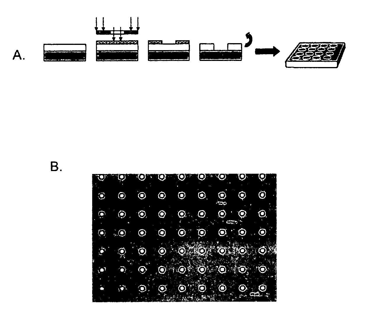
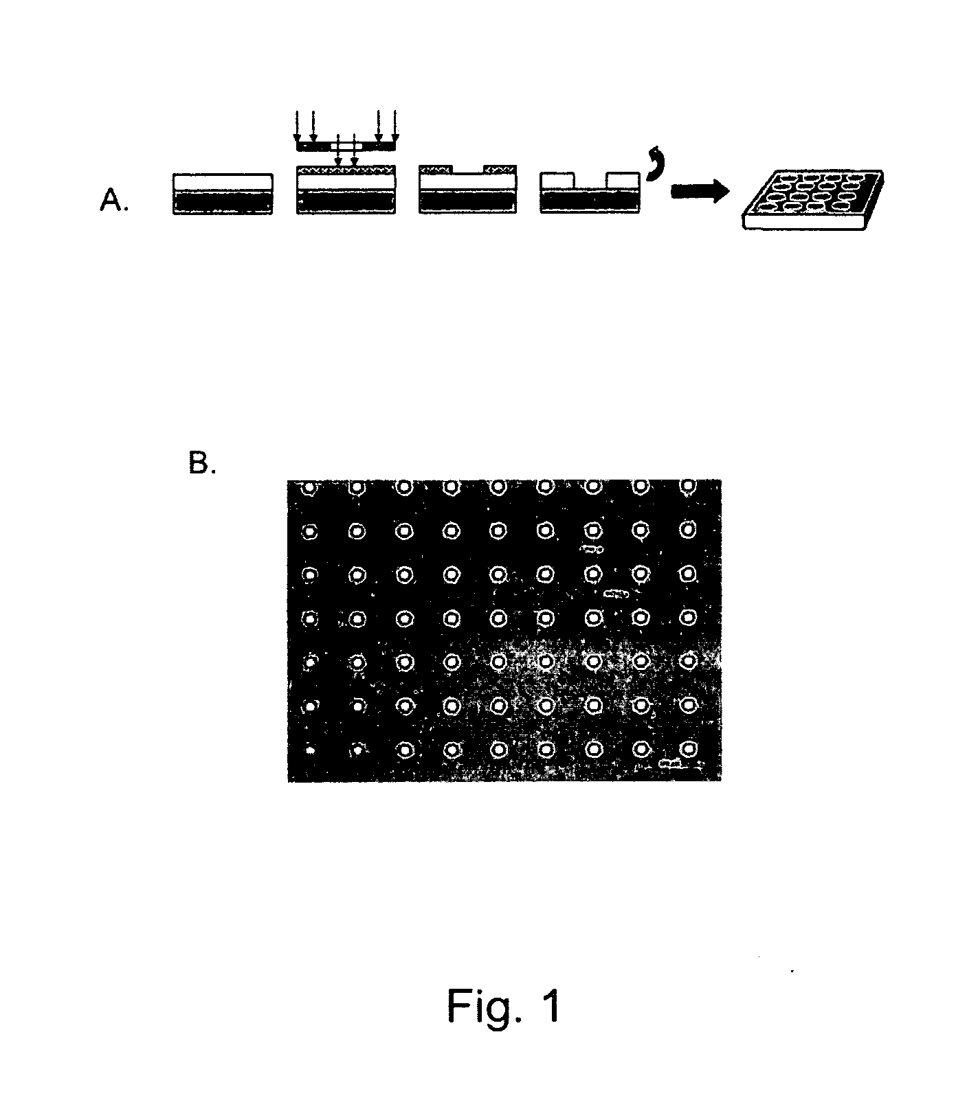
[0127]Micropatterned PMMA thin-film scaffolds were fabricated to contain through pores using a dual process of photolithography and reactive ion etching (FIG. 1, panel A). After the etching process, the diameter of the pores was found to be approximately 11 μm in diameter with an interpore distance of 63 μm (FIG. 1, panel B). The thickness of the ultra-thin film scaffold was measured to be approximately 6 μm in thickness by profilometry (Dektak 8, Veeco, Tucson, Ariz.). Unpatterned (non-porous) and micropatterned (porous) ultra-thin film PMMA scaffolds were separated from the silicon wafer in a single sheet by washing in sterile DI water to dissolve the water-soluble lift-off layer.

example 2

RPC Adherence In Vitro

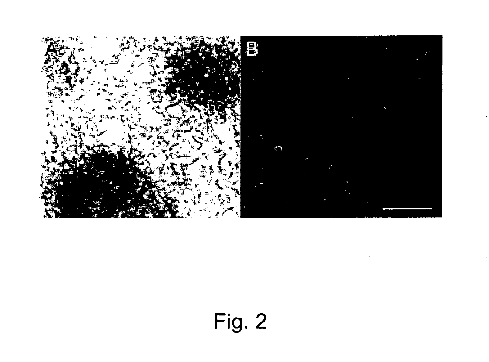
[0128]Non-porous PMMA scaffolds maintained their shape and structural integrity in culture with RPCs for the seven day culture period. Following incubation in poly-L-lysine and laminin, non-porous scaffolds retained RPCs in the primary culture well and through a transfer to a second culture well. Porous PMMA scaffolds retained RPCs with a consistency comparable to the non-porous type with no significant differences observed in culture. At one week in vitro, RPCs on both scaffold types proliferated uniformly across the surface forming an even monolayer radiating from relatively evenly spaced neurospheres (FIG. 2, panels A and B). RPCs were found to remain viable when cultured on both non-porous and porous PMMA. There was no observable difference in the survival or proliferation of RPCs when cultured in the presence or absence of 10×10 mm PMMA scaffolds.

example 3

RPC Survival and Adherence after Subretinal Transplantation

[0129]Although RPC adherence and survival were nearly identical in culture on both types of scaffolds, transplantation with non-porous scaffolds showed limited RPC retention. During the transplantation process non-porous scaffolds lost the majority of their RPCs. The failure of RPC adhesion and / or survival was established by a nearly complete absence of GFP+ RPC fluorescence upon microscopic examination at four weeks post-implantation. Cryosectioning followed by hemotoxalin and eosin staining allowed for localization of implanted non-porous PMMA scaffolds adjacent to host retinal tissue. Under fluorescent illumination non-porous scaffolds showed no visible GFP+ RPC signal.

[0130]Porous PMMA scaffolds demonstrated consistently higher retention of GFP+ RPCs. The porous topography allowed for RPC adherence through transplantation to the posterior eye for up to four weeks. Under microscopic examination many RPCs appeared closely ...


PUM

PropertyMeasurementUnit
lengthaaaaaaaaaa
diameteraaaaaaaaaa
pore diameteraaaaaaaaaa

Abstract

The present invention provides compositions including a cell contacting surface or film comprising nanotopography of nanofibers, nanotubes, nanochannels, microchannels or microwells, which are capable of enhancing or promoting cell differentiation or cell viability. The compositions are useful as medical implants, including orthopedic implants, dental implants, cardiovascular implants, neurological implants, neurovascular implants, gastrointestinal implants, muscular implants, and ocular implants. The present invention also provides methods of treating a patient in need of such an implant.

Description

CROSS-REFERENCE[0001]This application claims the benefit of U.S. Provisional Application No. 60 / 893,775, filed Mar. 8, 2007, and U.S. Provisional Application No. 60 / 911,439, filed Apr. 12, 2007, which applications are incorporated herein by reference in their entirety.BACKGROUND OF THE INVENTION[0002]One of the important challenges to designing better implant materials is to induce tissue growth on or at the implant surface. In a physiological environment, cells respond to nanometric topographies such as fibrous and porous materials formed by components of the extracellular matrix (e.g., callogen, hylauronic acid, laminin, fibronecton, etc.). For example, a number of obstacles remain to the development of a practical ocular implant therapy for the retina remain, including immediate reflux at the time of injection and massive death of donor cells following the standard bolus injection method. While photoreceptor loss is untreatable at present, one of the most promising therapies for ...

Claims


Application Information

Patent Timeline
Patent Type & Authority Applications(United States)
IPC IPC(8): A61F2/02
CPCA61L27/04A61L27/16A61L2400/12A61L2300/602A61L2300/416A61L27/18A61L27/38A61L27/50A61L27/54A61L27/56A61L2300/404A61L2300/41A61L2300/414C08L33/12C08L67/04
Inventor DESAI, TEJAL A.TAO, SARAHYOUNG, MICHAELKLASSEN, HENRY J.
Owner RGT UNIV OF CALIFORNIA
Why Eureka
Try Eureka