Synthesis of nanotopographic biomimetic membranes for tissue culture, engineering and prosthetics applications

Inactive Publication Date: 2013-08-15
RGT UNIV OF CALIFORNIA
View PDF0 Cites 7 Cited by
  • Summary
  • Abstract
  • Description
  • Claims
  • Application Information

AI Technical Summary

Problems solved by technology

Meanwhile, conventional cell culture ware and other commonly used biomedical devices comprise materials with essentially fore

Method used


Image

  • Synthesis of nanotopographic biomimetic membranes for tissue culture, engineering and prosthetics applications
  • Synthesis of nanotopographic biomimetic membranes for tissue culture, engineering and prosthetics applications
  • Synthesis of nanotopographic biomimetic membranes for tissue culture, engineering and prosthetics applications

Examples


example 1

Synthesis of Nanostructured Biomimetic Membrane Materials

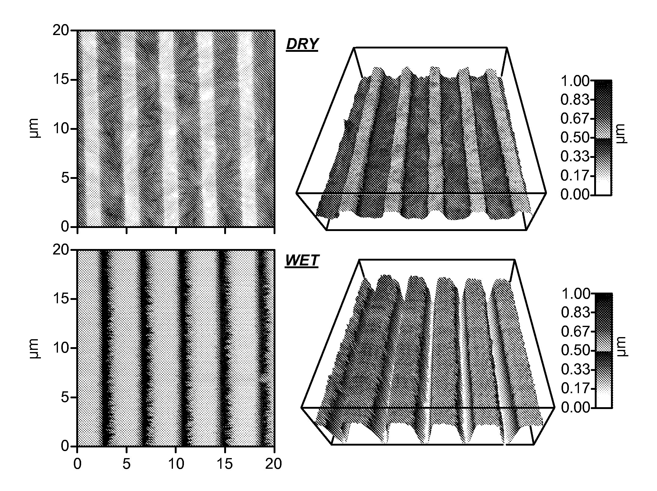
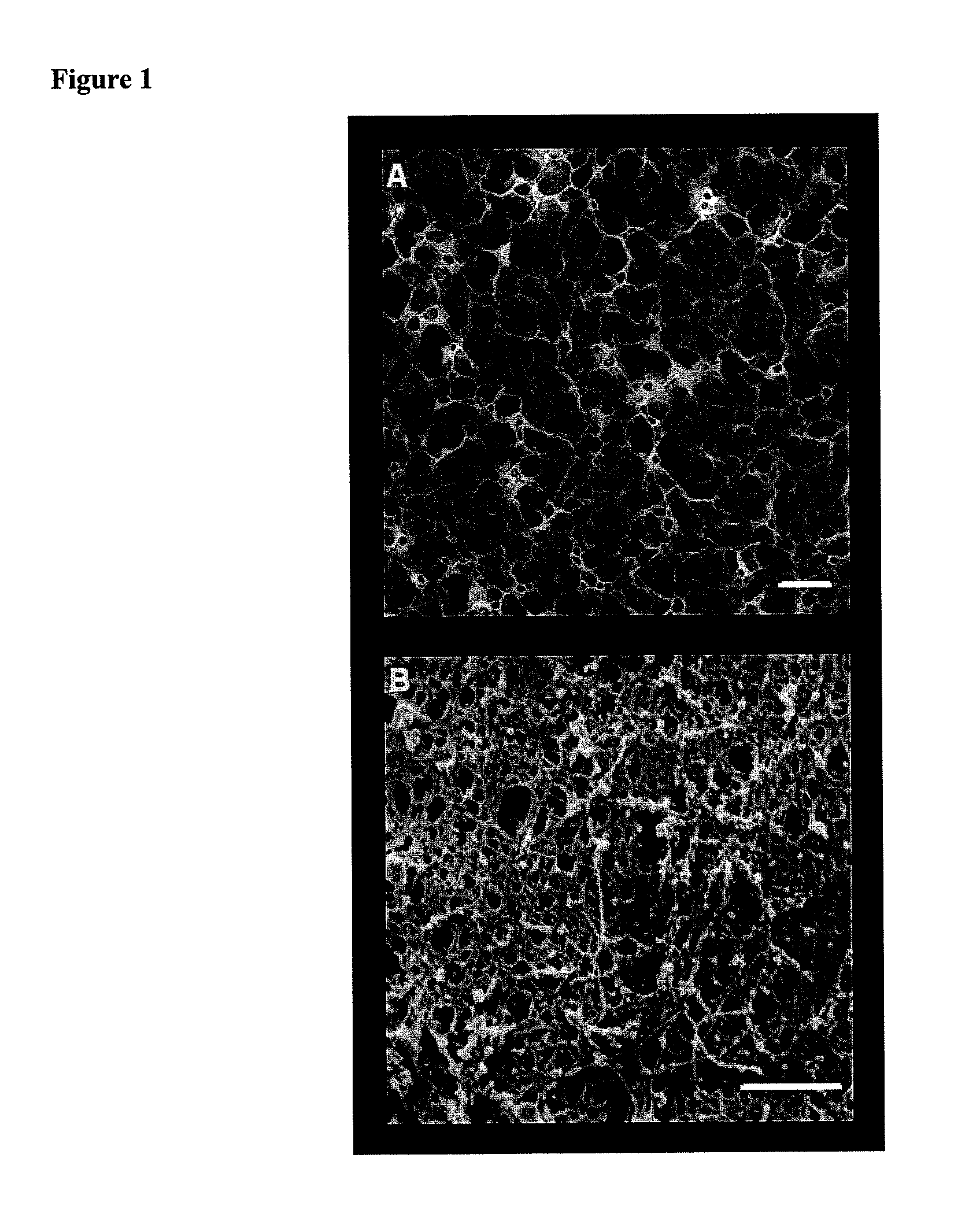
[0097]Nanostructured membrane fabrication. Stabilized nanoporous membranes were generated by forming porous polyelectrolyte membranes (PEMs) of poly(acrylic acid) (PAA, MW˜60,000, Polysciences, Inc., Warrington, Pa.) and poly(allylamine hydrochloride) (PAH, MW˜160,000, Alfa Aesar, Ward Hill, Mass.) on glass microscope slides. Solutions of PAA and PAH were prepared at 0.01 M (based on MW of monomer unit) at a pH of 3.5 and 7.5, respectively. Solutions were prepared using ultra-high purity water (18.2 MΩ.cm) and the pH was controlled with NaOH and HCl. Before PEM formation, the glass microscope slides were plasma treated (Harrick Plasma Cleaner, Hayrick Plasma, Ithaca, N.Y.) for one minute and then immediately silanized by exposure to 3-aminopropyltrimethoxysilane (APS, Sigma Aldrich) vapor for 1 hour under vacuum. After 1 hour in the presence of APS vapor, the APS was removed and the samples were left under vacuum for 24 hours ...

example 2

Study of Cell Proliferation and Migration on Nanostructured Biomimetic Membranes

[0105]Cell culture. Human umbilical vein endothelial cells (HUVECs) and human aortic endothelial cells (HAECs) were maintained in endothelial basal media supplemented with EGM-2 BulletKit (Lonza, Walkerville, Md.). The BulletKit contains GA-1000, hEGF, fetal bovine serum, heparin, ascorbic acid, R3-IGF, VEGF, hFGF-B and hydrocortisone. During culture and migration assays, cells were maintained at 37° C. and 5% CO2.

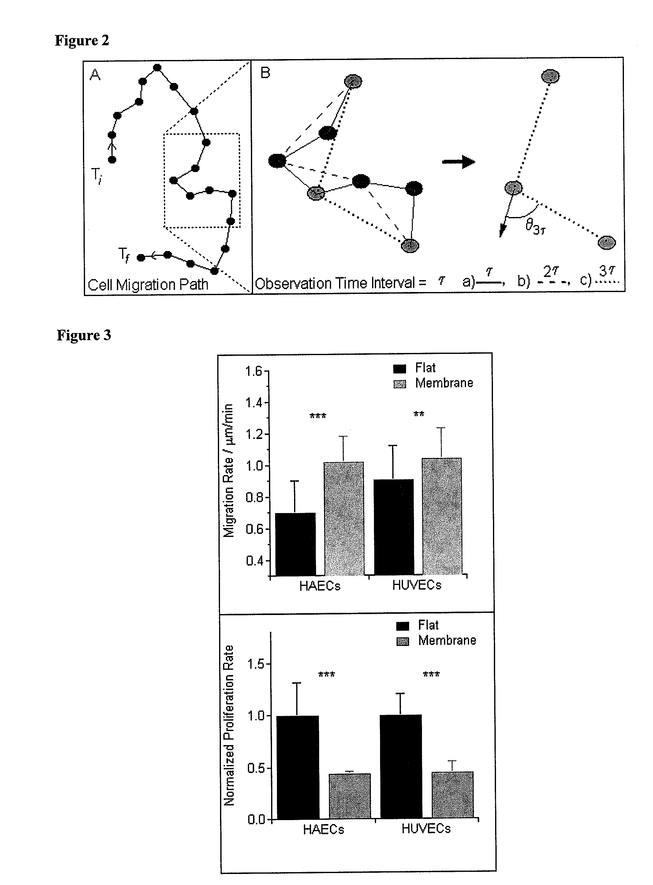
[0106]Cell Migration Analysis via a Custom Automated Cell Tracker. Time-lapse observations of cells were obtained on both the flat and membrane surfaces over a 12-hour time period, with a collection rate of one frame every eight minutes (90 frames). Cells were imaged in phase contrast using an inverted light microscope (Zeiss, Axio Observer Al, Carl Zeiss, Thornwood, N.Y.) with a 10× objective. Cells from a minimum of ten separate locations were imaged for each condition. Cells were plated at 7...

example 3

Analysis of Gene Expression in Endothelial Cells Cultured on Biomimetic Nanostructured Membranes

[0116]Gene expression assays. Both HUVECs and HAECs were plated at 100,000 cells per plate and allowed to culture for three days on the flat and membrane surfaces prior to RNA extraction. Expression of SPARC (secreted protein acidic and rich in cysteine), PECAM (platelet endothelial cell adhesion molecule), and MMP-2 (matrix metalloproteinase-2) was quantified for both HUVECs and HAECs. The influence of TNFα (tumor necrosis factor-alpha) on the expression of ICAM-1(intercellular adhesion molecule-1) in HAECs was also studied. For those experiments, HAECs were exposed to 10 ng / ml TNFα for 12 hours prior to RNA extraction. RNA was extracted using the Qiagen RNeasy kit according to the manufacturer's protocol (Qiagen, Valencia, Calif.). Semi-quantitative real-time PCR was performed using 75 ng of RNA per reaction and a one-step TaqMan kit with commercially available aptamers for SPARC, PECAM...


PUM

PropertyMeasurementUnit
Lengthaaaaaaaaaa
Nanoscale particle sizeaaaaaaaaaa
Phase separationaaaaaaaaaa

Abstract

The present invention provide methods for preparing nanostructured membranes. The methods include: providing a substrate with a charged silanized surface; forming a multilayered membrane containing at least two polyelectrolytes; inducing polyelectrolyte phase separation; crosslinking the multilayered membrane; and covalently linking the multilayered membrane to the silanized surface. Methods for fabricating membrane replicas are also disclosed, as well as devices such as cell- and tissue-culture substrates that contain the membranes and membrane replicas. Resulting materials exhibit topographic features and compliance of the extracellular matrix in vivo.

Description

CROSS-REFERENCES TO RELATED APPLICATIONS[0001]The present application claims priority to U.S. Provisional Patent Application No. 61 / 596,680, filed Feb. 8, 2012, and U.S. Provisional Patent Application No. 61 / 617,281, filed Mar. 29, 2012, the entirety of which are incorporated herein by reference.STATEMENT AS TO RIGHTS TO INVENTIONS MADE UNDER FEDERALLY SPONSORED RESEARCH AND DEVELOPMENT[0002]This invention was made with Government support under Grant No. RC2AR058971, awarded by the National Institutes of Health. The Government has certain rights in this invention.REFERENCE TO A “SEQUENCE LISTING,” A TABLE, OR A COMPUTER PROGRAM LISTING APPENDIX SUBMITTED ON A COMPACT DISK[0003]Not ApplicableBACKGROUND OF THE INVENTION[0004]It is well established that the physical attributes of the extracellular environment (e.g. topography and / or compliance) can inform internal cell processes and responses to external stimuli. The response of cells to these attributes is particularly relevant given ...

Claims


Application Information

Patent Timeline
IPC IPC(8): A61F2/02B29C59/02B29C35/08B05D3/10
CPCA61F2/02B82Y40/00B05D3/108B29C59/026B29C35/0805B29C59/022G03F7/0002B29C2035/0827B29C2059/023G03F7/027G03F7/0751B01D67/0002B01D67/0006B01D67/0034B01D69/125B01D71/40B01D71/48B01D71/60B01D71/70B01D71/82B01D2323/30B01D2323/345B82Y10/00B05D3/107
Inventor MCKEE, CLAYTONMURPHY, CHRISTOPHER J.RUSSELL, PAULGARLAND, SHAUN P.
Owner RGT UNIV OF CALIFORNIA
Why Eureka
Try Eureka