T1 rho magnetic resonance imaging for staging of hepatic fibrosis

a fibrosis and magnetic resonance imaging technology, applied in the field of rho magnetic resonance imaging, can solve the problems of high complication rate, mortality, and liver cirrhosis currently a significant cause of death

Inactive Publication Date: 2011-02-03
THE TRUSTEES OF THE UNIV OF PENNSYLVANIA
View PDF6 Cites 17 Cited by
  • Summary
  • Abstract
  • Description
  • Claims
  • Application Information

AI Technical Summary

Benefits of technology

[0018]The proposed technology offers a non-invasive MRI technique based on T1ρ contrast that can be sensitive enough to detect small changes in ECM protein concentration and architecture in all stages of hepatic fibrosis.

Problems solved by technology

Cirrhosis of the liver is currently a significant cause of death in the United States of America.
Liver biopsies, however, are invasive, associated with pain, have high complication rates, and may lead to morbidity and, in rare cases, mortality.
In addition, liver biopsies are inaccurate due to sampling variability (Talwalkar, J. A., et al., “Magnetic resonance imaging of hepatic fibrosis: Emerging Clinical Applications,”Hepatology 2008 47(1), pp.
This method can be used to monitor abnormal iron accumulation in the liver but is not used to diagnose or stage fibrosis.
Although magnetic resonance elastography (MRE) has shown promising in accurately classifying liver fibrosis based on liver stiffness, it was finally shown to be unable to distinguish between the early stages of fibrosis.
More recent studies, however, have shown that this is not necessarily the case during early stages of fibrosis.
These techniques above are all able to differentiate between mild and severe fibrosis, but are unable to make finer distinctions.
As a result, it is clear that these techniques do not offer a method for accurately staging early or intermediate fibrosis or for monitoring the progress of patients over short time periods.
It is a highly morbid and potentially fatal condition.
Multiple different non-invasive approaches have been developed for diagnosis and monitoring of hepatic fibrosis, however none are able to achieve sufficient sensitivity to accurately stage fibrosis, especially in its earliest stages.
Specifically, serum markers, ultrasound, T1- and T2-weighted MRI, and contrast-enhanced MRI were not able to differentiate the early and intermediate stages of fibrosis or could only detect late-stage fibrosis; they are unable to detect small differences from one time to the next in individual patients.
Similarly, studies using diffusion-weighted MR imaging and MR spectroscopy showed significant discrepancies and low sensitivities in staging the early and middle stages of fibrosis.
MR elastography can more accurately stage liver fibrosis based on stiffness, although it is ineffective in distinguishing between the early stages of fibrosis and is able to detect only significant differences in the amount of fibrosis.

Method used


Image

  • T1 rho magnetic resonance imaging for staging of hepatic fibrosis
  • T1 rho magnetic resonance imaging for staging of hepatic fibrosis
  • T1 rho magnetic resonance imaging for staging of hepatic fibrosis

Examples


example 1

[0049]T1ρ magnetic resonance scans were performed on collagen phantoms having different concentrations of Type I collagen, the main protein that increases during fibrosis. A 3T Siemens Trio MRI scanner was used. 1 cm×1 cm×1 cm liver explant samples were placed in a 42-well plate and the plate was mounted on a custom-built coil and imaged. The images are shown in FIG. 2.

[0050]A linear correlation between T1ρ relaxation time and collagen concentration was observed, as shown in FIG. 3. Relaxation times increased as collagen concentrations decreased as shown in FIG. 4.

example 2

[0051]In this example, T1ρ magnetic resonance scans were performed on several different liver explants including normal liver tissue (N), liver tissue with cirrhosis of undetermined cause (UC), liver tissue with cirrhosis caused by primary sclerosing cholangitis (PSC) and liver tissue with cirrhosis caused by biliary atresia. The results are shown in FIG. 5. Normal liver tissue exhibited a T1ρ relaxation time of about 25-30 ms, whereas the cirrhotic liver tissues each exhibited different T1ρ relaxation times thereby showing that the T1ρ relaxation time can differentiate between the degree of matrix abnormality even in cirrhotic livers that would be classified at the same stage (Metavir F4) by standard histological staging systems. FIG. 6 shows a plot of the T1ρ relaxation time versus the progression of liver fibrosis showing that the more extensive the liver fibrosis the longer the T1ρ relaxation time.

example 3

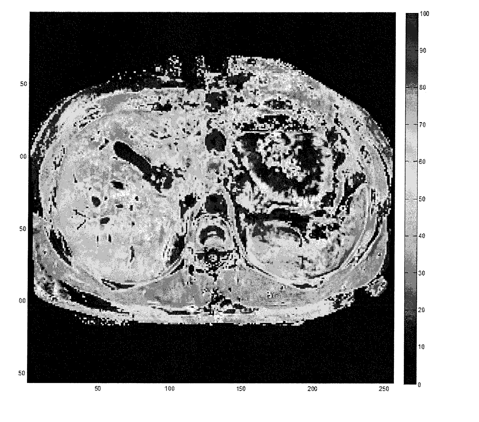
[0052]In this example, T1ρ magnetic resonance scans were performed in vivo on a human liver. An axial image of a human volunteer was performed at the thoracic level using a T1ρ imaging sequence at different spin lock amplitudes and a T1ρ map was generated (FIG. 7). This study showed that the human liver has a heterogeneous distribution of relaxations over its surface with T1ρ values ranging between 25 seconds and 50 seconds. This heterogeneity is most likely correlated with the normal micro-architecture of the liver. Regions of the liver with higher macromolecular content have higher relaxation times. In pathologic conditions, an increase in T1ρ relaxation times in the affected areas is expected, as compared to normal tissue. This example shows the feasibility of the technique in humans.

[0053]From these examples it can be concluded that T1ρ magnetic resonance scans can be employed to quantify changes in collagen concentrations and that the T1ρ signal can be directly correlated with ...


PUM


Abstract

Methods for diagnosis of fibrotic diseases, staging of fibrotic diseases and monitoring treatment of fibrosis. The presence of fibrotic tissue is detected. First, a T1ρ relaxation time of tissue is determined using magnetic resonance imaging. The determined T1ρ relaxation time is then compared to a baseline T1ρ relaxation time indicative of healthy tissue, and the presence of fibrotic tissue is then determined based on results of said comparison step. To determine a stage of fibrosis, a T1ρ relaxation time of tissue is determined and compared to one or more calibrated T1ρ relaxation times indicative of one or more stages of fibrosis, and the stage of fibrosis is determined based on results of said comparison step. The proposed technology offers a non-invasive MRI technique based on T1ρ contrast that is sensitive enough to detect small changes of ECM hepatic protein concentration and architecture in all stages of hepatic fibrosis.

Description

CROSS REFERENCE TO RELATED APPLICATIONS[0001]This application claims the benefit of U.S. Provisional Application No. 61 / 230,392, filed Aug. 1, 2009, the entirety of which is incorporated herein by reference.BACKGROUND OF THE INVENTION[0002]1. Field of Invention[0003]The present invention is directed to a method of using T1 Rho magnetic resonance imaging to detect and monitor the progression of fibrotic diseases.[0004]2. Brief Description of the Prior Art[0005]Cirrhosis of the liver is currently a significant cause of death in the United States of America. Wolf, D. C., “Cirrhosis,”eMedicine 2008. Despite the prevalence of liver disease, the primary method for diagnosing and monitoring the progress of patients afflicted with liver cirrhosis is liver biopsy. Manning, D. S. and Afdhal, N. H., “Diagnosis and Quantitation of Fibrosis,”Gastroenterology 2008, 134(6), pp. 1670-81. Liver biopsies, however, are invasive, associated with pain, have high complication rates, and may lead to morbi...

Claims


Application Information

Patent Timeline
Patent Type & Authority Applications(United States)
IPC IPC(8): A61B5/055
CPCG01R33/50A61B5/055
Inventor DAYE, DANIAREDDY, RAVINDERWELLS, REBECCA
Owner THE TRUSTEES OF THE UNIV OF PENNSYLVANIA
Why Eureka
Try Eureka