Process and apparatus for non-invasive, continuous in vivo measurement of hematocrit
a technology of in vivo measurement and non-invasive methods, applied in the field of non-invasive methods and apparatus for obtaining a continuous time record of relative blood hematocrit, can solve the problems of no means to obtain a continuous real-time record in order to log temporal changes, and difficult detection of internal bleeding, so as to reduce the standard deviation of hematocrit
- Summary
- Abstract
- Description
- Claims
- Application Information
AI Technical Summary
Benefits of technology
Problems solved by technology
Method used
Image
Examples
example 1
Simultaneous, Noninvasive Observation of Elastic Scattering, Fluorescence and Inelastic Scattering as a Monitor of Blood Flow and Hematocrit an Human Volar Side Fingertip Capillary Beds
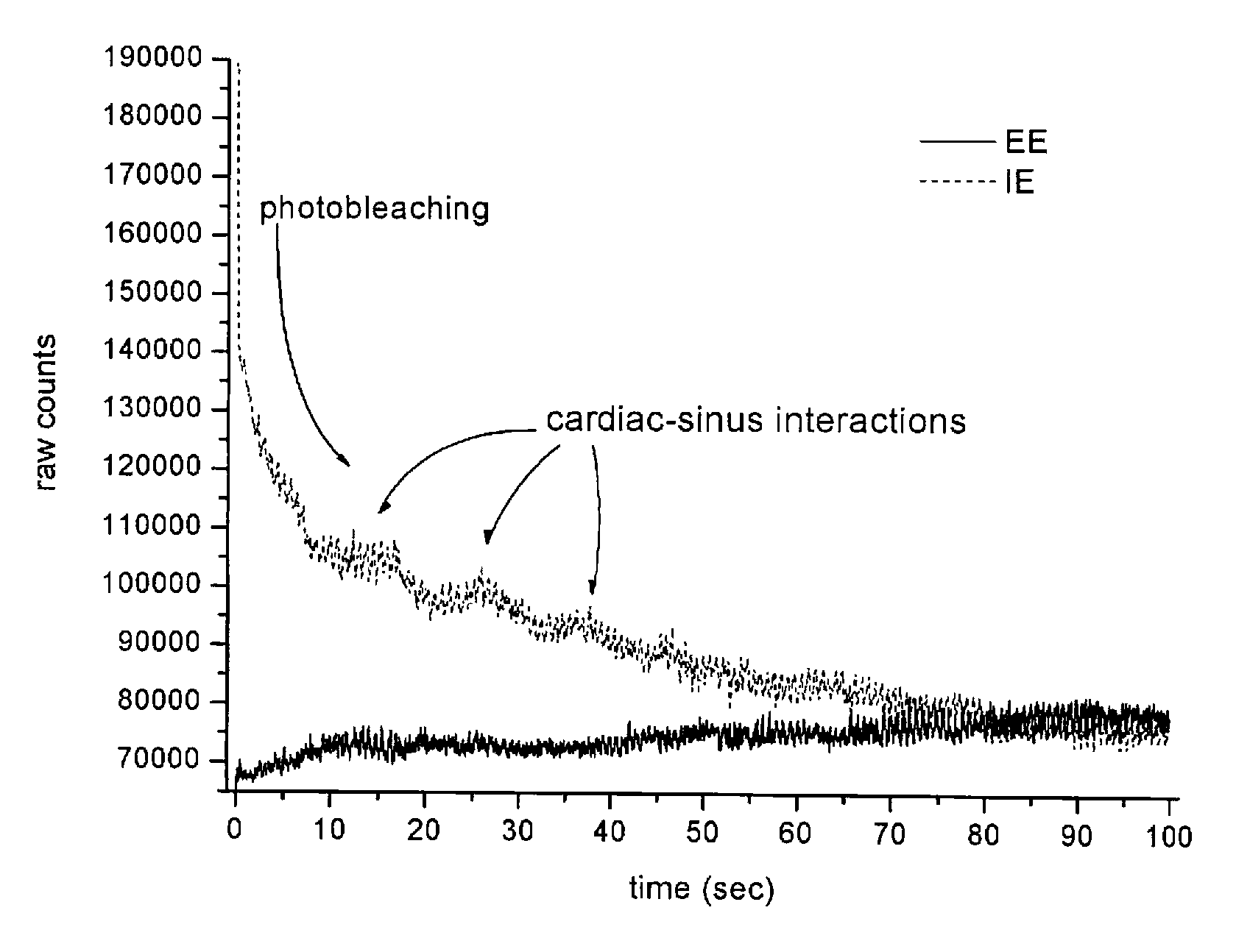
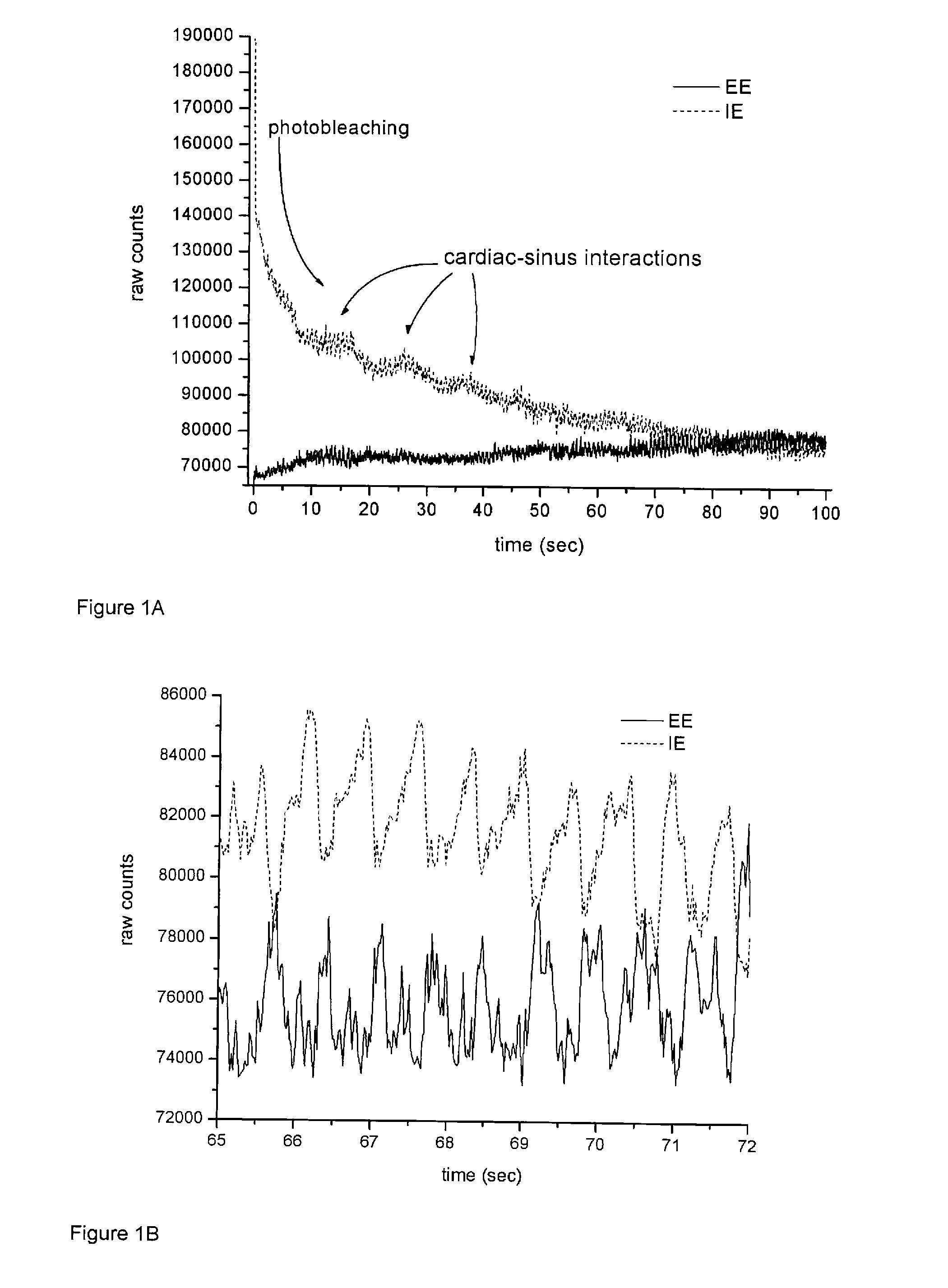
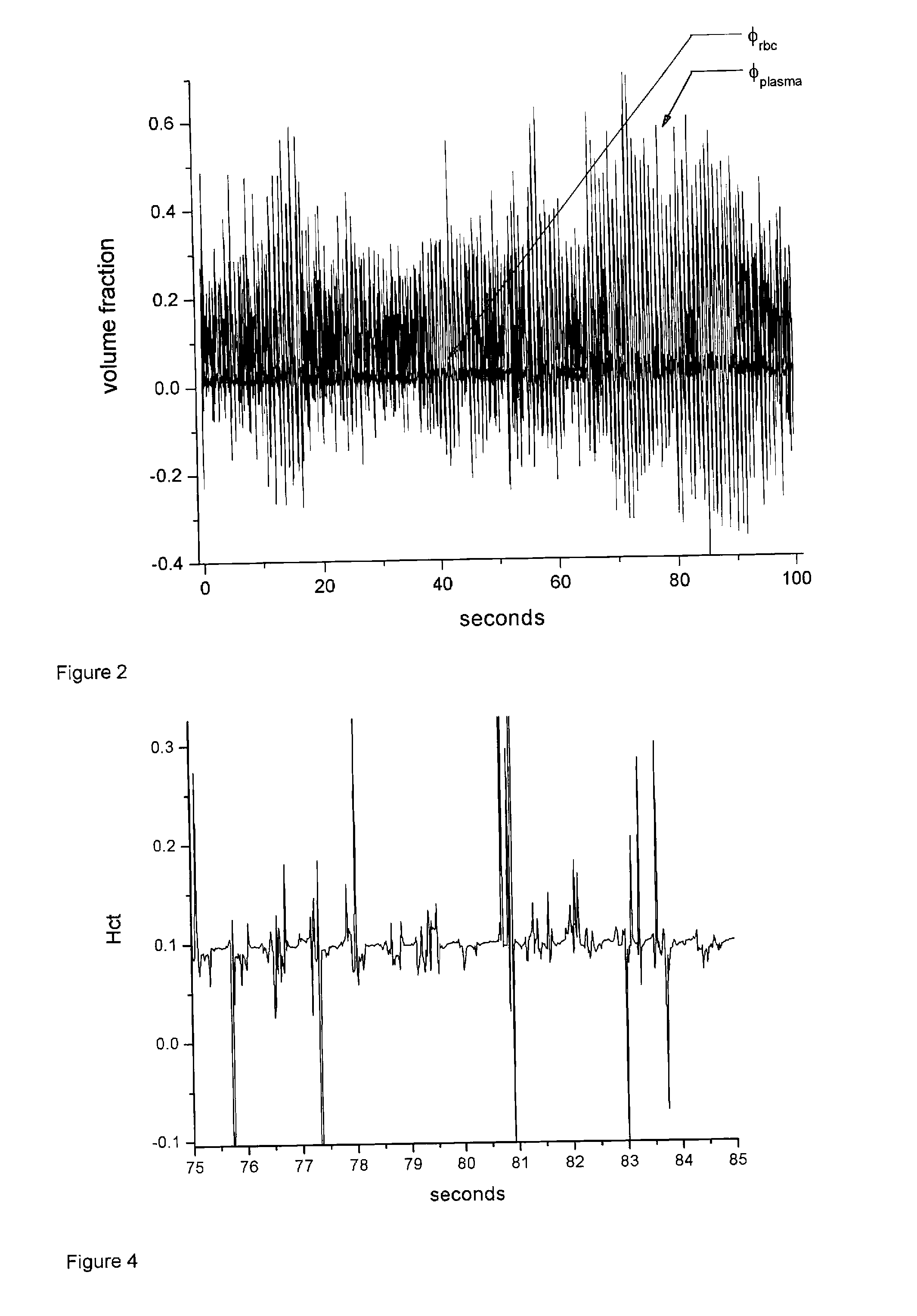
[0103]This Example describes simultaneous observation of elastic scattering, fluorescence and inelastic scattering from in vivo near infrared probing of human skin. Careful control of the mechanical force needed to obtain reliable registration of in vivo tissue with an appropriate optical system allows reproducible observation of blood flow in capillary beds of human volar side fingertips. Under continuous near infrared excitation, the time dependence of the elastically scattered light is highly correlated with that of the combined fluorescence and Raman scattered light. This is interpreted in terms of turbidity; i.e. the impeding effect of red blood cells on optical propagation to and from the scattering centers and the changes in the volume percentages of the tissues in the irradiated volume with no...
example 2
Probing Human Fingertips In Vivo Using Near Infrared Light: Model Calculations
[0120]The volar side fingertip capillary beds are probed with near infrared laser light and Raman, Rayleigh and Mie scattered light and fluorescence are collected. The results are interpreted using radiation transfer theory in the single-scattering approximation. The surface topography of the skin is modeled using the Fresnel Equations. The skin is treated as a three-layer material, with a mean-field treatment of tissue composition and related optical properties. The model, with a reasonable choice of tissue parameters, gives a remarkably accurate account of the features of actual measurements. It predicts the optimal values for the incident angle of the laser beam and the distance between beam and detector. It explains the correlated temporal changes in the intensities of elastically (EE) and inelastically (IE) scattered light caused by heart driven pulses, and why they are out of phase. With appropriate ...
example 3
Direct Noninvasive Observation of Near Infrared Photobleaching of Autofluorescence in Human Volar Side Fingertips In Vivo
[0208]Human transdermal in vivo spectroscopic applications for tissue analysis involving near infrared (NIR) light often must contend with broadband NIR fluorescence that, depending on what kind of spectroscopy is being employed, can degrade signal to noise ratios and dynamic range. Such NIR fluorescence, i.e. “autofluorescence” is well known to originate in blood tissues and various other endogenous materials associated with the static tissues. Results of recent experiments on human volar side fingertips in vivo are beginning to provide a relative ordering of the contributions from various sources. Preliminary results involving the variation in the bleaching effect across different individuals suggest that for 830 nm excitation well over half of the total fluorescence comes from the static tissues and remainder originates with the blood tissues, i.e. the plasma a...
PUM
 Abstract
Description
Claims
Application Information
 - R&D
- Intellectual Property
- Life Sciences
- Materials
- Tech Scout
- Unparalleled Data Quality
- Higher Quality Content
- 60% Fewer Hallucinations
Browse by: Latest US Patents, China's latest patents, Technical Efficacy Thesaurus, Application Domain, Technology Topic, Popular Technical Reports.
© 2025 PatSnap. All rights reserved.Legal|Privacy policy|Modern Slavery Act Transparency Statement|Sitemap|About US| Contact US: help@patsnap.com


