

Use of UTP for the diagnosis of stenoses and other conditions of restricted blood flow
- Summary
- Abstract
- Description
- Claims
- Application Information
AI Technical Summary
Benefits of technology
Problems solved by technology
Method used
Image

Examples
embodiments
[0091]The methods described herein are useful in determining compromised blood flow in an individual with suspected compromised blood flow. Such methods mimic the increased blood flow that occurs during exercise and are thus particularly useful in patients who cannot undertake exercise or for whom it is less desirable to undertake exercise.
[0092]The present invention relates to a method for determining whether blood flow is restricted in a blood vessel of an individual suspected of compromised blood flow in the vessel, the method comprising the steps of: (a) delivering UTP, a derivative thereof, or a salt thereof to said vessel, (b) assessing blood flow quantitatively in the vessel by obtaining a value that indicates or correlates to blood flow in said vessel, (c) comparing the obtained value with a reference value, and (d) whether the individual has compromised blood flow based on the results of the comparison.
[0093]The methods of the invention may be used to determine a suspected ...
example 1
Systemic Infusion of UTP for Myocardial Perfusion Imaging (Prophetic)
[0165]MPI is a form of functional cardiac imaging that may be used for the diagnosis of ischemic heart disease. The underlying principle is that under conditions of stress, diseased myocardium receives less blood flow than normal myocardium. MPI is one of several types of cardiac stress tests.
[0166]A cardiac specific radiopharmaceutical, such as 99mTc-tetrofosmin (Myoview, GE healthcare) or 99mTc-sestamibi (Cardiolite, Bristol-Myers Squibb) is administered. Following this, the heart rate and coronary blood flow is raised to induce myocardial stress by systemic infusion of UTP, a derivative thereof, or a salt thereof.
[0167]SPECT imaging performed after stress reveals the distribution of the radiopharmaceutical, and therefore the relative blood flow to the different regions of the myocardium. Diagnosis is made by comparing stress images to a further set of images obtained at rest (the reference value). As the radionu...
example 2
Local Infusion of UTP Via Guiding Catheter in the Coronary Arteries in Humans with Coronary Artery Disease
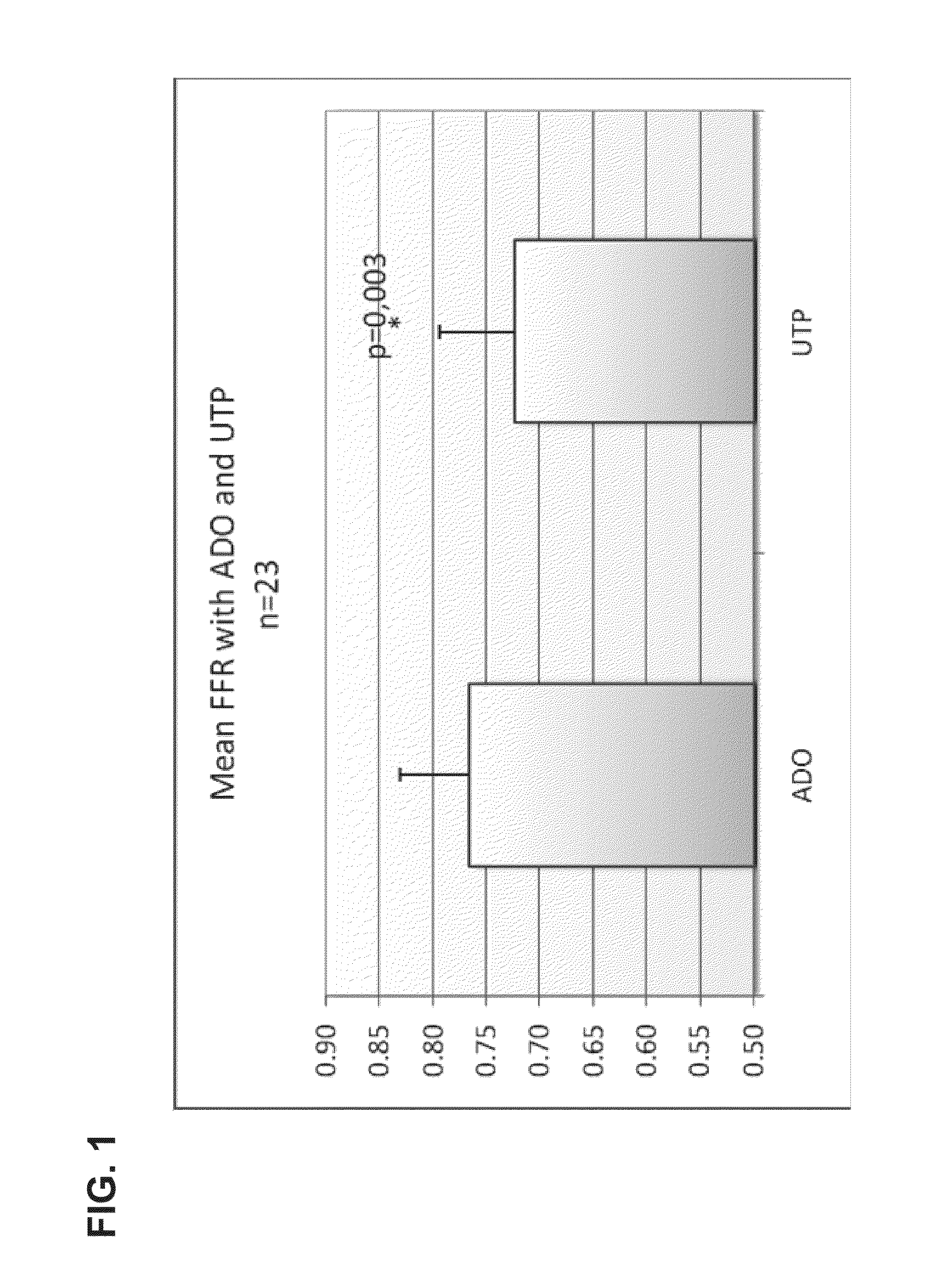
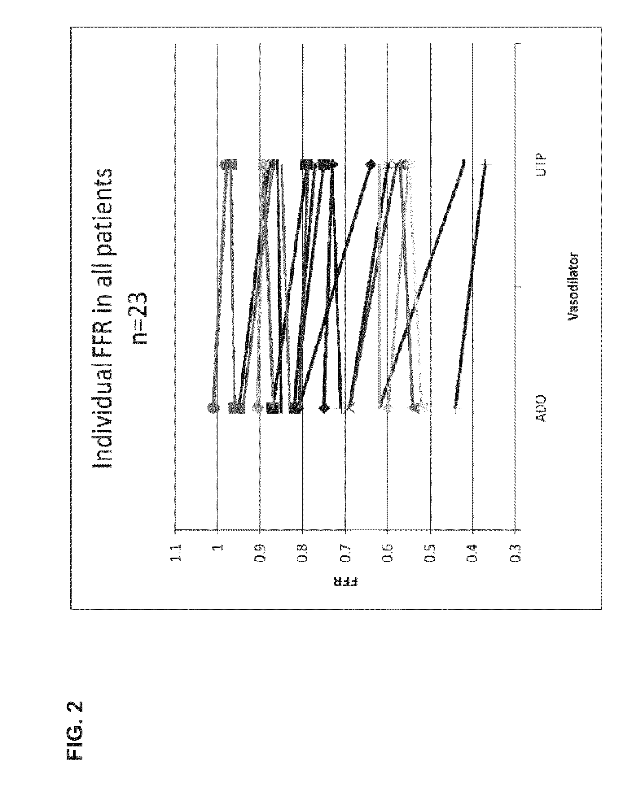
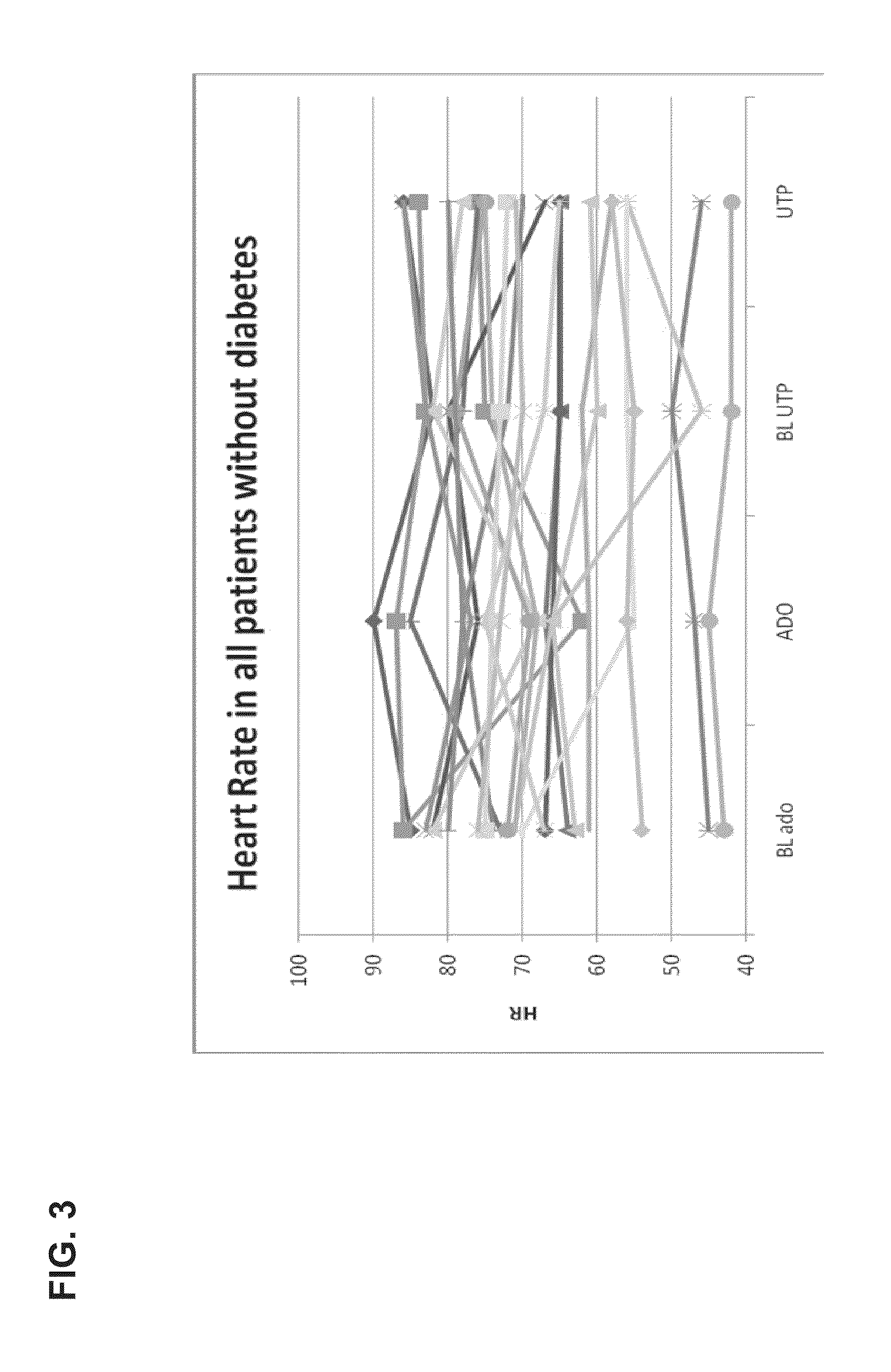
[0169]The study was performed in 23 patients undergoing elective coronary arteriography (CAG) due to repetitive episodes of typical ischemic chest pain or in patients with recent Non-ST Elevated Myocardial Infarction (>2 days from entry into study). In eligible patients with uni- or multivessel coronary artery disease, lesions with stenosis of at least 50% of their diameter and that were thought to require PCI on the basis of angiographic appearance and clinical data were identified.
Dose / Response Protocol
[0170]In the first part of the experiment, FFR and Coronary Flow (velocity, CFR) were measured after the induction of coronary hyperemia by a continuous intracoronary infusion of UTP (Jena Bioscience GmbH, Germany) and adenosine in random order in a guide catheter (5F catheter, Cordis Corp.). Two UTP doses of 240 and 360 μg / min were tested. UTP was prepared by dissolving 100 mg ...
PUM
| Property | Measurement | Unit |
|---|---|---|
| Mass | aaaaa | aaaaa |
| Mass | aaaaa | aaaaa |
| Density | aaaaa | aaaaa |
Abstract
Description
Claims
Application Information