Ultrasound guided optical coherence tomography, photoacoustic probe for biomedical imaging

a biomedical imaging and optical coherence tomography technology, applied in the field of intravascular imaging, can solve the problems of poor blood vessel imaging performance of conventional ultrasound imaging, medical safety concerns, and the fabrication of miniaturized ultrasound probes, and achieve the effects of high resolution, high resolution, and safe way to obtain intravascular oct images

Active Publication Date: 2011-04-28
RGT UNIV OF CALIFORNIA
View PDF5 Cites 127 Cited by
  • Summary
  • Abstract
  • Description
  • Claims
  • Application Information

AI Technical Summary

Benefits of technology

[0019]The purpose of this system is to provide a means for high resolution imaging of biomedical tissue. The guidance of ultrasound imaging allows the area of interest to be found and thus a relatively smaller amount of flush agent will be needed, which provides a safer way to obtain intravascular OCT images. The combination of the two imaging modalities yields high resolution thanks to OCT and deep penetration depth due to ultrasound imaging.
[0020]The

Problems solved by technology

Therefore, the fabrication of a miniaturized ultrasound probe is another important issue for IVUS imaging.
Nevertheless, one of drawbacks of OCT is that it needs to use saline water to flush blood away from the probe in order to remove the interference received from the blood.
However, both solutions have medical safety concerns

Method used


Image

  • Ultrasound guided optical coherence tomography, photoacoustic probe for biomedical imaging
  • Ultrasound guided optical coherence tomography, photoacoustic probe for biomedical imaging
  • Ultrasound guided optical coherence tomography, photoacoustic probe for biomedical imaging

Examples


Embodiment Construction

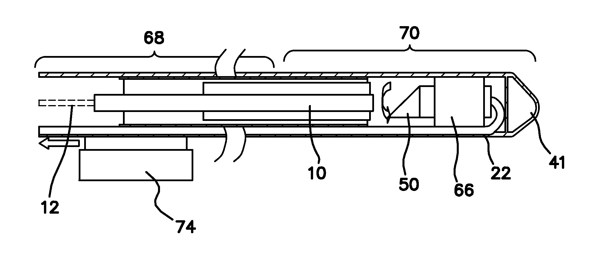
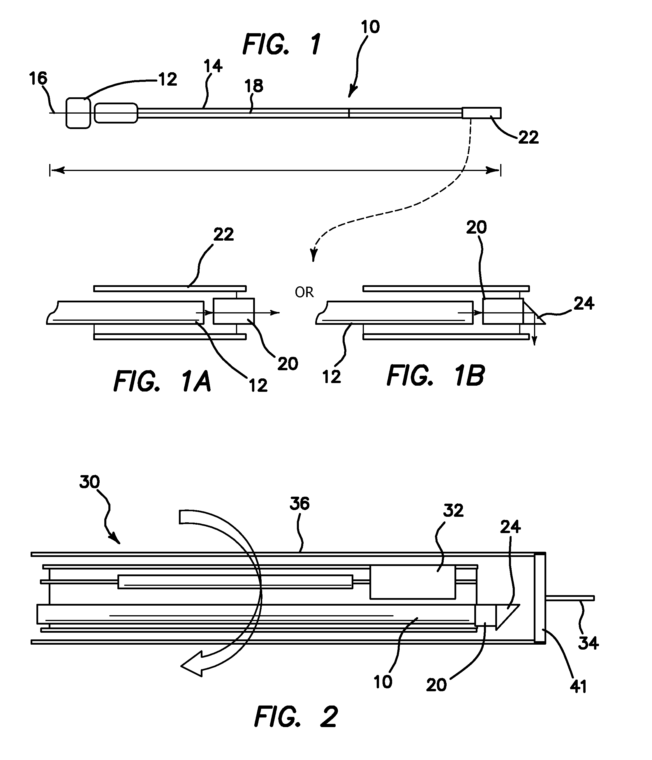
[0077]An optical coherence tomography (OCT) probe is shown in the side plan view of FIG. 1 and is generally represented by reference numeral 10. A single mode optical fiber 12 is used to transmit light from a light source to biomedical sample or target (both not shown). Any type of light source used or usable in optical coherence tomography may be employed. The single mode fiber 12 is protected by a steel tube 14, which helps increase the stiffness of the proximal portion of the fiber 12 that is near an adaptor 16 where mechanical rotation is introduced. A flexible coil wire 18 is disposed through and beyond the distal end of the steel tube 14 to give flexibility to the distal tip of the probe 10 contained within a polyimide tube 22. Light from the distal tip of the single mode fiber 12, which may be an angle polished fiber, is focused by a gradient-index (GRIN) lens 20 into a focusing point in or on the target tissue as seen in FIGS. 1a and 1b. FIG. 1a is a side cross-sectional vie...


PUM


Abstract

An imaging probe for a biological sample includes an OCT probe and an ultrasound probe combined with the OCT probe in an integral probe package capable of providing by a single scanning operation images from the OCT probe and ultrasound probe to simultaneously provide integrated optical coherence tomography (OCT) and ultrasound imaging of the same biological sample. A method to provide high resolution imaging of biomedical tissue includes the steps of finding an area of interest using the guidance of ultrasound imaging, and obtaining an OCT image and once the area of interest is identified where the combination of the two imaging modalities yields high resolution OCT and deep penetration depth ultrasound imaging.

Description

RELATED APPLICATIONS[0001]The present application is related to U.S. Provisional Patent Application, Ser. No. 60 / 109,146, filed on Oct. 28, 2008, which is incorporated herein by reference and to which priority is claimed pursuant to 35 USC 119.GOVERNMENT SUPPORT[0002]This invention was made with Government support under Grant No. EB000293, awarded by the National Institutes of Health. The Government has certain rights in this invention.BACKGROUND OF THE INVENTION[0003]1. Field of the Invention[0004]The invention relates to the field of intravascular imaging, in particular to an integrated ultrasound guided optical coherence tomography, photoacoustic probe used in intravascular or biomedical imaging and a method of using the same.[0005]2. Description of the Prior Art[0006]Intravascular ultrasound (IVUS) is a medical imaging methodology that has been used to show the anatomy of the wall of blood vessels in living animals and humans by using a miniaturized ultrasound probe. IVUS can he...

Claims


Application Information

Patent Timeline
IPC IPC(8): A61B8/14
CPCA61B5/0062A61B5/0066A61B5/0084A61B8/4461A61B5/0095A61B5/02007A61B8/12A61B5/0086A61B5/0035
Inventor CHEN, ZHONGPINGYIN, JIECHENZHOU, QIFAHU, CHANGHONGYANG, HAO-CHUNGCHIANG, HUIHUA KENNYSHUNG, KIRK K.
Owner RGT UNIV OF CALIFORNIA
Why Eureka
Try Eureka