
Transducer arrangement and method for acquiring sono-elastographical data and ultrasonic data of a material

a technology of sono-elastographical data and material, applied in the field of transducer arrangement, can solve the problems of difficult evaluation of lesions' quality, inability to specify, and limited palpation, and achieve the effect of low cos

Inactive Publication Date: 2011-06-09
KONINKLIJKE PHILIPS ELECTRONICS NV
View PDF52 Cites 13 Cited by
  • Summary
  • Abstract
  • Description
  • Claims
  • Application Information

AI Technical Summary

Benefits of technology

[0039]By means of a transducer arrangement according to the first aspect of the present invention it may be possible to obtain information of e.g. both, the topographical anatomy of a tissue and its elastic properties by one and the same transducer arrangement. Preferably, the different information can be acquired simultaneously. Therein, the transducer is realised e.g. in flexible form and, therefore, may be adjusted to the tissue's surface with high accuracy.
[0043]The “semiconductor layer” may be a layer of the transducer element which comprises e.g. semiconductor materials such as silicon and / or semiconductor components or which is a semiconductor component itself. In other words, the transducer elements may be fabricated using well established silicon technology. For example, the transducer elements may be made based on a thin silicon wafer or a silicon thin film in order to obtain sufficient flexibility. The semiconductor layer may comprise the controlling unit, the evaluation unit, the analyzing unit and / or the driving electronics. The inclusion of the semiconductor layer in the transducer elements is advantageous because it may help in significantly reducing the size of the transducer arrangement e.g. by including the control electronics directly in the semiconductor layer. The reduction of the size may in turn lead e.g. to greater patient comfort.
[0053]In this way the deformation or changes in the shape of a whole transducer element or parts of a transducer element (e.g. membranes) may be easily detected, by using the piezoresistive effect of the semiconductor layer. The layer acts thus as a strain gauge. It can also be placed in the flexible joints between transducer elements.

Problems solved by technology

Unfortunately, palpation is usually limited to the detection of lesions near the tissue surface and to lesions with high stiffness contrast.
Even if a lesion is palpable, in general it is not possible to specify its localisation and extension exactly because digital palpation does not provide any real-time image information of the topographical anatomy in parallel.
Moreover, it is usually very difficult to evaluate the lesions' quality (i.e. malignity, benignity) clearly, because this evaluation depends on the physician's subjective sensation and experience.
Two widely used medical imaging modalities, magnetic resonance imaging (MRI) and ultrasound (US) have reported accuracy levels for detecting prostate cancer, the accuracy levels being not high enough such that a significant portion of the cancerous lesions may not be detected.
Regardless of the different accuracy levels, these imaging modalities may not provide any direct information about the elastic tissue properties.
These conventional ultrasound heads are usually rigid, i.e. inflexible, and relatively large-sized.
That's why patients in general feel very uncomfortable when they are examined with such ultrasound heads intraluminally, e.g. in the rectum for detecting prostate lesions.
Furthermore, some regions that should be examined are only accessible with difficulty or there is only little room in such regions that makes it difficult for the examiner to place and handle a large-sized ultrasound head correctly.
A higher pressure on the tissue surface may cause deformation of the tissue's structure during the examination which may mask lesions of interest.

Method used


Image

  • Transducer arrangement and method for acquiring sono-elastographical data and ultrasonic data of a material
  • Transducer arrangement and method for acquiring sono-elastographical data and ultrasonic data of a material
  • Transducer arrangement and method for acquiring sono-elastographical data and ultrasonic data of a material

Examples


Embodiment Construction

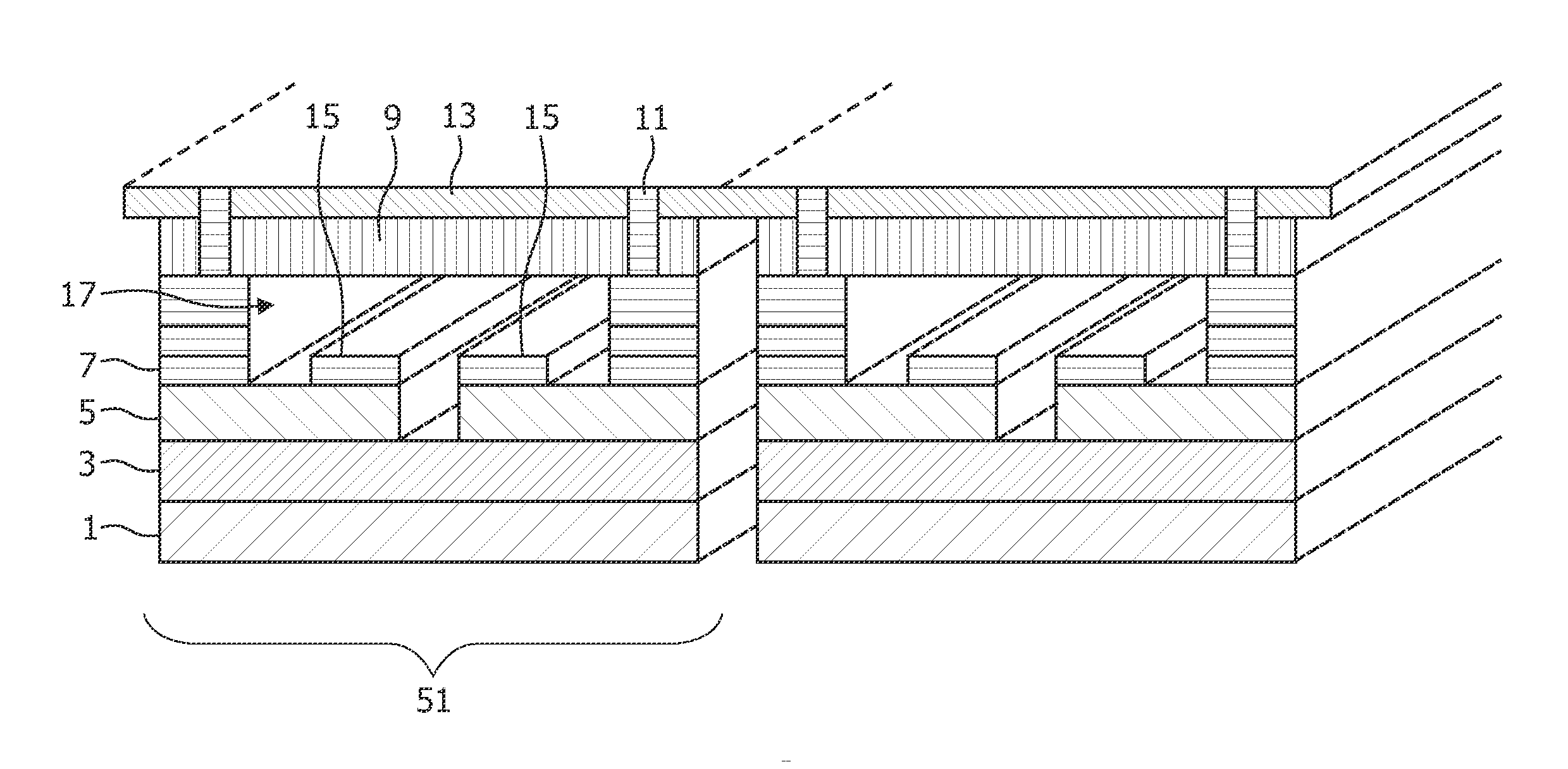
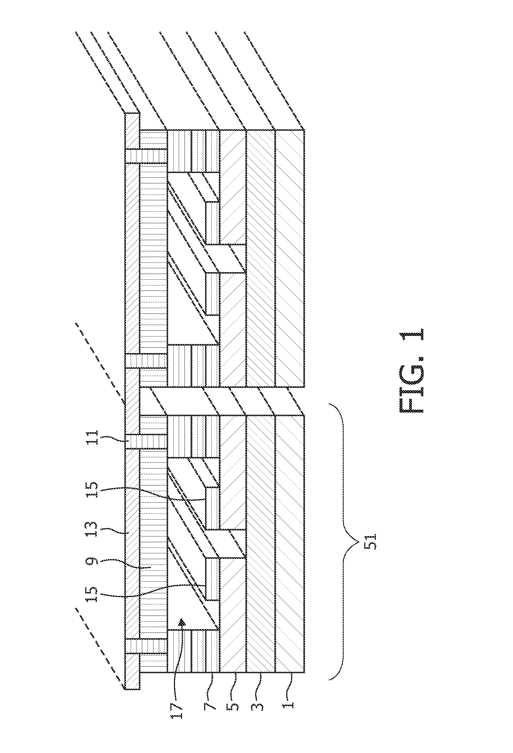
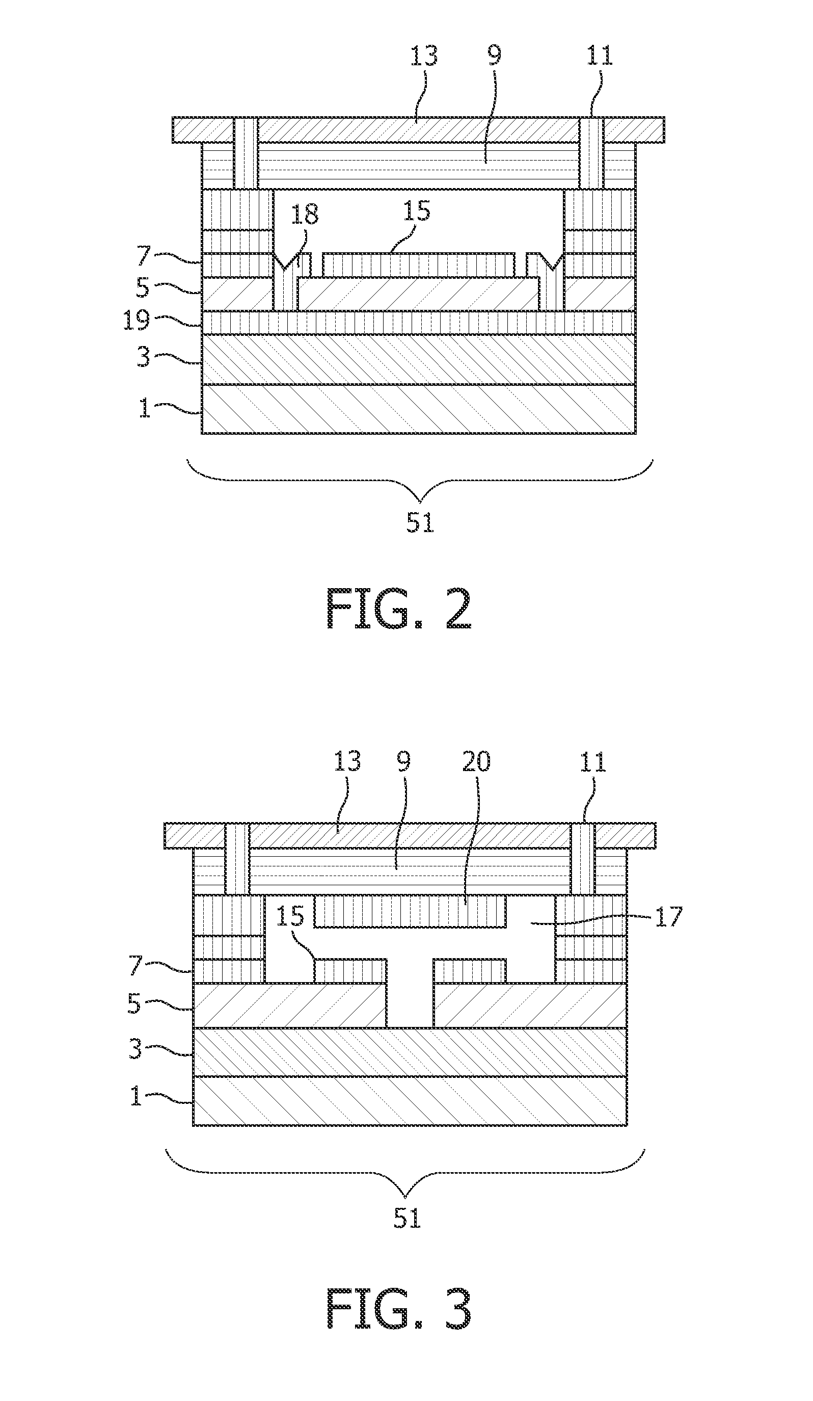
[0081]In FIG. 1 a flexible thin film ultrasound transducer arrangement according to an embodiment of the present invention is shown schematically.

[0082]It is a thin film flexible ultrasound transducer arrangement operating in the d33 mode.

[0083]In the d33 mode, also called longitudinal mode, the elongation of the piezoelectric layer is arranged in parallel to the direction of the applied voltage.

[0084]The figure shows two transducer elements 51, but the principle may be extended to 1D as well as 2D arrangements with numerous elements.

[0085]The piezoelectric transducer includes a membrane 1 and 3 formed on a substrate which is removed after formation of the transducer to allow movement of the membrane. The membrane is an inorganic material for example be composed of silicon nitride (e.g. membrane 1) and silicon oxide (e.g. membrane 3). Also a stack comprising the inorganic membrane and a barrier layer such as titanium oxide or aluminium oxide or zirconium oxide can be applied. Piezoe...


PUM


Abstract

The present invention relates to a transducer arrangement, particularly a transducer arrangement for acquiring tissue information, a method for using a transducer arrangement for acquiring tissue information and a glove which comprises a transducer arrangement. The transducer arrangement 21 for analysing material 40 comprises: a first transducer element 51 for inducing and receiving mechanical displacements in the material to be analysed 40; and an analysing unit 30. The transducer arrangement is arranged such as to be flexible in order to conform with a curved surface of the material to be analysed 40; and the transducer arrangement 21 is adapted to derive a first signal from a low frequency spectrum of mechanical displacements which first signal correlates to sono-elastographical properties of a material to be analysed 40; and the transducer arrangement 21 is adapted to derive a second signal from a high frequency spectrum of mechanical displacements received by the first transducer element 51 which second signal correlates to ultrasonic properties of a material to be analysed 40. With a transducer arrangement according to the invention it may be possible to generate information about the topographical anatomy and information about elastical properties of the material to be analyzed in parallel, whereby the transducer arrangement may be adapted to the unevenness of the material's surface optimally due to its flexibility which may allow the examiner or user of the transducer arrangement to analyze regions which normally may have an uneven surface profile, which may only be reached with difficulty or whose examination may cause inconvenience to the examiner as well as to the person that is being examined.

Description

FIELD OF THE INVENTION[0001]The present invention relates to a transducer arrangement, particularly a transducer arrangement for acquiring tissue information, a method for using a transducer arrangement for acquiring tissue information and a glove that comprises a transducer arrangement.BACKGROUND OF THE INVENTION[0002]Many forms of cancer manifest as hard lesions in soft tissue and because of this, physicians use palpation to detect presence of hard tumours within a human body.[0003]As one example for cancer tissue detection prostate cancer is discussed here.[0004]To screen for prostate cancer, digital rectal examination of the prostate is routinely performed on men who have reached middle age. Unfortunately, palpation is usually limited to the detection of lesions near the tissue surface and to lesions with high stiffness contrast. Even if a lesion is palpable, in general it is not possible to specify its localisation and extension exactly because digital palpation does not provid...

Claims


Application Information

Patent Timeline
Patent Type & Authority Applications(United States)
IPC IPC(8): A61B8/00
CPCA61B8/4281A61B8/4416A61B8/4472A61B8/4483G10K9/125A61B2562/0204A61B2562/164B06B1/0292G01H11/06A61B8/485A61B8/4494
Inventor KLEE, MAREIKEKUHN, MICHAEL HARALDTROVATO, KAREN IRENEVAN HEESCH, CHRISTIANUS MARTINUSMAUCZOK, RUEDIGERDE WILD, MARCOSREEDHARAN NAIR, BIJU KUMARREIMANN, KLAUS
Owner KONINKLIJKE PHILIPS ELECTRONICS NV
Learn More