
Particle-Based Electrostatic Sensing and Detection

a particle-based electrostatic and sensing technology, applied in the field of electrostatic imaging and electrostatic-based sensing, measurement and detection of charges and charged materials, can solve the problems of high cost, time-consuming and costly chemical labeling, reverse transcription, and high-power excitation sources, and none of these have gained widespread us

Inactive Publication Date: 2011-06-16
RGT UNIV OF CALIFORNIA
View PDF2 Cites 20 Cited by
  • Summary
  • Abstract
  • Description
  • Claims
  • Application Information

AI Technical Summary

Benefits of technology

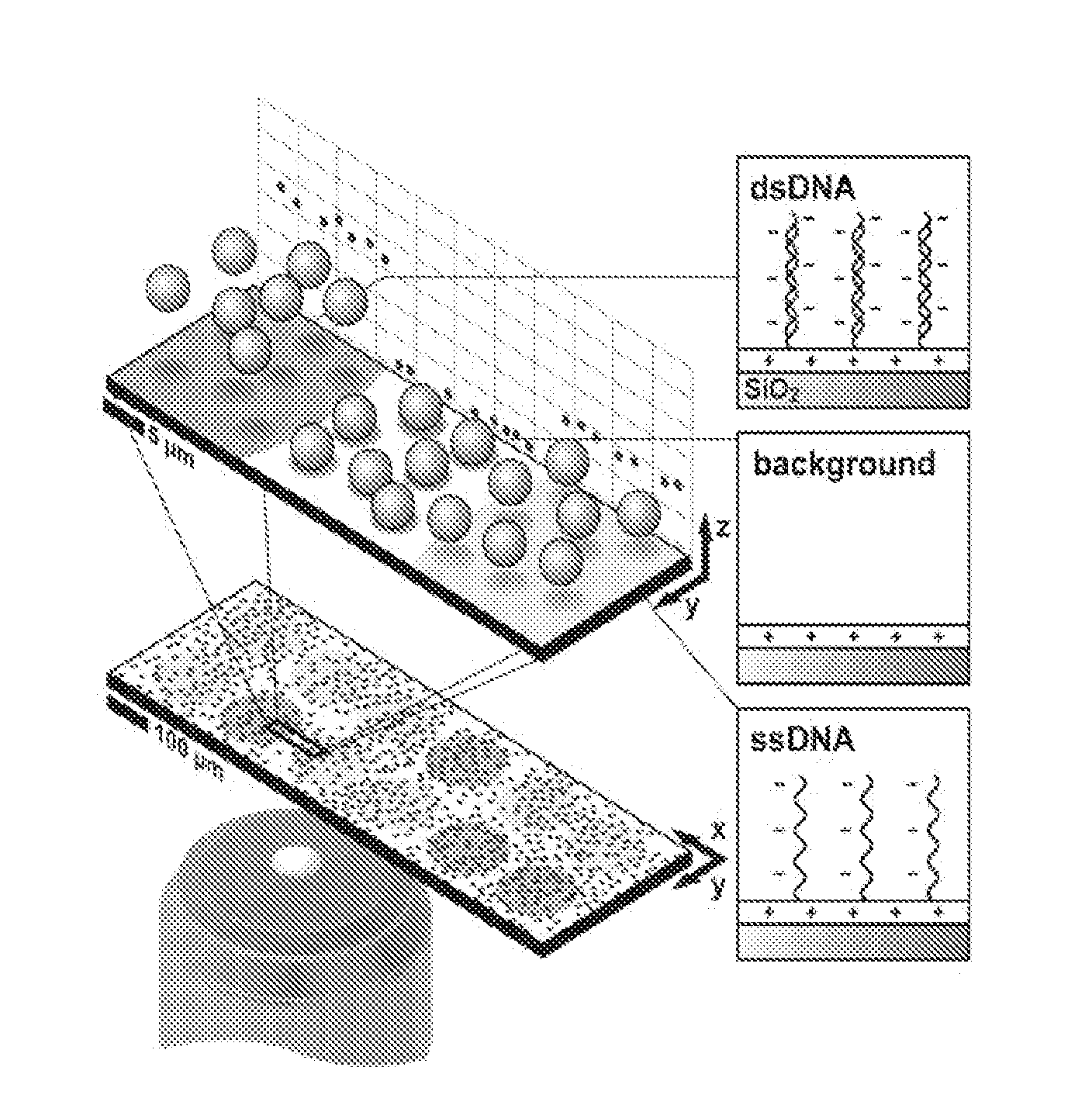
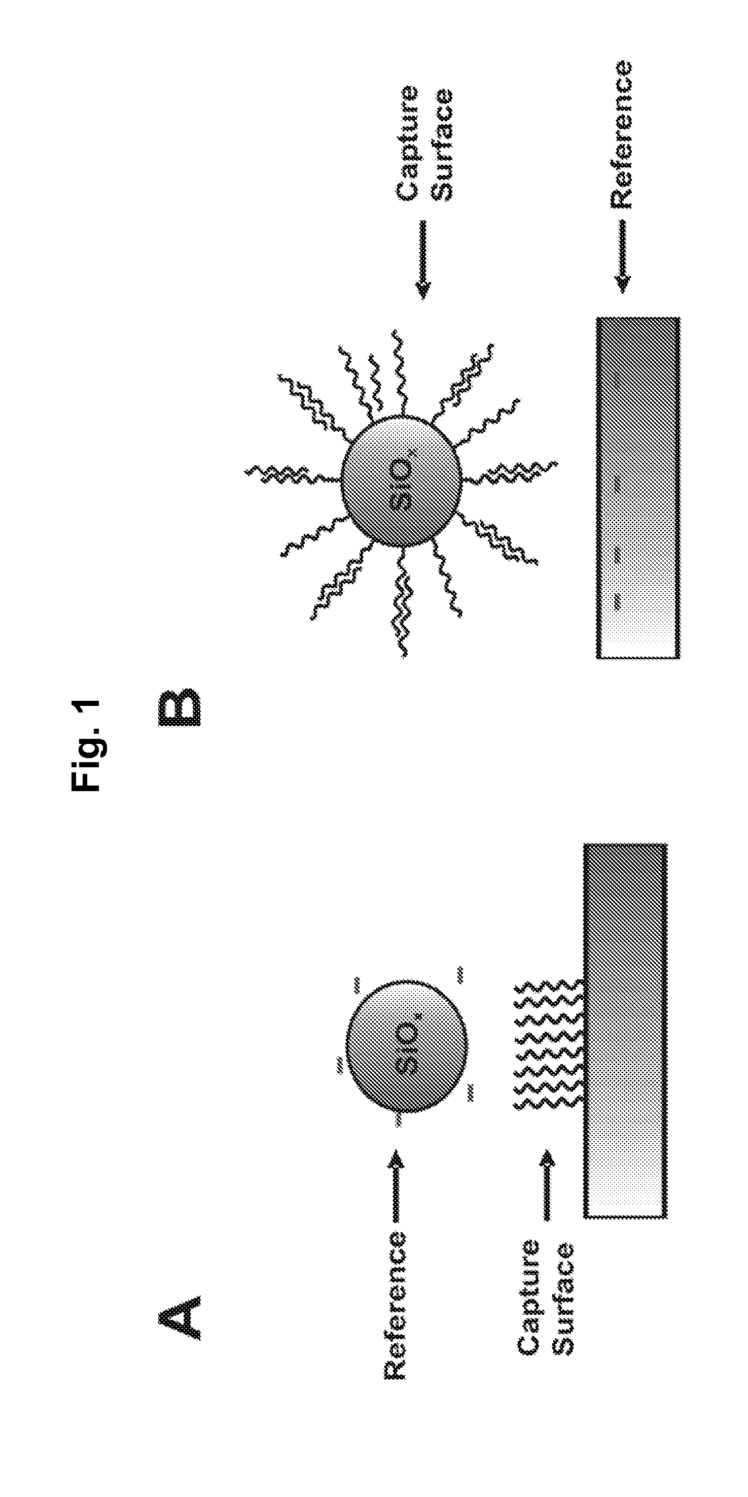
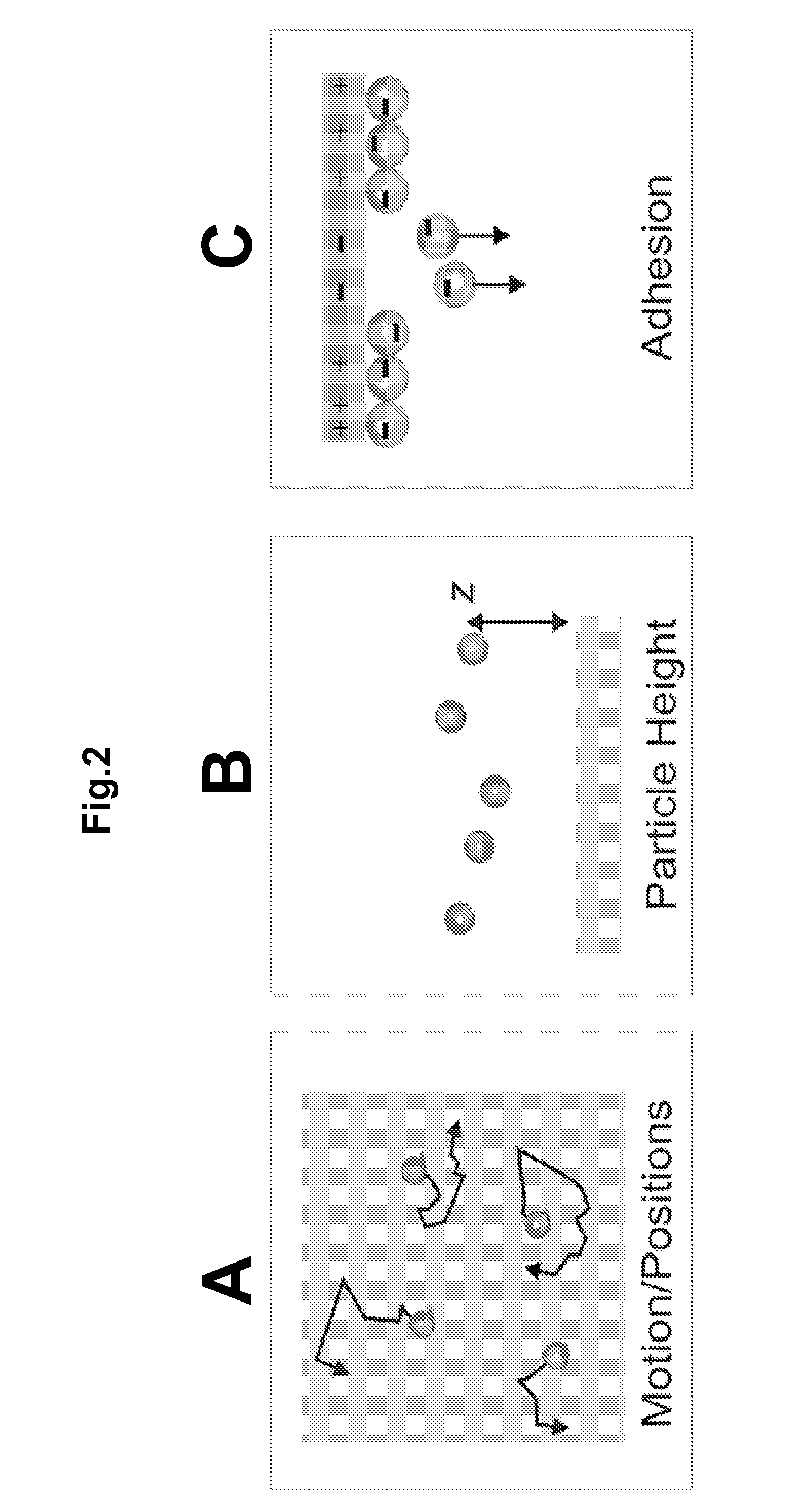
[0020]Another aspect of the invention provides for a method for detecting charge on a surface, comprising the steps of: a) providing a capture surface displaying capture molecules or materials, wherein the capture surface is a particle; b) providing a solution suspected of containing an analyte that binds to a specific one of said capture molecules or materials displayed on the capture surface, c) contacting said solution with the capture surface and allowing said binding to occur; d) applying an electric field to the capture surface, such that the capture surface migrates in the electric field; and e) determining the positions and motions of the particles under the electric field at a specific loci to determine the charge density at said loci. the present invention also provides a method for sensitive and label-free electrostatic readout of DNA or RNA hybridization in a microarray format. The electrostatic properties of the microarray are measured using the positions and motions of charged microspheres randomly dispersed over the surface. This approach enables non-destructive electrostatic imaging with 10 μm lateral resolution over centimeter length-scales, which is four orders of magnitude larger than that practically achievable with conventional scanning electric force microscopy. Changes in surface charge density as a result of specific DNA hybridization can be detected and quantified with 50 pM sensitivity, single base-pair mismatch selectivity, and in the presence of a complex background. Moreover, no more than a magnifying glass is needed to read out the microarray, potentially enabling the broad application of inexpensive genome-scale assays for point-of-care applications.

Problems solved by technology

Effective medical care is often limited by the failure to diagnose diseases in resource-limited settings.
However, microarray assays typically rely on fluorescence detection, which requires time-consuming and costly chemical labeling, reverse transcription, high-power excitation sources, and sophisticated instrumentation for scanning.
However, none of these have gained widespread use because each requires either complex device fabrication or sophisticated instrumentation for readout.
Additionally, none are compatible with conventional DNA microarrays where up to one million sequences can be interrogated in a single experiment.
The vertical deflection of an electrostatic force microscope (EFM) tip is used to report local electrostatic surface properties, however, EFM is a serial technique practically limited to a field of view of 100 μm2.

Method used


Image

  • Particle-Based Electrostatic Sensing and Detection
  • Particle-Based Electrostatic Sensing and Detection
  • Particle-Based Electrostatic Sensing and Detection

Examples


example 1

Electrostatic Response Using a Microarray

[0094]To examine the electrostatic response across a range of DNA densities, a series of spots was printed with binary mixtures formed from ratios of A and B on a microarray (FIG. 5A). In each series, the total ssDNA density was maintained while linearly adjusting the mole fraction of A from 0 to 1. Since both A and B strands are electrostatically and sterically identical, the hybridization efficiency at each spot remains constant. (Peterson, A., Heaton, R. & Georgiadis, R. The effect of surface probe density on DNA hybridization. Nucleic Acids Research 29, 5163-5168 (2001)). Therefore, after hybridization, the density of A′ is linearly related to the density of A strands at each spot, and the total DNA density varies linearly.

[0095]The charge density map of two of these series, printed with total ssDNA concentrations of 5 and 6 μM respectively, show a gradual increase in charge density as a function of the mol fraction of A (FIG. 5A). The ch...

example 2

Multiplexing Electrostatic Response Using Microarray

[0107]To demonstrate that this method is truly massively parallel and can be used to readout conventional microarrays, DNA spots were printed on a standard 1″×3″ glass microscope slide at a density >1000 spots / cm2. Arrays were imaged after hybridizing with 50 nM Cy3-B′ over a 1 sq. in. area by fluorescence and dark field scattering from electrostatically adsorbed 2.34 μm-diameter silica microspheres. Both fluorescence and dark field (negative contrast) images reveal specific hybridization to complimentary spots (FIG. 6D).

example 3

Electrostatic Readout in Gene Expression Profiling

[0108]Since gene expression profiling is the most widely implemented application of DNA microarray technology, it is important to demonstrate that electrostatic readout can be applied to physiological samples with complex background. To demonstrate this we focused on detection of the human β-actin mRNA in purified but unamplified poly(A)-RNA extracted from human breast adenocarcinoma (MCF-7) cells. The β-actin housekeeping gene served as a positive control to demonstrate a specific transcript could be identified in the complex background of cellular mRNA. Prior to measurements, the unamplified poly(A)-RNA was randomly fragmented to 60-200 bp in length to better match probe lengths, and hybridizations were performed for 20 min. with 50 ng of RNA in 30 μl of 1×SSC heated to 60° C. (FIG. 7A). Dark field imaging of arrays interrogated with 2.34 μm diameter silica spheres indicates the electrostatic response of the β-actin probe spot rela...


PUM

PropertyMeasurementUnit
Lengthaaaaaaaaaa
Lengthaaaaaaaaaa
Lengthaaaaaaaaaa

Abstract

An apparatus and methods for electrostatic-based sensing and detection of charges and charged materials displayed on a surface. In a general embodiment, a method for electrostatically sensing charges or charged materials by comparing the electrostatic interaction between a capture surface and a reference surface. Assays to detect binding or interactions between a capture surface and a material to be detected are also described. We also describe a sensitive and label-free electrostatic readout of DNA or RNA hybridization in a microarray format and using a microfluidic device. The electrostatic properties of the hybridized particles are measured using the positions and motions of charged microspheres. This approach enables sensitive, non-destructive electrostatic imaging. Changes in surface charge density as a result of specific molecular interaction can be detected and quantified with great sensitivity, and in the presence of a complex background.

Description

CROSS-REFERENCE TO RELATED APPLICATIONS[0001]This application claims priority to U.S. Provisional Patent Application No. 61 / 126,905, filed on May 7, 2008, and International Patent Application PCT / US09 / 043196, filed on May 7, 2009, both of which are hereby incorporated by reference in their entirety for all purposes.STATEMENT OF GOVERNMENTAL SUPPORT[0002]This invention was made with government support under Contract No. DE-AC02-05CH11231 awarded by the U.S. Department of Energy. The government has certain rights in the invention.REFERENCE TO SEQUENCE LISTING[0003]This application incorporates by reference the attached sequence listing in paper form.BACKGROUND OF THE INVENTION[0004]1. Field of the Invention[0005]The present invention relates to electrostatic imaging and electrostatic-based sensing, measurement and detection of charges and charged materials displayed on a surface.[0006]2. Related Art[0007]Effective medical care is often limited by the failure to diagnose diseases in re...

Claims


Application Information

Patent Timeline
IPC IPC(8): G01N27/60G01D7/02
CPCB01L3/502761C12Q1/6825C12Q1/6837G01N27/60G01N33/5308G01N33/54313C12Q2565/629C12Q2565/607C12Q2563/149C12Q2565/601
Inventor GROVES, JOHN T.CLACK, NATHANIEL G.SALAITA, KHALID S.WU, HUNG-JEN
Owner RGT UNIV OF CALIFORNIA
Learn More