
Iron oxide nanoparticles as MRI contrast agents and their preparing method

a technology of contrast agent and iron oxide nanoparticle, which is applied in the direction of iron oxide/hydroxide, in-vivo radioactive preparation, granular delivery, etc., can solve the problems of insufficient mri tsub>2 /sub>contrast agent, and inability to use tsub>2 /sub>contrast agent, etc., to achieve superior mri t2 contrast effect, low toxicity to human body

Inactive Publication Date: 2011-07-07
KOREA BASIC SCI INST
View PDF2 Cites 27 Cited by
  • Summary
  • Abstract
  • Description
  • Claims
  • Application Information

AI Technical Summary

Benefits of technology

[0004]Therefore, it is an object of the present invention to provide iron oxide nano contrast agents which use Fe as a metal having low toxicity and pre-existing in human body.
[0006]It is another object of the present invention to provide iron oxide nano contrast agents which have superior MRI T2 contrast effect and can be used as a storage or a carrier for drugs and so on, and a method for preparing the same.
[0010]The iron oxide nano contrast agents of the present invention have low toxicity to human body because it is prepared with Fe which exists in human body in a relatively large amount. The iron oxide nano contrast agents have relatively high iron content while there is an interior space or crack-shaped grooves in it. Therefore, the iron oxide nano contrast agents have superior MRI T2 contrast effect, and can be used as a storage or carrier for drugs and so on.

Problems solved by technology

However, the prepared iron oxide nanoparticles include less amount of Fe, and not preferable in r2 value (relaxivity, a value representing contrast effect) and T2 contrast effect, and thus insufficient as MRI T2 contrast agents.
However, the Mn3O4 nanoparticles are prepared with highly toxic Mn, and thus are not preferable for humans' use, and also cannot be used as T2 contrast agents.

Method used


Image

  • Iron oxide nanoparticles as MRI contrast agents and their preparing method
  • Iron oxide nanoparticles as MRI contrast agents and their preparing method
  • Iron oxide nanoparticles as MRI contrast agents and their preparing method

Examples


example 1

Preparation of Iron Oxide Nano Contrast Agent

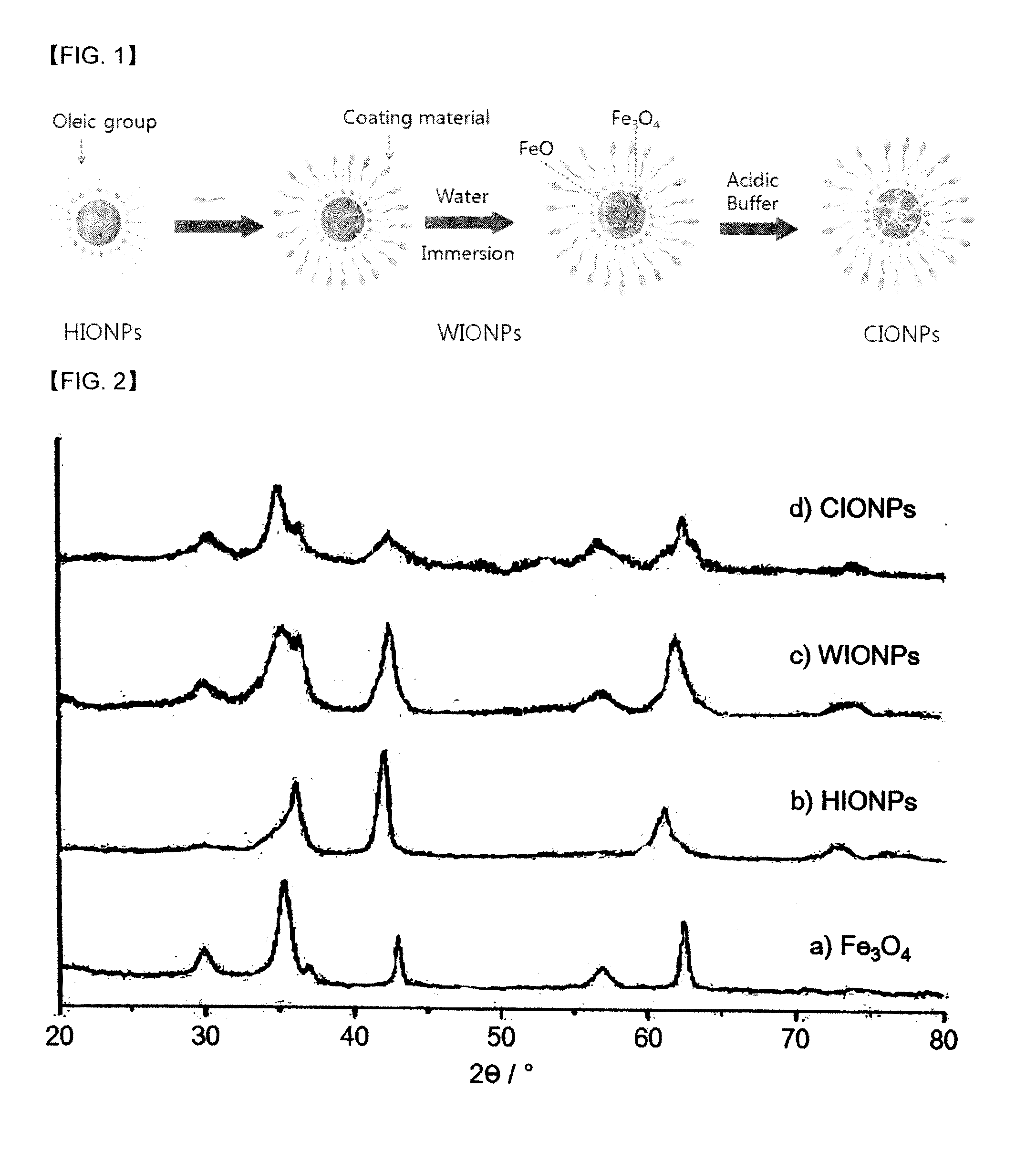
[0029]1. Synthesis of Hydrophobic FeO Nanoparticles (HIONPs)

[0030]Fe(acac)3(iron(III)(acetyl acetonate)3, 1.4 g, 4 mmol, precursor), oleic acid (8 mL, 25 mmol) and oleylamine (12 mL, 35 mmol) were added into a 3-neck round-bottom flask, and heated to 120° C. The reactants were stirred under reduced pressure and at the same temperature for 2 hours to remove water and oxygen in the reactants. After 2 hours, the reaction temperature was raised to 220° C. while feeding Ar gas in the reactor. After further reacting for 30 minutes, the reaction temperature was raised to 300° C. (heating rate: 2° C. / min). After further reacting for 30 minutes at 300° C., the temperature of the reactor was dropped to room temperature quickly, and excess ethanol was added to the reaction solution. The reactants were centrifuged to obtain precipitated solid (hydrophobic FeO). A small amount of hexane was added to disperse the obtained solid, and excess ethanol was ...

example 2

Preparation of Iron Oxide Nano Contrast Agents

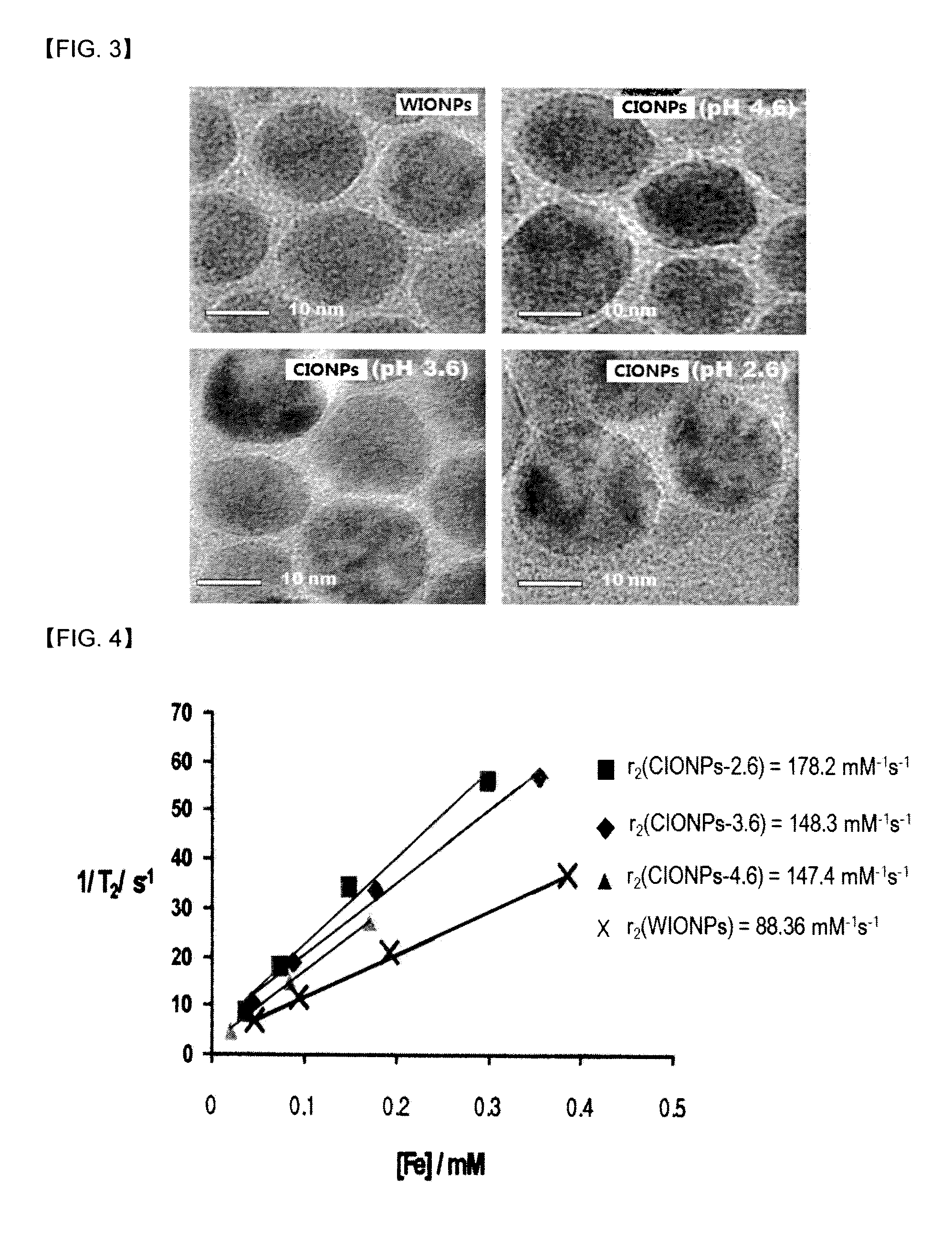
[0038]Except for using dimercaptosuccinic acid (DMSA) instead of polyethylene glycol-phospholipid conjugate (PEG-phospholipid) of Step 2, and increasing the reaction time to 12 hours instead of 1 hour of Step 2, iron oxide (mostly Fe3O4) nanoparticles having interior space (CIONPs) was prepared by the same manner as Example 1. The hydrophilic iron oxide (FeO) nanoparticles (WIONPs) of Step 2 were dispersed in water for 1 day, and contacted to acidic buffer (pH=3.6 and 2.6) for 7 days. The concentrations of Fe were measured with ICP-AES, r2 (relaxivity) values of CIONPs were calculated by T2 values of CIONPs according to the concentrations of Fe measured by MRI (4.7 T). The results are shown in Table 2.

TABLE 2nanoparticlescontact time in acidic bufferT2[ms]r2[s−1mM−1]WIONPs—53167.8CIONPs-3.6(7)[a]7 days50167.9CIONPs-3.6(7)[b]7 days44185.7CIONPs-2.6(7)[a]7 days40216.4CIONPs-2.6(7)[b]7 days27345.7In Table 2, T2[ms] were measured at the conc...

example 3

Preparation and Test of Iron Oxide (Fe3O4) Nanoparticle Drug Carrier

[0040]A. Preparation of Iron Oxide (Fe3O4) Nanoparticle Drug Carrier

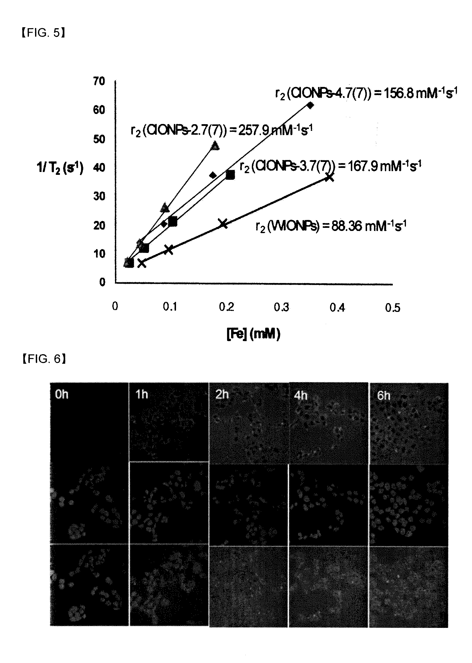
[0041]An iron oxide (mostly Fe3O4) nanoparticle drug carrier (DOX-CIONPs) was prepared by mixing an aqueous solution of the iron oxide nano contrast agents (Fe3O4 nanoparticles having interior space (CIONPs)) prepared in Example 2 and an aqueous solution of anti-cancer medicines (Doxorubicine), and thereby absorbing (loading) the hydrophobic anti-cancer medicines (Doxorubicine) to the interior space of the iron oxide nano contrast agents. The Doxorubicine has a fluorescence property, and thus the loading of the anti-cancer medicines was confirmed by measuring the fluorescence of the DOX-CIONPs.

[0042]B. Test of Iron Oxide Fe3O4 Nanoparticle Drug Carrier

[0043]The iron oxide nanoparticle drug carriers (DOX-CIONPs) prepared in Step A were introduced into colon cancer (HT-29) cells, and the degree of absorption according to the incubation time (0, 2, 4, ...


PUM

PropertyMeasurementUnit
particle diameteraaaaaaaaaa
diameteraaaaaaaaaa
particle diameteraaaaaaaaaa

Abstract

Iron oxide nano contrast agents for Magnetic Resonance Imaging which have superior T2 contrast effect, and also can be used as a storage or a carrier for drugs and so on, are disclosed. The iron oxide nano contrast agents can be prepared by the steps of: coating surfaces of hydrophobic FeO nanoparticles with a coating material selected from the group consisting of polyethylene glycol-phospholipid conjugate, dextran, chitosan, dimercaptosuccinic acid and mixtures thereof in an organic solvent to form hydrophilic FeO nanoparticles having hydrophilic surfaces and dispersibility in water; dispersing the hydrophilic FeO nanoparticles in water to oxidize FeO; and exposing the oxidized hydrophilic FeO nanoparticles to an acidic buffer to dissolve and remove interior unoxidized FeO portions, and thereby to form Fe3O4 nanoparticles having an interior space.

Description

[0001]This application claims the priority benefit of Korean Patent Application No. 10-2010-0001184 filed on Jan. 7, 2010. All disclosure of the Korean Patent application is incorporated herein by reference.FIELD OF THE INVENTION[0002]This invention relates to iron oxide nano contrast agents and a method for preparing the same, and more particularly to novel iron oxide nano contrast agents for Magnetic Resonance Imaging (MRI) which have superior T2 contrast effect for magnetic resonance image, and also can be used as a storage or a carrier for drugs and so on.BACKGROUNDS OF THE INVENTION[0003]Nanoparticles having hollow or cracked structures are used in various fields such as catalysis, nanoreactor, chemical storage or drug delivery system for drugs, lubrication, nanoelectronics, and so on (Angew. Chem. Int. Ed. 2008, 47, 9504-9508; Angew. Chem. Int. Ed. 2004, 43, 1540-1543; Adv. Mater. 2000, 12, 808-811; Science 1999, 285, 1537-1539, 5; Nature 1997, 387, 791-793; Nat. Mater. 2005, ...

Claims


Application Information

Patent Timeline
Patent Type & Authority Applications(United States)
IPC IPC(8): A61B5/055A61K9/50B05D3/10C01G49/02B82Y5/00
CPCY10S977/773C01G49/04A61B5/055Y10T428/2991C01P2004/34Y10S977/81A61K9/5115C01P2004/04A61K49/1863B82Y30/00C01G49/08A61K9/51Y10S977/788B82Y5/00Y10S977/906C01P2002/72A61K49/1836C01P2004/64A61K49/186Y10S977/93B82Y15/00C09C1/24
Inventor LEE, CHULHYUNJEON, SUNG LANCHAE, MIN KYUNGCHO, JEE-HYUNJANG, EUN JU
Owner KOREA BASIC SCI INST
Learn More