Percutaneous Methods for Apparatus for Creating Native Tissue Venous Valves

a technology of venous valve and percutaneous method, which is applied in the direction of catheters, incision instruments, applications, etc., can solve the problems of varicose vein formation, retrograde blood flow in the veins, and distended veins typically in the leg

Active Publication Date: 2011-10-27
MEDTRONIC VASCULAR INC
View PDF6 Cites 26 Cited by
  • Summary
  • Abstract
  • Description
  • Claims
  • Application Information

AI Technical Summary

Problems solved by technology

Incompetent valves can also lead to reflux of blood from the deep venous system to the superficial venous system and the formation of varicose veins.
Veins typically in the leg can become distended from prolonged exposure to excessive pressure and due to weaknesses found in the vessel wall causing the natural venous valves to become incompetent leading to retrograde blood flow in the veins.
Such veins no longer function to help pump or direct the blood back to the heart during normal walking or use of the leg muscles.
As a result, blood tends to pool in the lower leg and can lead to leg swelling and the formation of deep venous thrombosis, phlebitis, and varicose veins.
The formation of thrombus in the veins can further impair venous valvular function by causing valvular adherence to the venous wall with possible irreversible loss of valvular function.
Continued exposure of the venous system to blood pooling and swelling of the surrounding tissue can lead to post phlebitic syndrome with a propensity for open sores, infection, and may lead to possible limb amputation.
Such patients are at increased risk for development of soft tissue necrosis, ulcerations, pulmonary embolism, stroke, heart attack, and amputations.
After a vein segment becomes incompetent, the vessel wall dilates such that the fluid velocity decreases within the incompetent vein segment, which may lead to flow stasis and thrombus formation in the proximity of the venous valve.
Repair and replacement of venous valves presents a formidable problem due to the low blood flow rate found in native veins, the very thin wall structure of the venous wall and the venous valve, and the ease and frequency of which venous blood flow can be impeded or totally blocked for a period of time.
However, such prostheses have not been devoid of problems leading to thrombosis and / or valve failure due to leaflet thickening / stiffening, non-physiologic flow conditions, non-biocompatible materials and / or excessive dilation of the vessels with a subsequent decrease in blood flow rates.
However, methods of in situ venous valve formation according to Drasler et al. are surgical in nature and involve re-shaping a distended, diseased vein, which carries with it the risk of rupture or tearing of the thin-walled structure.
Further, such percutaneous methods and apparatus should attend to biocompatibility and thrombosis issues that current approaches do not adequately address.

Method used


Image

  • Percutaneous Methods for Apparatus for Creating Native Tissue Venous Valves
  • Percutaneous Methods for Apparatus for Creating Native Tissue Venous Valves
  • Percutaneous Methods for Apparatus for Creating Native Tissue Venous Valves

Examples


Embodiment Construction

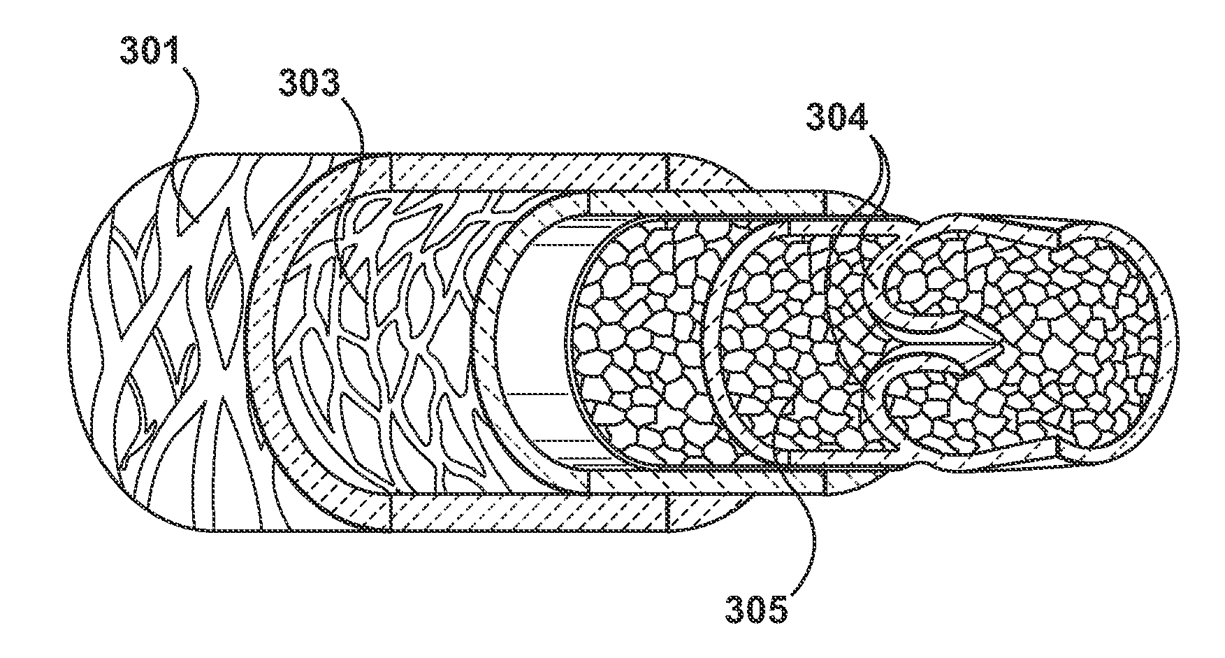
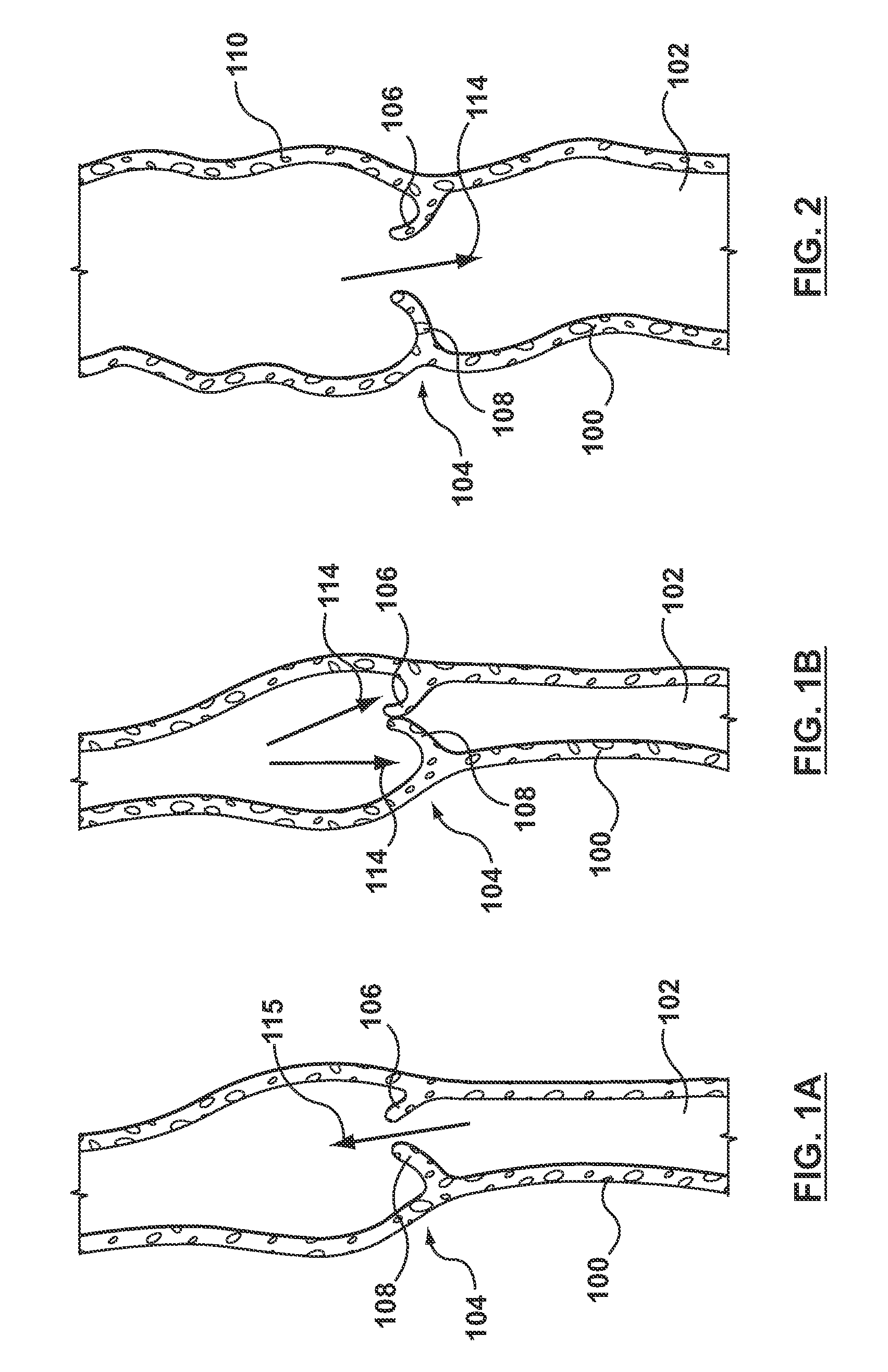
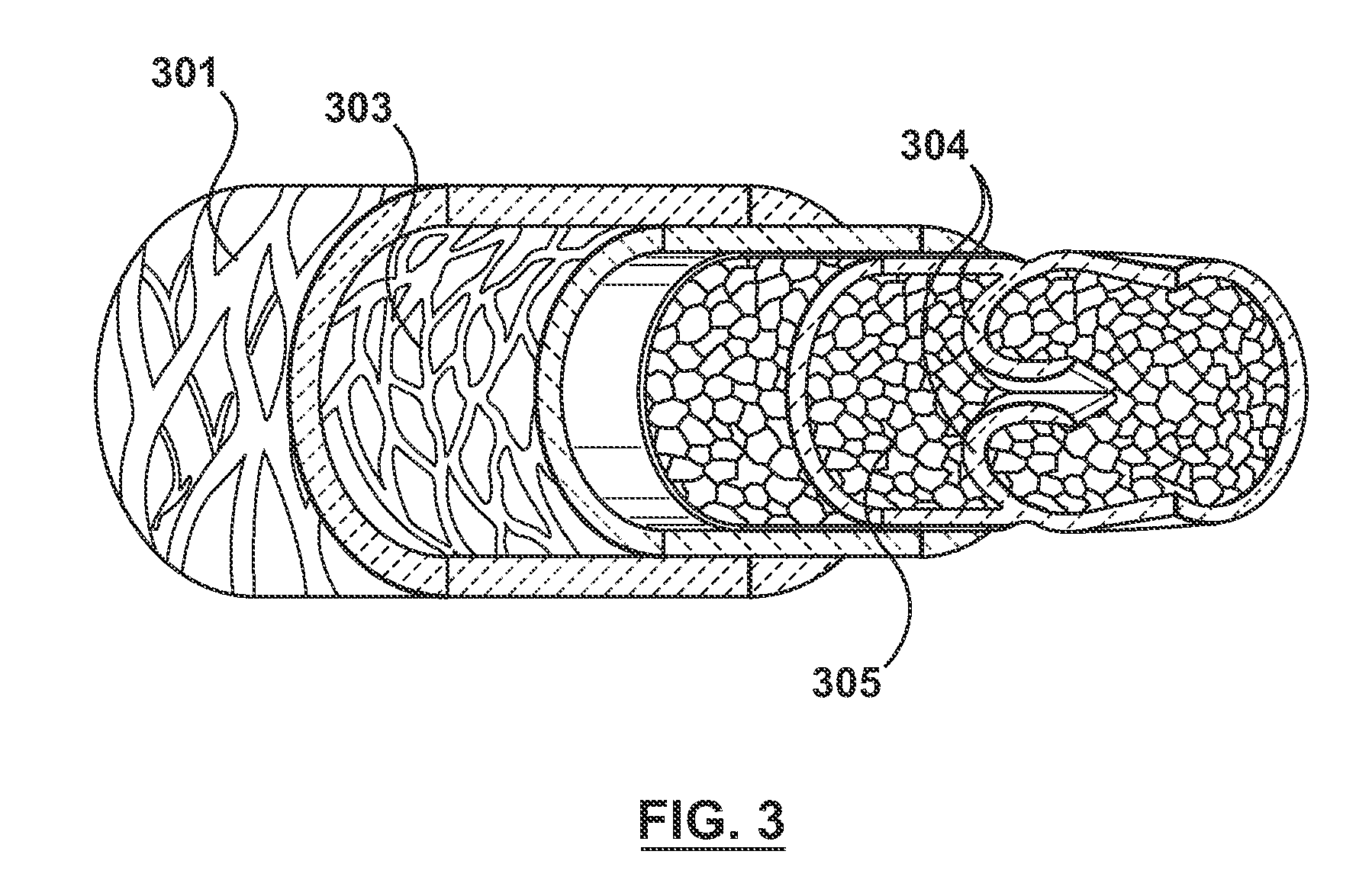
[0040]Specific embodiments hereof are now described with reference to the figures, wherein like reference numbers indicate identical or functionally similar elements. The terms “distal” and “proximal” are used in the following description with respect to a position or direction of the balloon catheter and the retractable dissecting system relative to the treating clinician. “Distal” or “distally” are a position distant from or in a direction away from the clinician. “Proximal” and “proximally” are a position near or in a direction toward the clinician. With reference to venous valves and / or the flaps of tissue that form a venous valve, the term “proximal” refers to an end or portion in a direction toward the heart by way of blood flow path while the term “distal” refers to an end or portion in a direction away from the heart by way of blood flow path.

[0041]The following detailed description is merely exemplary in nature and is not intended to limit the invention or the application a...


PUM


Abstract

Percutaneous methods and apparatuses for forming a venous valve from autologous tissue. A catheter having a retractable dissecting system received therein is delivered to a target location where a new venous valve is to be created. A distal balloon or other radially-expandable component mounted on the catheter is expanded against the vein wall, and the dissecting system is proximally retracted to deploy one or more dissecting components that dissect a subintimal layer of the vein wall. Radial expansion of the dissecting component(s) within the vein wall creates one or more leaflets and corresponding pocket / sinuses in the vein that collectively act as a venous valve, and / or the radially-expandable component of the catheter is subsequently collapsed such that the dissecting component(s) each pull a flap of the dissected tissue towards the vein lumen to create one or more leaflet(s) and corresponding pocket / sinuses in the vein.

Description

FIELD OF THE INVENTION[0001]The invention relates to apparatus and methods for percutaneously creating a one-way venous valve in vivo from autologous tissue.BACKGROUND OF THE INVENTION[0002]Venous valves are found within native venous vessels and are used to assist in returning blood back to the heart in an antegrade direction from all parts of the body. The venous system of the leg includes the deep venous system and the superficial venous system, both of which are provided with venous valves that are intended to direct blood toward the heart and prevent backflow or retrograde flow, which can lead to blood pooling or stasis in the leg in the setting of incompetent valves. Incompetent valves can also lead to reflux of blood from the deep venous system to the superficial venous system and the formation of varicose veins. Superficial veins, which include the greater and lesser saphenous veins, have perforating branches in the femoral and popliteal regions of the leg that direct blood ...

Claims


Application Information

Patent Timeline
Patent Type & Authority Applications(United States)
IPC IPC(8): A61B17/32
CPCA61B8/0841A61B8/12A61B17/320725A61B17/3209A61B2017/00783A61B2017/22061A61M2025/1081A61F2/2475A61M25/0105A61M25/10A61M25/104A61M2025/1075A61F2/2463
Inventor MAUCH, KEVINARREGUIN, MARIA
Owner MEDTRONIC VASCULAR INC
Why Eureka
Try Eureka