
Detection of Nucleic Acids

a nucleic acid and detection method technology, applied in the field of detection of nucleic acids, can solve the problems of difficult to adjust the length of the oligonucleotide probe to provide difficult to discriminate closely related sequences, and difficult to achieve the desired specificity and sensitivity. achieve the effect of easy modification

Inactive Publication Date: 2012-03-01
AFFYMETRIX INC
View PDF11 Cites 30 Cited by
  • Summary
  • Abstract
  • Description
  • Claims
  • Application Information

AI Technical Summary

Benefits of technology

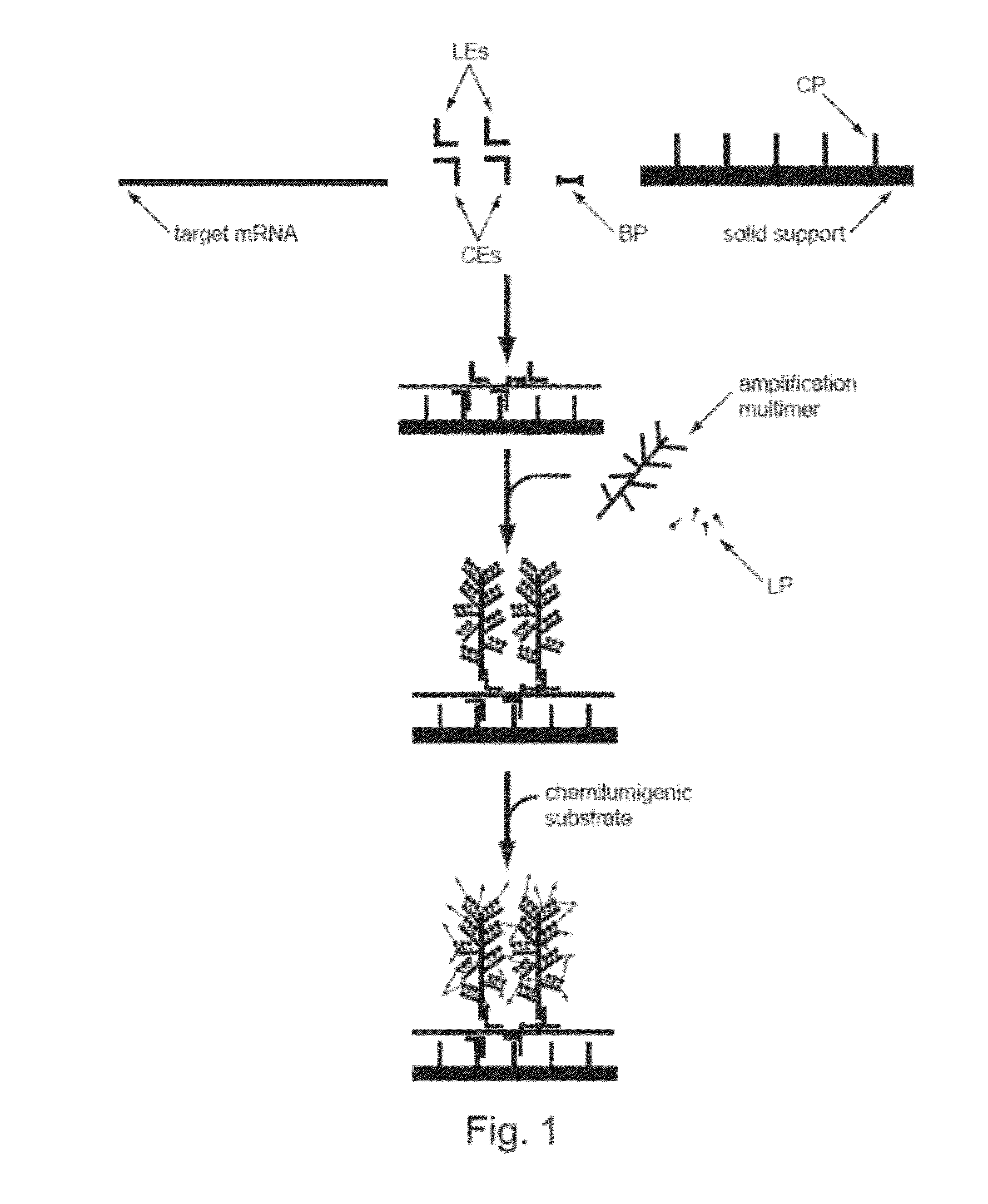
[0022]The assays, embodiments and systems disclosed may be easily altered for multiplex reactions, i.e. wherein multiple targets are detected in a single sample. The sample may comprise a target nucleic acid comprising at least two single nucleotide polymorphisms, or multiple target nucleic acids. Due to the inherent flexibility of the present assays, methods, embodiments, compositions and systems, it is shown that the label extenders may be designed to operate in any one of a number of different structural orientations, such as the cruciform orientation.

Problems solved by technology

SNPs are also evolutionarily stable—not changing much from generation to generation—making them easier to follow in population studies.” However, it is pointed out that SNPs do not cause disease and they are not absolute indicators of disease development.
Use of longer probes can provide increased specificity, but it can also make discrimination of closely related sequences difficult.
Adjusting the length of the oligonucleotide probe to provide the desired specificity and sensitivity often proves extremely difficult.
However, these approaches are time-consuming and have limited sensitivity, and the data generated are more qualitative than quantitative in nature.
Greater sensitivity and quantification are possible with reverse transcription polymerase chain reaction (RT-PCR) based methods, such as quantitative real-time RT-PCR, but these approaches have low multiplex capabilities.
Microarray technology has been widely used in discovery research, but its moderate sensitivity and its relatively long experimental procedure have limited its use in high throughput expression profiling applications (Epstein and Butow, (2000) “Microarray technology—enhanced versatility, persistent challenge,”Curr. Opin. Biotechnol., 11:36-41).
Each of these steps has the potential of introducing variability in yield and quality that often leads to low overall assay precision.
Although this assay has the advantage of measuring mRNA transcripts directly from cell lysates, limited assay sensitivity and reproducibility were reported.
However, this assay requires the use of a specially designed and synthesized set of eTagged signal probes, complicated capillary electrophoresis equipment, and a special data analysis package.
Thus, aberrant expression of miRNA can lead to various disease states.
Thus, the FISH analysis, though the golden standard today in cytogenetics, is time consuming and requires many steps, and in the end only provides 60 bp to 100 kb resolution.
However, these types of instruments also have limitations, governed by the ability of the oligonucleotide on the bead to specifically and sensitively bind to the target molecule.
If the target is too short, such techniques are unable to detect the target.
Additionally, the number of targets able to be assayed in a single test is limited by the types of beads, microparticles or substrate used.
The QUANTIGENE® technology allows unparalleled signal amplification capabilities that provide an extremely sensitive assay.
However, in practice the limit of detection, due to the variability in the assay, is generally found to be around 50-60 copies of message per cell.
This limit of detection limits the field of research since 80% of mRNAs are present at fewer than 5 copies per cell and 95% of mRNAs are present in cells at fewer than 50 copies per cell.
As mentioned above, to arrive at this sensitivity, other approaches are very time consuming and complicated.

Method used


Image

  • Detection of Nucleic Acids
  • Detection of Nucleic Acids
  • Detection of Nucleic Acids

Examples


example 1

SNP Detection

[0273]In the present experiment, SNPs were detected in RNA targets of approximately 20 base pairs, in situ using the QUANTIGENE® ViewRNA protocol. In these assays, no capture probes or capture extenders are utilized since there is no need to capture the target nucleic acids onto a solid surface. The samples are placed onto a solid surface, such a slide or well but not directly covalently attached to the surface. Each target contained 0, 1 or 2 SNPs. The RNA targets were synthesized using known protocols and purified to homogeneity. RNA target probes were then transfected at 15 nM into HeLa cells using known transfection protocols and reagents, such as DeliverX (Panomics, Santa Clara, Calif., see website for transfection protocols, incorporated herein by reference for all purposes). Label extenders were designed in the “Double Z” (ZZ) format as depicted, for instance, in the right panel of FIG. 9A. Cell were prepared as in the published QUANTIGENE® ViewRNA User Manual pr...

example 2

siRNA and mRNA Detection In Situ

[0277]In the present experiment, HeLa cells were plated at a density of 4000 cells / well on 96-well plastic bottom plates (Thermo Scientific Nalge Nunc, Int'l., Rochester, N.Y.) which were pre-treated with Poly-L-Lysine. Cells were allowed to grow for 2 days under standard tissue culture conditions for HeLa cells. Using a non-residual volume protocol, cells were fixed in 4% formaldehyde and cross-linked with 0.16 M EDC. Briefly, the non-residual volume protocol calls for aspiration of all but a few microliters of assay solution from sample wells throughout the entire assay. This prevents drying of the cells between manipulations and minimizes cell loss due to over-aspiration / dispension manipulations of samples. Cells were then treated with protease to permeabilize the membranes. The type of protease used for this protocol was Protease XXIV (Sigma-Aldrich, MO, also known as alkaline protease, protease from Bacillus licheniformis), at a concentration of ...

example 3

GAPDH siRNA Detection in HeLa Cells

[0283]Protocol was similar to that described for Example 3. Briefly, HeLa cells were plated at a density of 8000 cells per well on 96-well plates which were pretreated with Poly-L-Lysine. Cells were allowed to incubate overnight under typical tissue culture conditions. The following day, the HeLa cells were transfected with FAM-labeled or unlabelled GAPDH siRNA at a final concentration of 15 nM using the QUANTIGENE DeliverX product and protocol (Panomics, CA). Cells were again incubated overnight at standard HeLa cell tissue culture conditions.

[0284]Target sequences and probe sequences utilized in this example are as follows, where capital letters in the Probeset sequences indicate the use of cEt nucleic acid analogs and lower case letters indicate use of normal or wild type nucleic acids and “LE1” and “LE2” indicate the LE L-1 sequences, one for each LE:

[0285]GAPDH-siRNA Sequence:

CAUCAUCCCUGCCUCUACUTT-sense(SEQ ID NO: 116)AGUAGAGGCAGGGAUGAUGTT-ant...


PUM

PropertyMeasurementUnit
volumesaaaaaaaaaa
diameteraaaaaaaaaa
diameteraaaaaaaaaa

Abstract

Methods of detecting various types of nucleic acids, including methods of detecting two or more nucleic acids in multiplex branched-chain DNA assays, are provided. Detection assays may be conducted at least in vitro, in cellulo, and in situ. Nucleic acids which are optionally captured on a solid support are detected, for example, through cooperative hybridization events that result in specific association of a label probe system with the nucleic acids. Various label probe system embodiments are provided. Compositions, kits, and systems related to the methods are also described.

Description

RELATED APPLICATIONS[0001]The present application claims priority to U.S. Provisional Patent Application Ser. No. 61 / 360,887, filed on Jul. 1, 2010, the entire disclosure of which is incorporated herein by reference for all purposes.FIELD OF THE INVENTION[0002]Disclosed are methods, compositions and kits for detection of nucleic acids, including methods for detecting the presence of two or more nucleic acids simultaneously in a single sample. Detection may be, for instance, in vivo, in cellulo or in situ. Detection may include or be directed towards detection and quantitation of single nucleotide polymorphisms, i.e. SNP detection, copy number, micro-RNA, siRNA, transcription level determination, and other similar genetic information.BACKGROUND OF THE INVENTION[0003]A variety of techniques for detection of nucleic acids involve a first step of capturing or binding of the target nucleic acid or nucleic acids to a surface through hybridization of each nucleic acid to an oligonucleotide...

Claims


Application Information

Patent Timeline
Patent Type & Authority Applications(United States)
IPC IPC(8): C12Q1/68
CPCC12Q1/682C12Q1/6827C12Q2525/313C12Q2537/143C12Q2563/149C12Q2565/101
Inventor NGUYEN, QUAN N.MCMASTER, GARY K.MA, YUNQINGWU, YEN-CHIEHLIN, AUDREYLAI, CHUNFAINGUYEN, CUNG-TUONG
Owner AFFYMETRIX INC
Learn More