
Device and Methods for the Detection of Cervical Disease

Inactive Publication Date: 2012-06-21
MILAGEN
View PDF8 Cites 13 Cited by
  • Summary
  • Abstract
  • Description
  • Claims
  • Application Information

AI Technical Summary

Benefits of technology

[0030]The present invention also provides a novel immunostaining method of patient cervical samples using the multiwell device. Resorting to the mixture of cervical cancer cells and normal cervical cells to mimic a patient cervical sample, a specific antibody targeting an antigen that is overexpressed in cervical cancer cells is used to stain the latter among normal cervical cells. The present invention takes advantage of the use of specific antibodies to pinpoint a few cancer cells in a mixture of normal cells, thus increasing diagnostic accuracy over the visual evaluation of cell morphologies by the pathologist.
[0031]Encompassed by the present invention is the use of other antibodies or combination of antibodies against biomarkers or combination of biomarkers that are associated to preneoplastic, neoplastic or dysplastic cervical cells. Use of antibodies against such biomarkers would greatly improve current Pap screening sensitivity.
[0032]Finally the present invention details a novel method for cervical disease detection and screening which is practiced on a cell suspension in solution. Said cell suspension is a mixture of normal cervical cells and cells from a cervical cancer cell line, as described above, an experimental condition mimicking a patient sample for cervical screening Authentic ThinPrep samples are encompassed in the present invention. In this preferred embodiment, cell staining is described with an anti-p16(INK4a) antibody, which localizes to the cell nucleus and cytoplasm in the cancer cell type used. Other antibodies or combination of antibodies against biomarkers or combination of biomarkers that are associated to preneoplastic, neoplastic or dysplastic cervical cells are encompassed by the present invention, and would greatly improve current Pap screening sensitivity.
[0033]The latter embodiment further provides the ability to quantify the specific antigen-antibody reaction, in turn facilitating and potentially automating the whole process of cervical cancer screening. In the present example a colorimetric reaction is used, but other detection systems are encompassed by the present invention. Immunostaining based on colorimetric reading of cell reactivity represents an accurate, cost-effective, and user-friendly improvement over visual evaluation of staining intensities and cell morphologies by the pathologist.
[0036]In conclusion, the present invention provides several improvements and utility applications thereof over current cervical screening procedures: i) single to multiple high-throughput capability, ii) increased accuracy in diagnostic interpretation as compared to evaluation of cell morphology alone, due to further use of specific antibodies, and finally iii) cost-effective, user-friendly and quantitative measurement of antigen-antibody reaction.

Problems solved by technology

These tests require highly trained staff and adequate laboratories, making the tests labor-intensive and expensive.
So, in addition to limited sensitivity, Pap test is also a relatively costly procedure requiring infrastructure and pathology expertise.
The relatively low sensitivity results in a high false-negative rate, mostly due to inadequate sampling and improper slide preparation.
Nonetheless, while improving test accuracy, these technologies contribute to increased high-tech infrastructure and cost required for cervical screening, clearly not to the advantage of low infrastructure settings.
However, according to the NACB recommendations (Gaarenstroom, 2007), SCC is not a sufficiently sensitive biomarker for cervical cancer screening, and its clinical utility in prognosis and monitoring response to treatment needs further evaluation.

Method used


Image

  • Device and Methods for the Detection of Cervical Disease
  • Device and Methods for the Detection of Cervical Disease
  • Device and Methods for the Detection of Cervical Disease

Examples


example 1

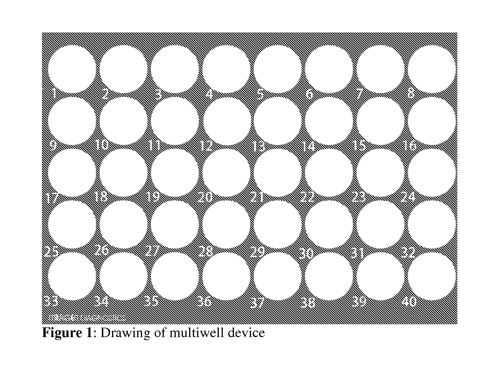
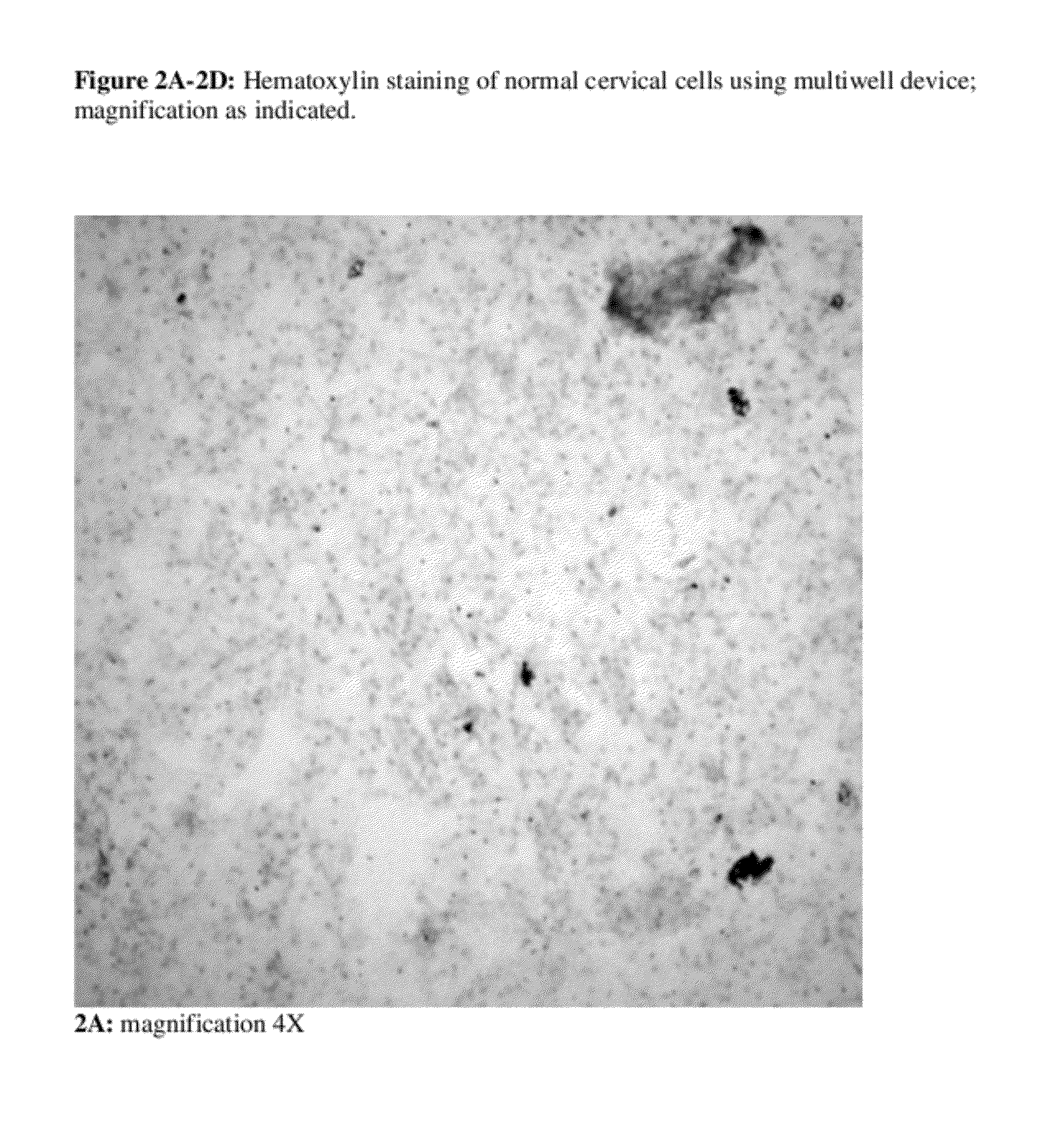
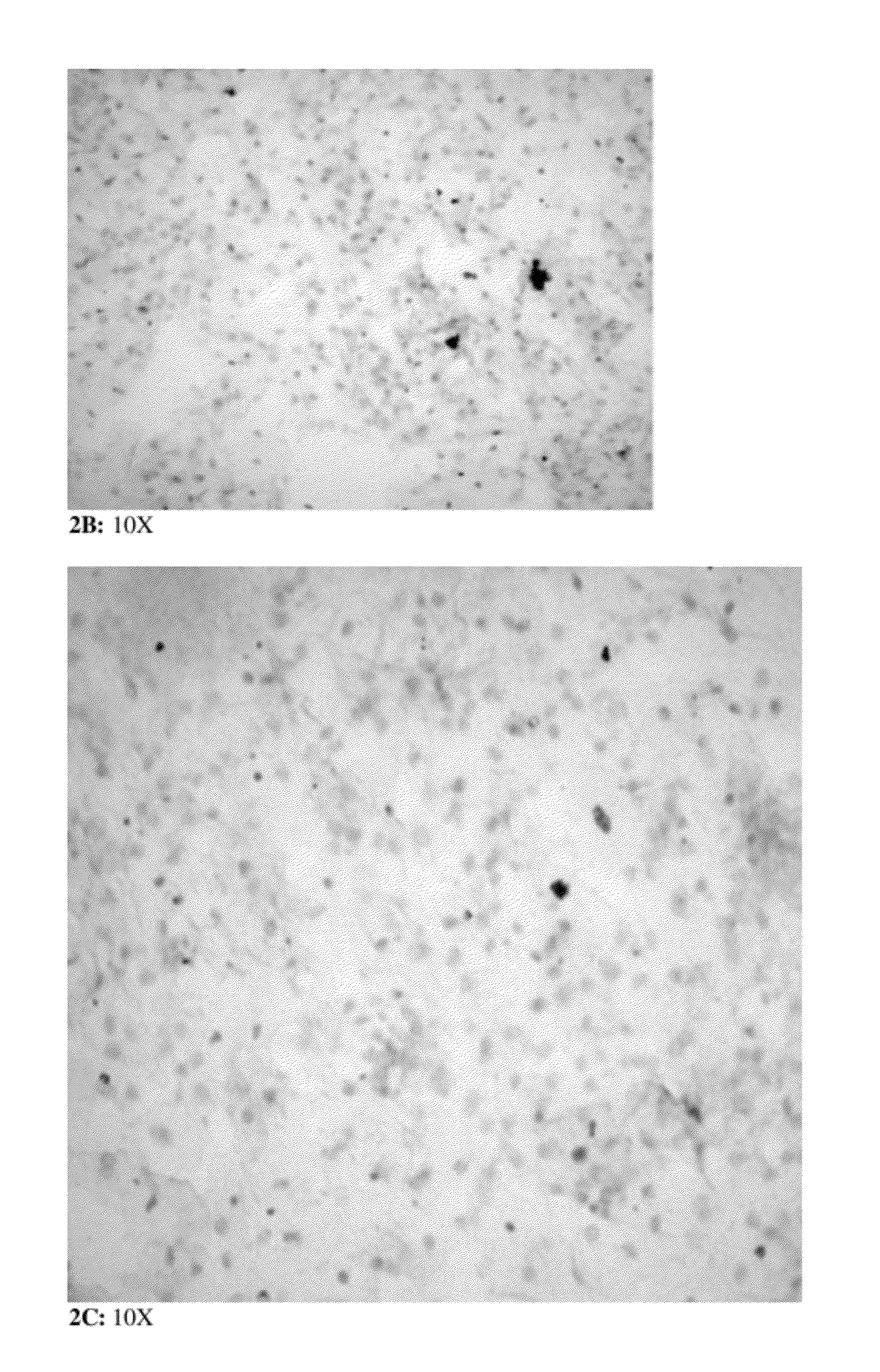
[0102]Hematoxylin staining of patient cervical samples using multiwell device. A fraction of an individual Thin Prep sample (i.e. 250 μl volume, approximately 50-100,000 cells) is layered on a single circular area of the multiwell device. Multiple samples can thus be handled and evaluated simultaneously. The device has been previously UV irradiated to enable cell attachment. However, alternate methods for cell attachment can be utilized depending on the material the device is made of. Samples are air dried either at RT or in a 37-40° C. chamber, then rinsed with PBS. Cleared from excess cell debris, cells are ready for any cytological stain.

[0103]In this specific embodiment, hematoxylin staining is performed. For hematoxylin staining, multiwell device is rinsed with ddH2O, and covered with a few drops of weak Mayer's hematoxylin solution for 1-2 min. Multiwell device is rinsed twice with tap H2O or bluing solution (Scott's tap water, or sodium or lithium carbonate solution to ensure...

example 2

[0104]Co-culture and hematoxylin staining of patient cervical cells and cancer cells in multiwell device, in an assay condition mimicking Pap smear screening. First, 250 μl of an individual Thin Prep sample, previously diagnosed as normal, is layered on a single circular area of the multiwell device. Multiple samples can thus be handled and evaluated simultaneously. Samples are air dried then rinsed with PBS, and stained with hematoxylin, as described in Example 1. The multiwell is rinsed with 95% ethanol prior to the addition of cancer cells.

[0105]The cervical cancer cell line SiHa (ATCC #HTB-35), a human squamous cervix carcinoma cell line, reported to contain one to two copies per cell of integrated HPV16 genome, is used in the co-culture experiment together with cervical cells from Thin Prep samples. Cell line is cultured according to ATCC or provider's recommendations, trypsinized and counted according to standard procedures. A few SiHa cells are deposited on each individual ci...

example 3

[0106]Immunostaining of patient cervical sample using multiwell device and commonly used monoclonal antibodies. This example describes how immunostaining can be performed in the multiwell device using commercially available and commonly used monoclonal antibodies. This example thus further demonstrates the utility of immunostaining as an adjunct to microscopic evaluation of cell morphologies in cervical screening.

[0107]Because preneoplastic, dysplastic and cancerous Thin Prep samples are rare, we resorted to an experimental condition mimicking a patient sample for cervical cancer screening, ie. mixing normal cervical cells with cells from a cancer cell line that are grown ON in co-culture, as described in Example 2. In this experimental model, a constant amount of normal patient cervical cells derived from a previously diagnosed normal Thin Prep (250 μl) is layered in each circular area of the multiwell device, air dried at RT or in a 37-40° C. chamber then rinsed with PBS. Subseque...


PUM


Abstract

The present invention discloses a novel device and methods thereof with utility application in the high-throughput detection, screening and disease management of cervical disease. The “multiwell” device of the present invention consists of a solid support featuring multiple well-separated areas, each accommodating a patient sample, leading to simultaneous evaluation of patient samples. The methods of the present invention comprise conventional cytological staining, cervical Pap staining, and immunochemical staining using antibodies or combination of antibodies which are capable of binding to biomarkers that are overexpressed in cancer including in cervical carcinoma and dysplasia, as compared to normal controls. The device and methods of the present invention can be practiced in either manual or automated mode, and applied to any biological fluid or cell suspension from any biological specimen in view of a variety of cell biology assays, and in view of detection and screening of cervical and other diseases.

Description

FIELD OF INVENTION[0001]The field of the invention is cervical disease detection and screening, using a novel multiwell device and methods thereof.BACKGROUND[0002]Cervical cancer incidence and mortally. Cervical cancer is the second most commonly diagnosed malignancy and the third leading cause of cancer death in women worldwide. There were 555,100 new cases and 309,800 deaths estimated in 2007, 83% of which occurred in the developing world (American Cancer Society, ACS Global Cancer Facts and Figures, 2007).[0003]In the US in 2010 there were an estimated 12,200 new cases of invasive cervical cancer and an estimated 4,210 deaths from cervical cancer (American Cancer Society, ACS Cancer Facts & Figures, 2010; Jemal A, et al., Cancer statistics, 2010, CA Cancer J Clin 60:277-300, 2010). Since its establishment in 1957, Pap smear has become a routine screening test in the US. As a result, cervical cancer incidence rates have decreased in both white and African American women, pre-invas...

Claims


Application Information

Patent Timeline
IPC IPC(8): G01N33/574C12M1/34
CPCB01L3/5085G01N33/57411G01N33/5008G01N33/5304
Inventor JENDOUBI, MONCEF
Owner MILAGEN
Learn More