Cell concentration, capture and lysis devices and methods of use thereof

a cell concentration and cell technology, applied in the field of microfluidic diagnostic devices, can solve the problems of inability to easily afford to automate in a cost effective manner, increase in sensitivity or decrease in assay time, and can have life or death consequences for patients

Active Publication Date: 2012-07-26
QVELLA CORP
View PDF9 Cites 56 Cited by
  • Summary
  • Abstract
  • Description
  • Claims
  • Application Information

AI Technical Summary

Problems solved by technology

For example, in the case of sepsis diagnosis, even a moderate increase in sensitivity or a decrease in assay time can have life or death consequences for a patient.
Unfortunately, the processes require several time consuming manual steps and are not easily amenable to automation in a cost effective manner.
While some solutions have proposed the use of electric fields for the concentration and capture of species, such methods typically still require complex sample preparation steps in order to obtain a precisely controlled ionic environment.
A failure to address these effects results in problems associated with the difficulty of establishing an electric field inside a raw or minimally treated aqueous sample due to screening effects of the dissolved ions, and the onset of electrochemical reactions, such as water electrolysis, at the electrode-electrolyte interfaces.
Such limitations impair the utility of electrical sample concentration approaches due to the onerous and costly pre-processing steps.

Method used

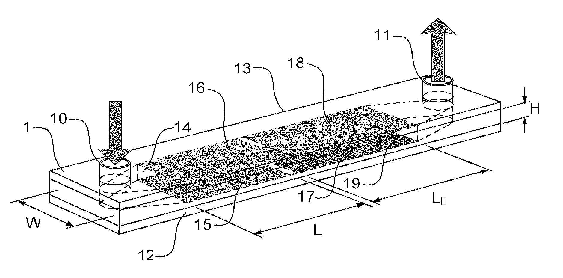
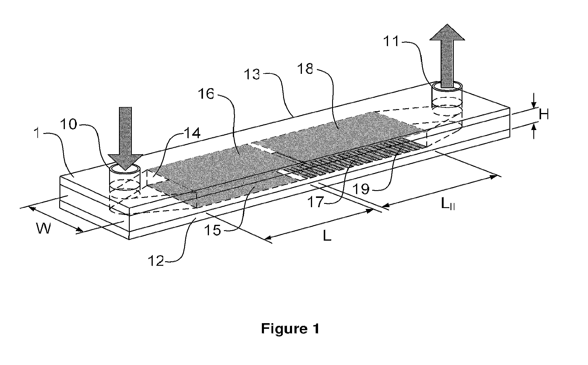
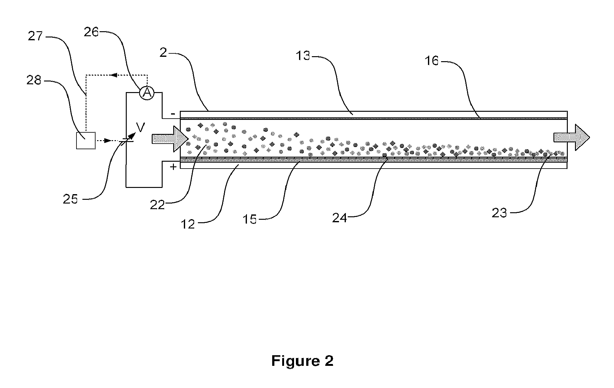

Image

  • Cell concentration, capture and lysis devices and methods of use thereof
  • Cell concentration, capture and lysis devices and methods of use thereof
  • Cell concentration, capture and lysis devices and methods of use thereof

Examples


example 1

Preparation of Immobilization Regions Comprising Antibodies or Nucleic Acid Probes

[0303]In one example, a solid support is prepared for the immobilization of either an antibody recognizing a cell surface or a nucleic acid probe for binding intracellular nucleic acids. Polished aluminum bottom plates were cleaned with water then rinsed twice with methanol and air-dried. 2% 3-Aminopropyl Triethoxysilane was prepared in 95% Methanol 5% water and the plates were immersed in silane for 5 min. Then, the plates were rinsed in methanol twice, air-dried and baked at 110° C. for 10 min. After cooling, the plates were immersed in 2.5% glutaraldehyde homobifunctional crosslinker in phosphate buffered saline, pH 7.4 for 1 hour, thoroughly rinsed in water and air-dried. The reaction zone of the microfluidic channel was defined by applying a double-sided adhesive spacer on the treated surface of the aluminum plates. Amino-labeled capture oligonucleotide probe of 1 uM final concentration which reco...

example 2

Preparation of Immobilization Regions Comprising Antibodies and Nucleic Acid Probes

[0304]In a second example, the above protocol was adapted to support the co-immobilization of antibody and nucleic acid probes within a common immobilization region. The method is schematically illustrated in the flow chart shown in FIG. 14, as henceforth described. 1 μM final concentration of biotin-labeled capture oligonucleotide probe was mixed with 20 μg / mL of Streptavidin (Sigma) in 10 mM carbonate buffer pH 9 for 5 min, and then goat anti-E.coli antibody (Abcam) of 50 ug / mL final concentration was added. The antibody and probe mixture was spotted on the reaction zone of prepared aluminum plates, therefore forming a common immobilization region, and incubated in a humidified chamber at 4° C. overnight. Unbound materials were washed from the surface with water and the reaction zone was blocked with 0.2% bovine serum albumin and 0.1% Tween-20 in PBS pH 7.4 at room temperature for 1 hr. After washin...


PUM

PropertyMeasurementUnit
thicknessaaaaaaaaaa
dielectric constantaaaaaaaaaa
ionic strengthaaaaaaaaaa

Abstract

The present invention provides a microfluidic devices and methods of use thereof for the concentration and capture of cells. A pulsed non-Faradic electric field is applied relative to a sample under laminar flow, which results to the concentration and capture of charged analyte. Advantageously, pulse timing is selected to avoid problems associated with ionic screening within the channel. At least one of the electrodes within the channel is coated with an insulating layer to prevent a Faradic current from flowing in the channel. Under pulsed application of a unipolar voltage to the electrodes, charged analyte within the sample is moved towards one of the electrodes via a transient electrophoretic force.

Description

CROSS-REFERENCE TO RELATED APPLICATION[0001]This application claims priority to U.S. Provisional Application No. 61 / 230,740, titled “DIAGNOSTIC METHODS AND DEVICES INCORPORATING ELECTRO-LYSIS OF BOUND CELLULAR ARRAYS” and filed on Aug. 2, 2009, the entire contents of which are incorporated herein by reference; U.S. Provisional Application No. 61 / 230,738, titled “LATERAL FLOW DEVICES AND METHODS FOR THE DETECTION OF CELLULAR ANALYTE” and filed on Aug. 2, 2009, the entire contents of which are incorporated herein by reference, and U.S. Provisional Application No. 61 / 287,253, titled “CELL CONCENTRATION AND CAPTURE DEVICE AND METHOD OF USE THEREOF” and filed on Dec. 17, 2009, the entire contents of which are incorporated herein by reference.FIELD OF THE INVENTION[0002]This invention relates to in-vitro diagnostic methods and devices for the concentration and / or detection of cellular analytes. More particularly, the invention relates to microfluidic diagnostic devices involving the conce...

Claims


Application Information

Patent Timeline
Patent Type & Authority Applications(United States)
IPC IPC(8): G01N33/53G01N21/17C12M1/42G01N21/64C12N13/00C12M1/34
CPCB01L3/502753C12M23/16C12M25/00C12M25/04C12M29/10C12M35/02B01L3/502715C12N1/066C12N11/14C12N13/00G01N33/5005G01N33/5438C12M41/38B01L3/502761B01L2200/0647B01L2300/0645B01L2300/0809
Inventor TALEBPOUR, SAMADKHINE, AYE AYELEONARD, STEPHEN W.MAASKANT, ROBERTALAVIE, TINO
Owner QVELLA CORP
Why Eureka
Try Eureka