Traceable retinoid acid for imaging, disease prevention and therapy
- Summary
- Abstract
- Description
- Claims
- Application Information
AI Technical Summary
Problems solved by technology
Method used
Image

Examples
example 1
Methods and Materials
[0065]Cell Lines.
[0066]The human colon (HT-29), nasal (CCL-30) and lung (A549) cancer cell lines were purchased from the American Type Culture Collection (ATCC, Manassas, Va.). Human osteosarcoma (F5M2, OS-9901, SOSP-S607, F4, CM319) and nasopharyngeal carcinoma cell lines (CNE-2) were generated in-house. The cells were cultured in Dulbecco's Modified Eagle Medium supplemented with high glucose and F12 nutrient (DMEM / F12, Invitrogen, Carlsbad, Calif.) with 10% fetal bovine serum (Hyclone, Logan, Utah) in a humidified incubator maintained at 37° C. with 5% CO2.
[0067]Tumor Xenografts:
[0068]Four- to six-week-old male athymic nude mice (18-22 g) were housed and fed sterilized pellet chow and provided sterilized water. Animals were maintained in a pathogen-free mouse colony. The facility is accredited by the American Association for Laboratory Animal Care, and all experiments were performed in accordance with the guidelines of the Institutional Animal Care and Use Co...
example 2
Synthesis of Retinoic Acid Derivative
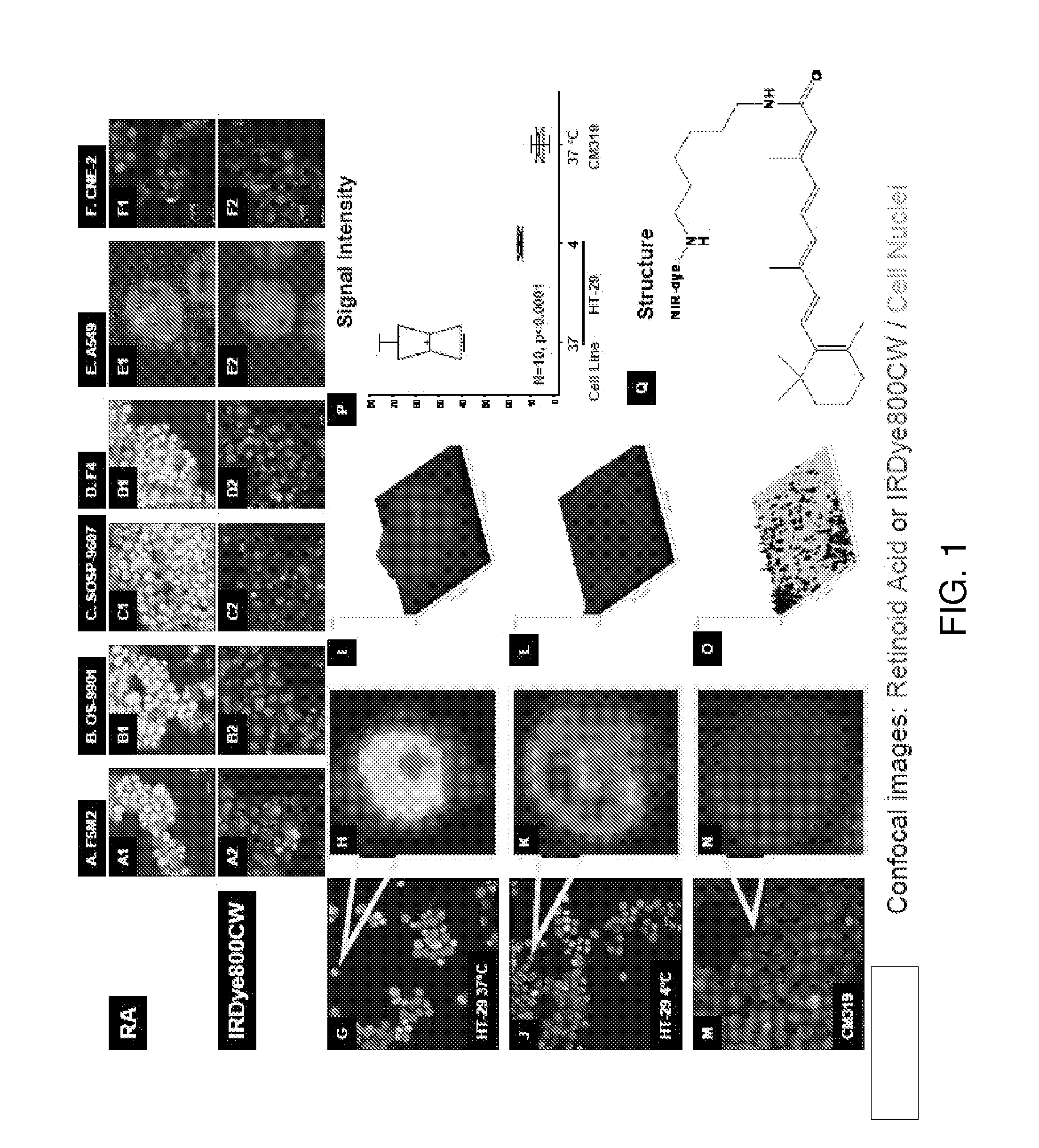
[0081]The reaction scheme and structure of retinoic acid derivative is shown in Scheme 1. Retinoic acid (compound 1) was reacted with 1-ethyl-3-(3′ dimethyl-aminopropyl)-carbodimide (EDC), N-hydroxysuccinimide (HOSu) and mono-tirtyl-1,6-diaminohexane acetic acid salt (H2N(CH2)6NH-Trt) under basic conditions, followed by acidic deprotection to form compound 2. This compound was conjugated to IRDye800CW to result in the NIR optical imaging retinoic acid derivative (compound 3).
Synthesis of N-(aminohexyl)retinoicamide (RA-NH(CH2)6NH2)
[0082]Retinoic acid (120 mg, 0.4 mmol), EDC (120 mg, 0.6 mmol), HOSu (50 mg, 0.43 mmol), DIPEA (200 μL, 1.12 mmol) and DMAP (20 mg, 0.16 mmol) were dissolved in 5 mL of DCM After 3 hr stirring H2N(CH2)6NH-Trt (150 mg, 0.4 mmol) was added to the solution. The mixture was stirred overnight at room temperature. The solvents were evaporated under vacuum, and the residue in ethyl acetate was washed with 5% NaHCO3, 2% KHSO4 a...
example 3
Retinoid Imaging Agent, In Vitro Cell Binding, and Effect of Temperature
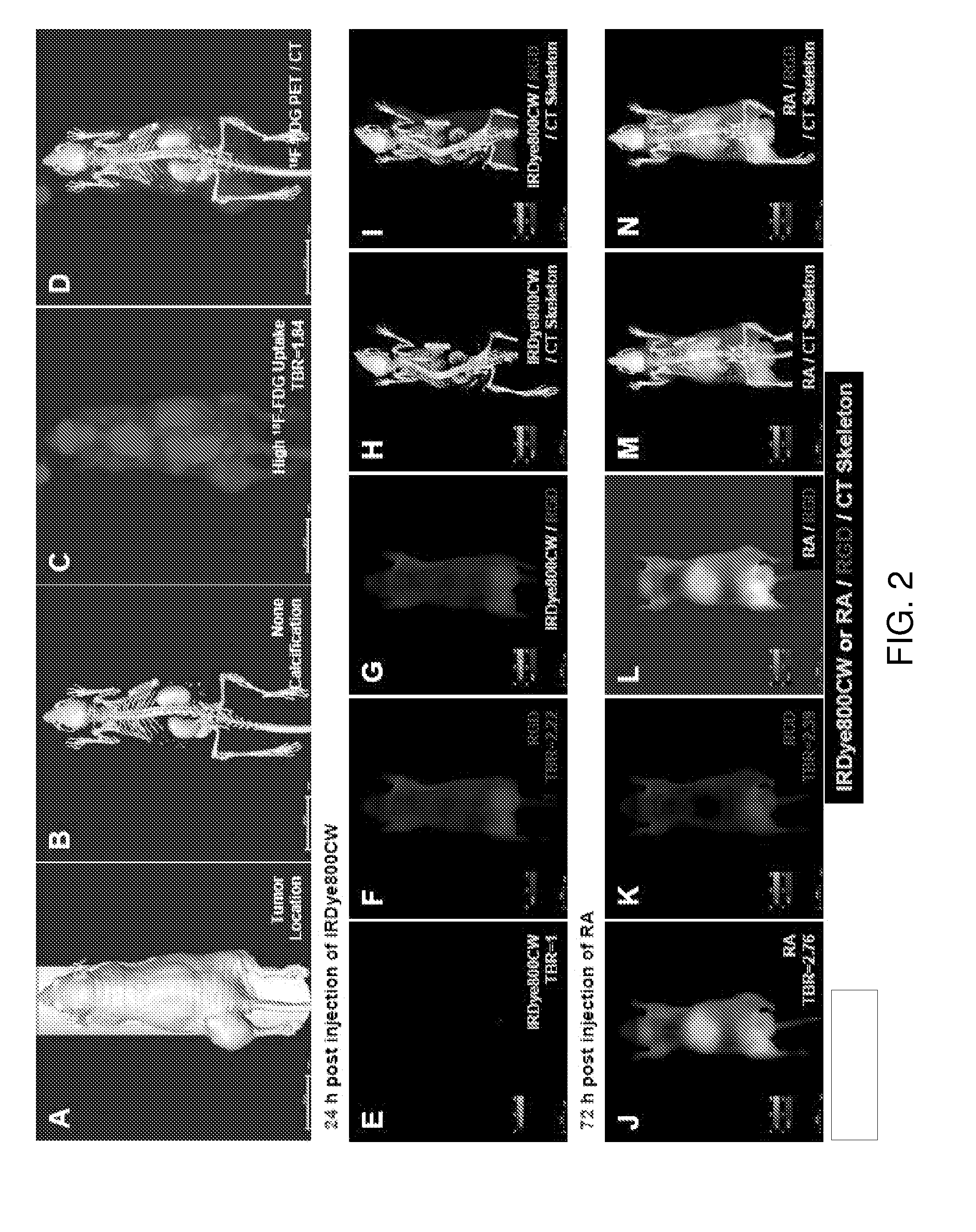
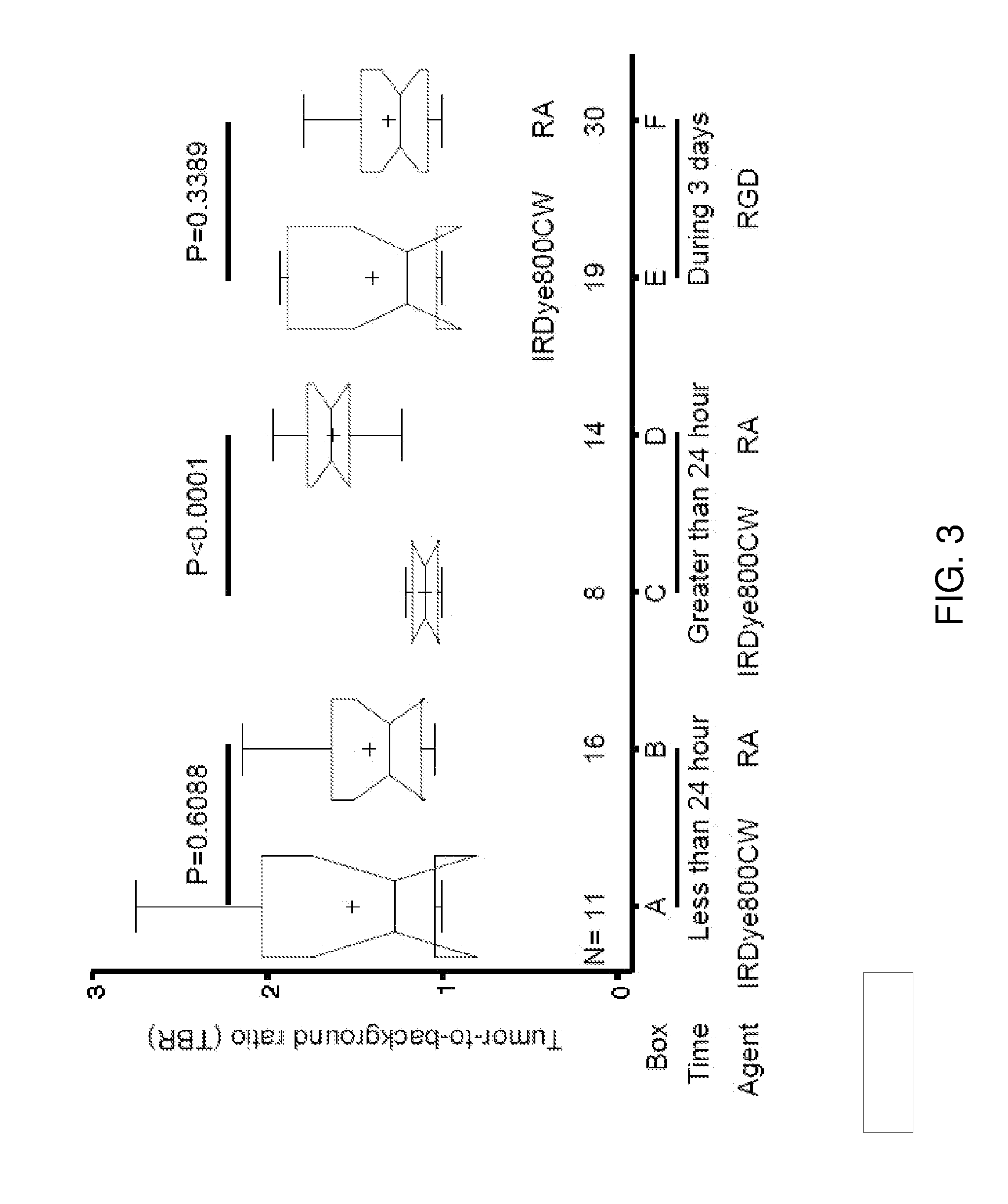
[0084]The uptake of NIR-labeled retinoid agent by human cancer cells was verified by confocal microscopy (FIG. 1A to 1H). The cell population image shows that this agent bound to human osteosarcoma (F5M2, OS-9901, SOSP-9607, and F4), human lung cancer (A549), human nasopharyngeal cancer (CNE-2), and human colon cancer (HT-29) cells (FIG. 1A1 to FIG. 1F1, FIG. 1G and FIG. 1H, red). In contrast, there was no detectable signal when these cell lines were incubated with free dye under the same conditions (FIG. 1A2 to FIG. 1F2). The retinoid signals were not only detected in the cytoplasm but also in the nuclei. The high magnification of a single cell confocal image shows that the retinoid signals localized to the cytoplasm (FIG. 1H, red). The nuclear internalization was temperature-dependent (FIG. 1J and FIG. 1K). The signal intensity was stronger in cells incubated at 37° C. (FIG. 1I) than in those incubated at 4° C...
PUM
| Property | Measurement | Unit |
|---|---|---|
| Composition | aaaaa | aaaaa |
| Structure | aaaaa | aaaaa |
| Toxicity | aaaaa | aaaaa |
Abstract
Description
Claims
Application Information
 - Generate Ideas
- Intellectual Property
- Life Sciences
- Materials
- Tech Scout
- Unparalleled Data Quality
- Higher Quality Content
- 60% Fewer Hallucinations
Browse by: Latest US Patents, China's latest patents, Technical Efficacy Thesaurus, Application Domain, Technology Topic, Popular Technical Reports.
© 2025 PatSnap. All rights reserved.Legal|Privacy policy|Modern Slavery Act Transparency Statement|Sitemap|About US| Contact US: help@patsnap.com