
Apparatus and method for real-time imaging and monitoring of an electrosurgical procedure

an electrosurgical and real-time imaging technology, applied in the field of apparatus and methods for real-time imaging and monitoring of electrosurgical procedures, can solve the problems of system inability to provide two-dimensional information, difficult judgment of incision depth, etc., and achieve the effect of improving imaging speed and resolution, and profound effect on ophthalmic imaging and diagnosis

Active Publication Date: 2012-12-06
VANDERBILT UNIV
View PDF2 Cites 48 Cited by
  • Summary
  • Abstract
  • Description
  • Claims
  • Application Information

AI Technical Summary

Benefits of technology

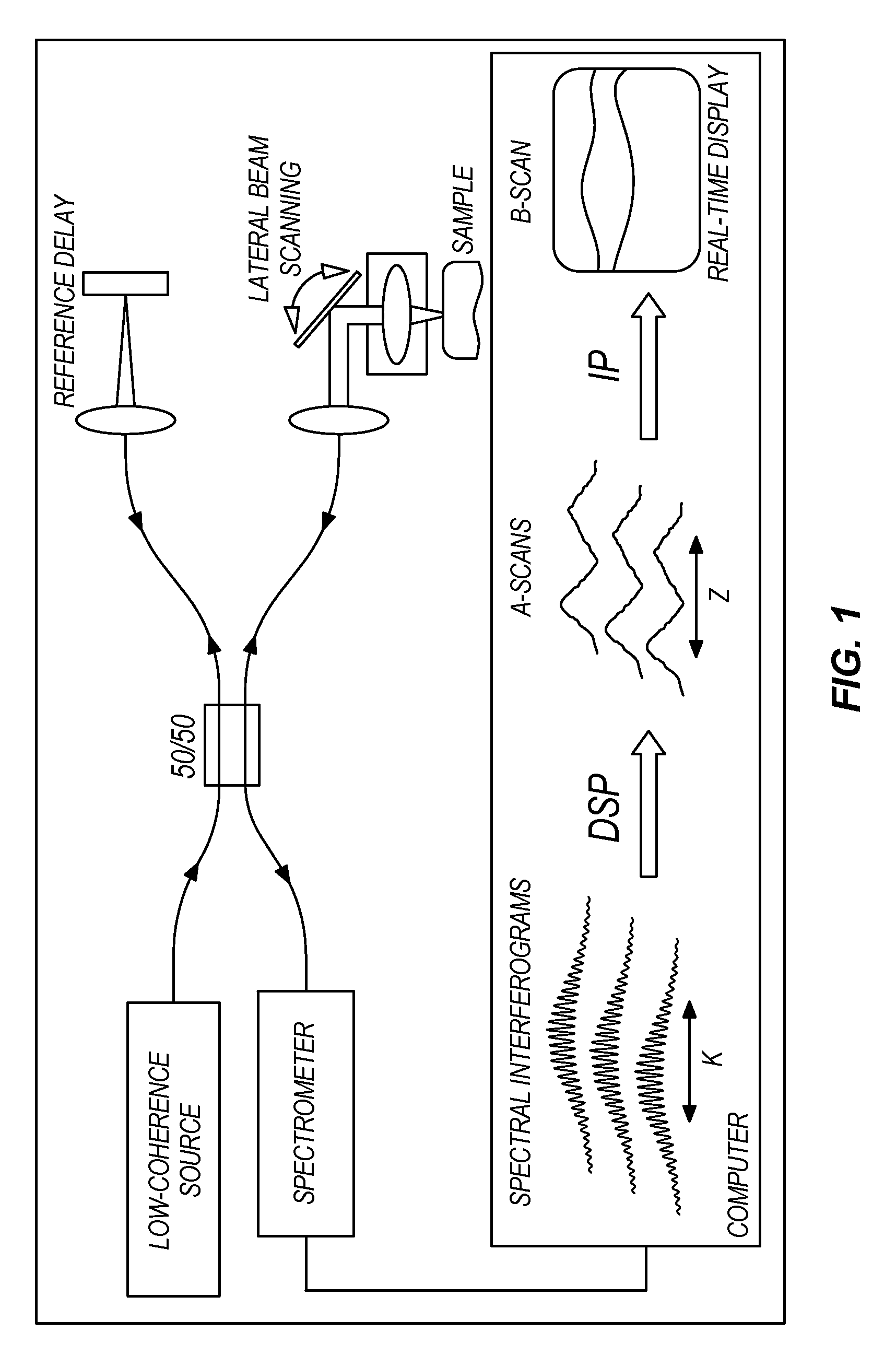
[0008]OCT technology has had a profound effect upon ophthalmic imaging and diagnosis. Its capabilities are also being embraced by gastroenterology, urology, oncology, and other specialties. The OCT B-scan is used daily in ophthalmology clinics to evaluate the delicate structures within the eye for evidence of macular edema, macular holes, subtle retinal lesions, glaucomatous retinal nerve fiber thinning, etc. As noted in the Photonik Article, OCT has evolved with improved imaging speed and resolution especially of the retinal layers in research investigations.
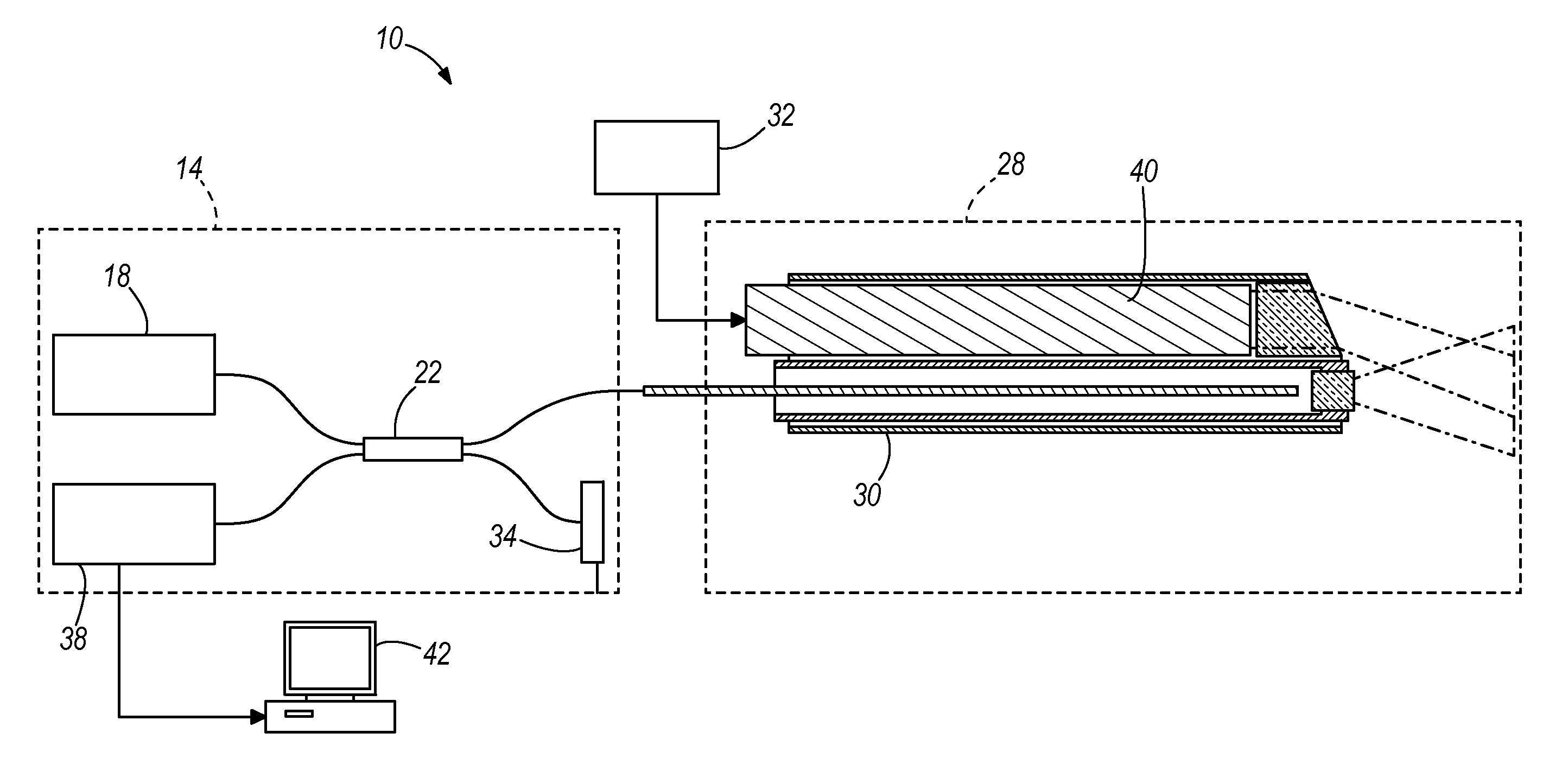
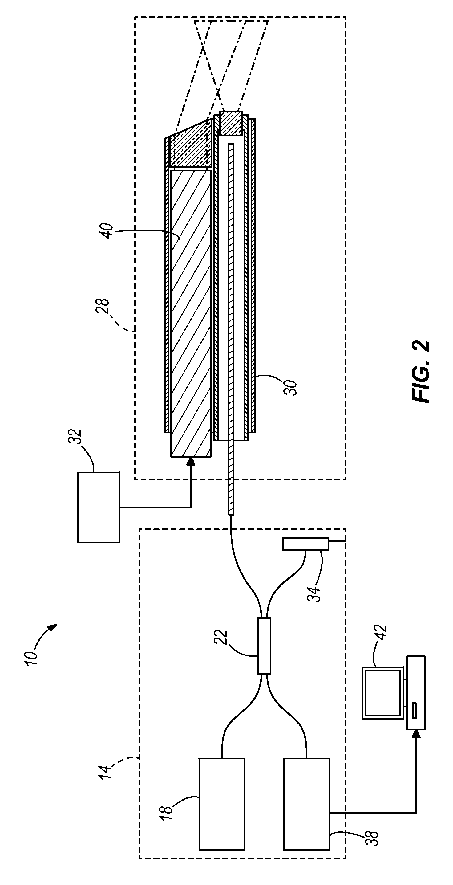
[0011]Accordingly, in one construction, the invention is related to a OCT probe miniaturized for insertion into a working channel of an endoscope for imaging tissue. In another construction, the invention is related to the combination of OCT technology and laser technology that is reduced to a miniature probe that can be inserted into the working channel of an endoscope for detecting incision of a tissue layer as it occurs so that the underlying tissues are protected from injury. High-resolution OCT forward-imaging alone could be used to evaluate sub-surface structures during endoscopic procedures. In addition, it can precisely guide the depth of an endoscopic laser incision with real-time monitoring. This is likely to advance therapies within small spaces, such as the space behind the eye. This endoscopic-capable device has the potential for adoption in multiple surgical specialties with or without the laser.

Problems solved by technology

Individual OCT A-scan components alone would permit miniaturization of the sensing probe, but the system would be unable to provide two-dimensional information.
We successfully have performed endoscopic optic nerve sheath fenestration, but judgment of the incision depth is challenging.

Method used


Image

  • Apparatus and method for real-time imaging and monitoring of an electrosurgical procedure
  • Apparatus and method for real-time imaging and monitoring of an electrosurgical procedure
  • Apparatus and method for real-time imaging and monitoring of an electrosurgical procedure

Examples


example i

[0075]INTRODUCTION: Several groups, including ours, have identified a wavelength at 6.1 mm produced by an experimental tunable free electron laser (FEL) as capable of ablating tissue with a minimal amount of collateral damage. This is desirable for precise incisions of tissue. This laser wavelength is at a water-absorption peak and is near the 6.0 mm Amide I protein peak in the collagen spectrum. Tissues which have been treated with this wavelength include articular cartilage, fibro-cartilage, skin, cornea, and optic nerve sheath.

[0076]Previously, we developed a robust hollow-glass waveguide intraocular probe to deliver this mid-infrared energy. The waveguides also were used to transect vitreous bands that were produced by injecting fibroblasts intravitreally in an animal model. Balanced salt solution (BSS) functioned well as the transmission medium. Another surgical medium, perfluorodecalin, also permitted retinal ablation by mid-infrared laser energy. However, the FEL is a cost-pr...

example ii

[0083]FIG. 41 illustrates real-time imaging which occurred with 6.1 μm laser ablation of retina with one construction of the device 700 as shown in the sequential images from a video.


PUM


Abstract

An optical coherence tomography probe and laser combination device configured for real-time z-directional guidance of the incisional depth of a surgical procedure. It can be used alone or placed within the working channel of an endoscope. The device includes an OCT single mode fiber, and a laser fiber or laser hollow waveguide or electrical surgical wire positioned adjacent to the OCT single mode fiber. The single mode fiber is configured to move laterally when activated by an actuator to scan light data reflected from a sample that is positioned in front of a distal end of the device. The light data can be processed to generate a B-scan image. The device can collect data in real-time during lasing, or immediately prior to and following the cutting. The surgical tool, when coupled to a processor, can deactivate when the B-scan image identifies that the incision is within a predefined tolerance.

Description

STATEMENT REGARDING FEDERALLY SPONSORED RESEARCH OR DEVELOPMENT[0001]This invention was made with government support under FA9550-04-1-0045 awarded by the Air Force Office of Scientific Research—DOD. The government has certain rights in the invention.BACKGROUND OF THE INVENTION[0002]Advances in Optical Coherence Tomography (OCT) technology have made it possible to use OCT in a wide variety of applications. One application of OCT is in ophthalmology for imaging eye diseases due to the high transmittance of ocular media. OCT technology was invented in the early 1990's to generate depth-resolved images of tissue level microstructures, in vivo, and without physical contact. Second generation imaging technology, such as frequency-domain, swept-source, and spectral-domain OCT, has improved the signal-to-noise ratio over first generation technology, translating to faster imaging. As a result of this speed increase, high resolution cross-sectional images (B-scans) can be acquired at video-r...

Claims


Application Information

Patent Timeline
IPC IPC(8): A61B1/07A61B18/20
CPCA61B18/14A61B18/20A61B18/201A61B18/18A61B2019/5234A61B3/102A61B18/22A61B2090/3735A61B2018/00982A61B2018/00958A61B90/37
Inventor JOOS, KARENSHEN, JIN HUIHUTSON, M. SHANEKOZUB, JOHN
Owner VANDERBILT UNIV
Learn More