
Method for isolating target cells

Inactive Publication Date: 2013-03-07
UNIVERSITAETSKLINIKUM HAMBURG EPPENDORF
View PDF2 Cites 11 Cited by
  • Summary
  • Abstract
  • Description
  • Claims
  • Application Information

AI Technical Summary

Benefits of technology

The present invention provides a method for processing large volumes of sample, which involves using a filtration step to concentrate target cells and remove unwanted components such as red blood cells and platelets that may interfere with the subsequent binding of the target cells to the affinity molecules on the separation surface. This leads to higher recovery rates of target cells and improved overall performance of the method compared to current methods that use magnetic beads which can only handle small volumes of fluid.

Problems solved by technology

Metastasis formation rather than progression of the primary tumor itself is the main cause of death in tumor diseases.
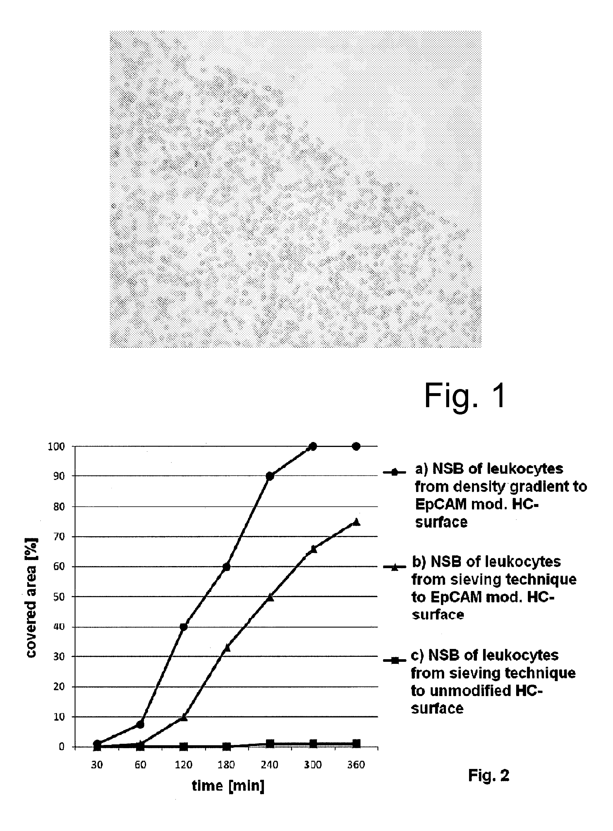
However, the current available techniques are associated with a high degree of unspecific cell adhesion to the beads as well as a low detection rate of tumor cells.
It is known that due to the high number of non-target cells—in particular red blood cells—in the sample, it cannot be ensured in the routinely used methods that each tumor cell in a sample can interact with the antibody-presenting surface of a bead for a sufficiently long time so that binding between the immobilized antibody and the target cell occurs.
This approach has the particular disadvantage that it is labor-intensive and requires highly experienced personnel for performing the separation.
Moreover, disseminated tumor cells may exhibit a broad density range which means that part of these cells may not be available for a subsequent affinity binding step.
The use of density gradient media also requires several washing steps of the tumor cells which are associated with several adverse effects, such as shear stress and unspecific adsorption of the cells to the wall of the reaction tubes, both of which may result in a loss of target cells.
Furthermore, density gradient centrifugation does not allow the processing of larger sample volumes, which is a clear disadvantage when considering that disseminated tumor cells are present in the blood or bone marrow in extremely low concentrations.
By adding lysis buffer the membranes of the red blood cells are disrupted which results in cell death and release of intracellular components.
However, it has been demonstrated that the use of lysis buffers regularly also disrupts a considerable portion of the disseminated tumor cells present in the sample.
Furthermore, it can be assumed that the use of these buffers will adversely affect the metabolism of the target cells, which could be disadvantageous for further processing steps (e.g. culturing the isolated cells).
In these methods, only small volumes of between 2-5 ml sample fluid can be incubated with the magnetic beads, because otherwise the likelihood of a contact between the target cells and the antibodies on the surface of the particles significantly decreases, thereby leading to low recovery rates.
Similarly, the material should also not allow for any substantial unspecific adhesion of target cells, for example, disseminated tumor cells, to the filter material, which would impede the subsequent transfer of the target cells to the separation surface.
These cells are present in the blood or bone marrow in very low concentrations, so that their detection and isolation is generally complicated.
The high number of red blood cells and platelets in the blood has the adverse effect that the binding of target cells, such as tumor cells floating in the blood stream, via specific binding molecules (e.g. antibodies) on a separation surface, for example, a magnetic particle or a coated support surface, is often inefficient, because most of the target cells do not sufficiently interact with their corresponding binding molecules to provide for a binding to the separation matrix.
In consequence many cells will be not identified due to a loss of the nucleus, a unregular plasma-nucleus ratio and the occurrence of cell debris which stains positively for markers for antibody detection thus leading to false positive results.

Method used


Image

  • Method for isolating target cells
  • Method for isolating target cells
  • Method for isolating target cells

Examples


example 1

Preparation of a Test Sample Containing Tumor Cells

[0098]Cultured MDA-468 cells derived from human breast carcinoma were pre-stained with 4′,6-Diamidino-2-phenylindol (DAPI, Pierce / ThermoScientific) without permeabilization. DAPI is a cationic fluorescent dye which binds to adenine-thymine-rich DNA. DAPI is regularly used for staining cell nuclei. The stained cells can be counted by visual inspection or by suitable automated devices. The cells were washed twice with Dulbecco's modified Eagle Medium (DMEM) to remove excessive dye. Each suspension of pre-stained cells was controlled for fluorescence emission. Aliquots of tumor cells were counted manually and spiked in human whole blood samples collected from healthy donors by venipuncture in collection tubes with EDTA to prevent coagulation. The blood samples were used within 4 h from collection. For recovery testing, the tumor cells were diluted to cell numbers between 50 and 500 cells / 100 μl to minimize dilution and counting errors....

example 2

Preparation of a Woven Fabric for Cell Sieving

[0099]The filter of a blood administration set (Sarstedt AG & Co; 74.4255) was separated and the woven filter removed. A sheet of a woven fabric (Sefar AG, Sefar Nitex 03-5 / 1, Sefar Petex 07-5 / 1) having a mesh size of 1 μm was fixed on the filter rack by use of an adhesive. After fixing the woven fabric to the filter rack, any remaining solvent deriving from the adhesive was removed in a vacuum for 30 min. A leak test was carried out with a cell suspension of monocytes from buffy coat obtained by centrifugation of 500 ml blood over the whole volume of the filter rack. The so prepared filter rack was fixed in the middle of a beaker by using a double-faced medical adhesive (50 μm thick) so that the closed bottom of the filter rack was in contact with the bottom of the beaker.

example 3

Removal of Red Blood Cells and Platelets from the Sample

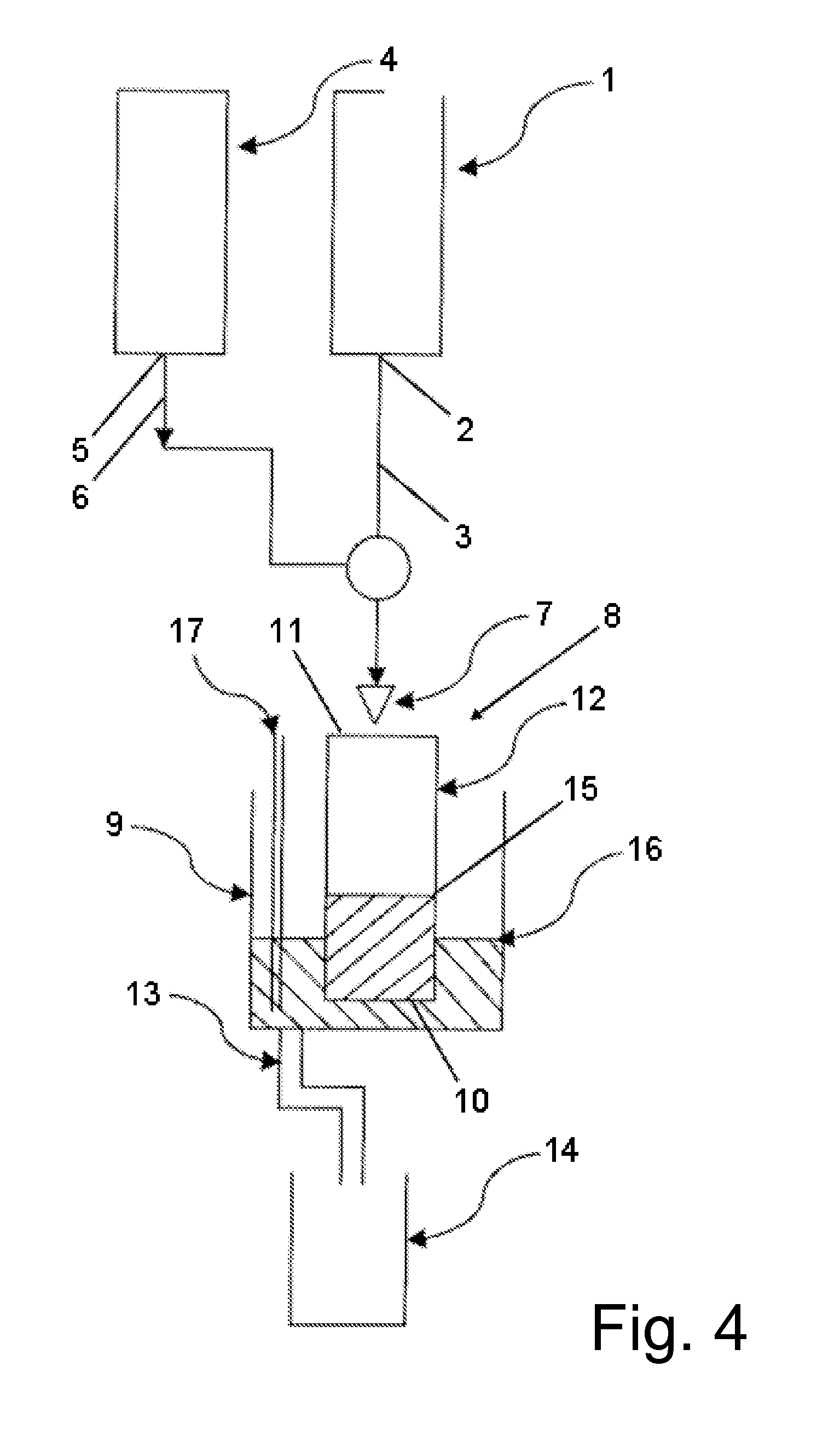
[0100]A pre-wetting of the filter rack prepared in example 2 is preferred to prevent initial unspecific adhesion of cells to the filter unit. Hence, a volume of 10-15 ml of a physiological and iso-osmolaric buffer (e.g. PBS) was added to the filter rack. The filtering was performed in an assembly as depicted in FIG. 4. The buffer was applied to the center of the cylindrical filter rack by a conduit which is located 1-2 cm above the opening of the filter rack. A flow rate of 1 ml / min was set with a peristaltic pump. A blood sample as prepared in example 1 (stored for 24 h under agitation at room temperature) containing between 10 and 120 MDA-468 cells per ml in spike-in experiments was applied to the filter rack by a second conduit with a flow rate of 100-200 μl / min. In this way, dilutions of the blood samples of in a ratio of about 1:5 to 1:10 could be obtained.

[0101]Due to the narrow mesh size of the woven filter element, the ...


PUM

PropertyMeasurementUnit
Lengthaaaaaaaaaa
Lengthaaaaaaaaaa
Pore sizeaaaaaaaaaa

Abstract

The present invention relates to a method for the enrichment and / or isolation of target cells in a sample, which sample comprises red blood cells and / or platelets, comprising (a) filtering the sample through a filter element having a pore or mesh size of between 0.5 and 5 μm, (b) contacting the cells retained by the filter element in step (a) with a separation surface, wherein said separation surface comprises affinity molecules which selectively bind to the target cells, (c) incubating the cells and the separation surface under conditions which allow for the binding of the affinity molecules to the target cells; and (d) separating the separation surface from any unbound cells and material.

Description

[0001]The present invention relates to a method for the enrichment and / or isolation of target cells in a sample, which sample comprises red blood cells and / or platelets, said method comprising (a) filtering the sample through a filter element having a pore or mesh size of between 0.5 and 5 μm, (b) contacting the cells retained by the filter element in step (a) with a separation surface, wherein said separation surface comprises affinity molecules which selectively bind to the target cells, (c) incubating the cells and the separation surface under conditions which allow for the binding of the affinity molecules to the target cells; and (d) separating the separation surface from any unbound cells and material.BACKGROUND OF THE INVENTION[0002]Disseminated tumor cells (DTC), i.e. tumor cells which are detectable in the peripheral blood or in the bone marrow of a cancer patient, have been shown in clinical studies to provide informative value in terms of prognosis of a cancer patient as ...

Claims


Application Information

Patent Timeline
IPC IPC(8): C12N5/09C12Q1/68G01N33/53C12N5/078
CPCC12Q1/6806G01N33/56966G01N33/57484C12Q2531/113
Inventor DANKBAR, NICOBRANDT, BURKHARDGEDIG, ERK TJALLING
Owner UNIVERSITAETSKLINIKUM HAMBURG EPPENDORF
Learn More