

Polymer scaffolds and their use in the treatment of vision loss
a polymer scaffold and vision loss technology, applied in the field of scaffolds for growing cells, can solve the problems of logistically impracticality and graft rejection
- Summary
- Abstract
- Description
- Claims
- Application Information
AI Technical Summary
Benefits of technology
Problems solved by technology
Method used
Image

Examples
example 1
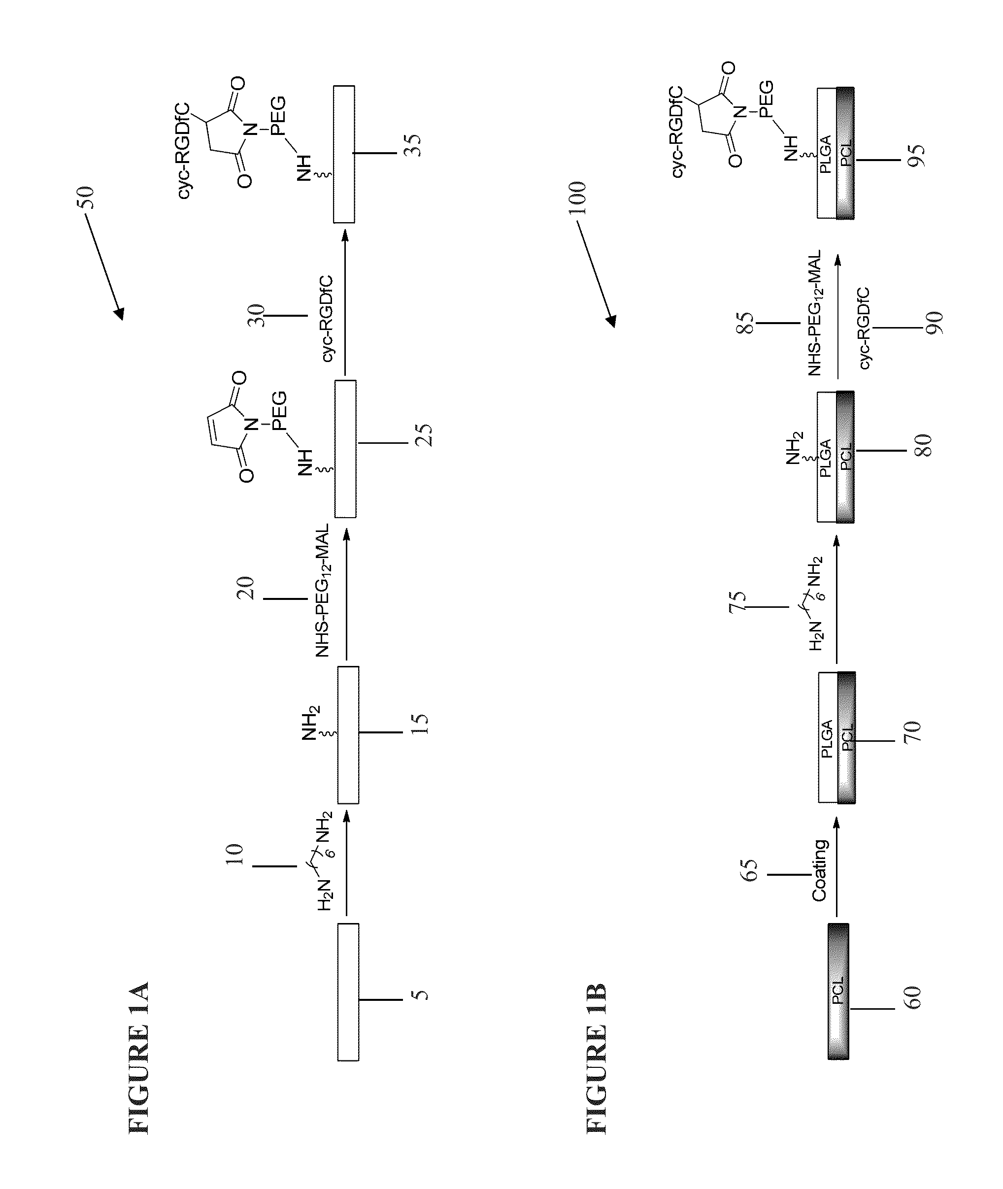
Formation of Cyclic-RGD Linked Peptide Coated Polymer Scaffold
[0121]A chloroform solution of PLGA copolymer (85:15) or PLGC (70:10:20) copolymer at a concentration of 25 mg / ml was casted onto a glass cover (70 μL onto 12 mm glass cover). The film was allowed to air dry, followed by drying completely under high vacuum condition. Typical thickness of film was around 5-10 μm, measured by profilometer. The polymer coated glass covers were placed in a tissue culture plate. To covalently modify the surface, a 1,6-hexamethylenediamine solution was added into the plate (1 mL solution from 1.0 mg / mL in isopropanol). The plate was shaken for 2 hour at room temperature, soaked in de-ionized water for 1 hour, and thoroughly washed with deionized water. The surface became colored during this step (blue or rainbow). The amine modified film was reacted with a cross linking agent for 5 hour while shaking at room temperature (crosslinking agent: NHS-PEG12-Maleimide from Thermo Scientific, 1 mL from ...
example 2
Use of Cyclic-RGD Peptide Linked Polymer Scaffolds for Culturing hESC-RPE Cells
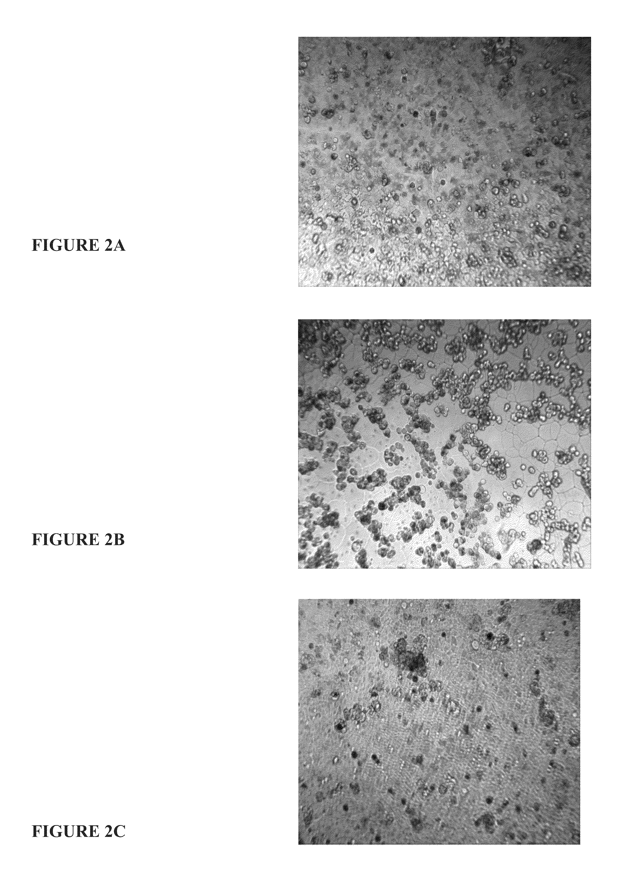
[0122]Cyclic-RGD peptide linked polymer scaffolds were cut into 6 mm diameter discs and placed into wells of a 96 well-plate. The discs were incubated at room temperature for 1 hour with 70% ethanol solution to prevent contamination. The discs were washed for two 15 min washes with PBS. hESC-RPE cells were trypsinized, washed twice with 10 mL of PBS, and adjusted to a concentration of 500,000 cell / mL. 100 μL of hESC-RPE was deposited to each disc containing well. The cells were incubated overnight at 37° C. at 5% CO2. The resulting cell attachment were observed and recorded.
Results
[0123]After 24 hours from the seeding time, hESC-RPE cells appeared to be attached to the modified PLGA scaffold with 80 to 90% confluence. The cells exhibited a flat cuboidal (cobblestone) morphology confirming cell attachment (FIG. 2A). Scaffolds without cyclic RGD were unable to retain cell attachment as shown by the spherica...
example 3
Immunocytochemistry of Polymer Scaffolds
PCL-PLGA-cRGD Linked Scaffolds
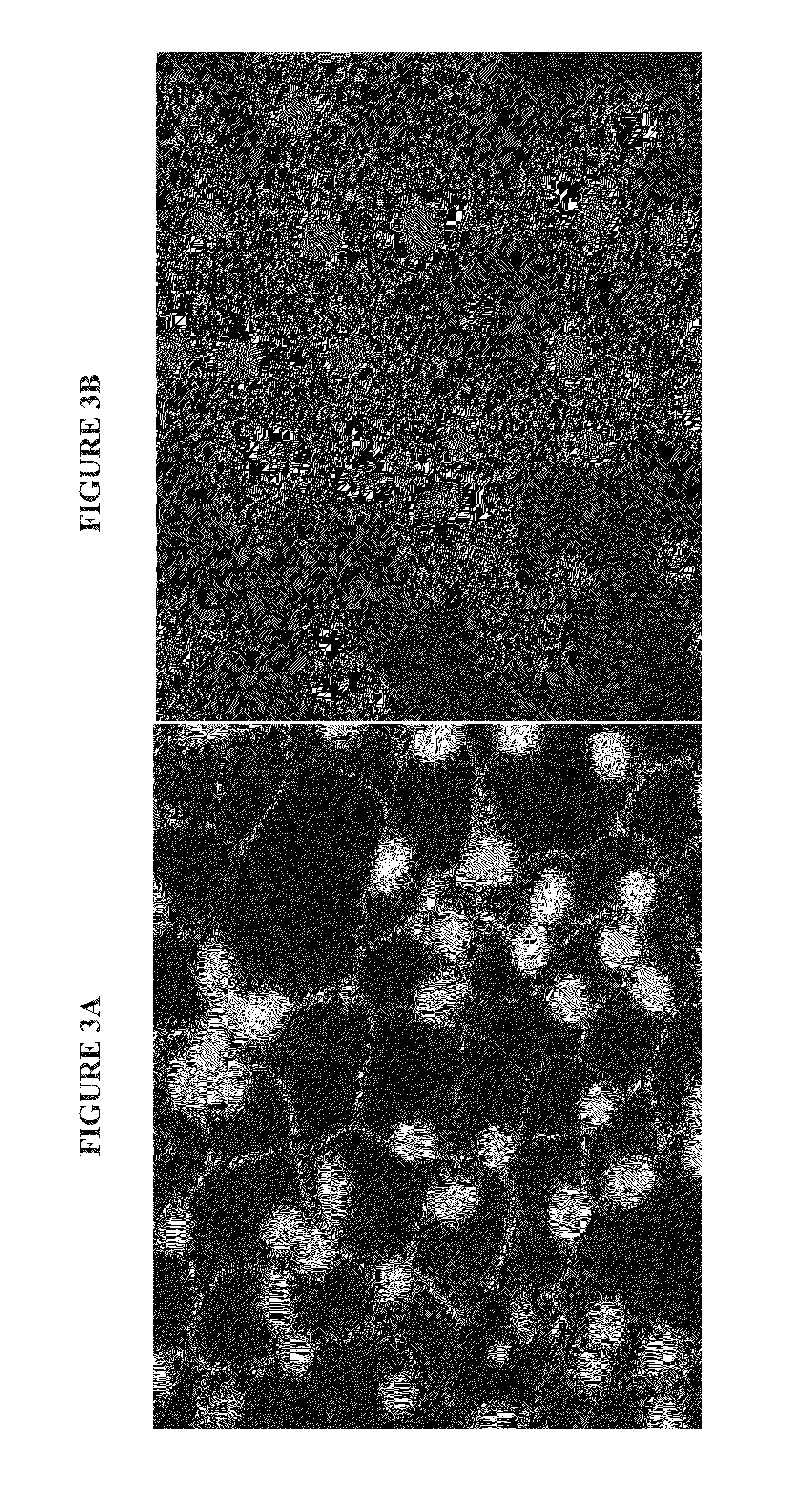
[0125]To observe the RPE phenotypes of the hESC-RPE cells seeded on a thin film of PCL-PLGA-cRGD peptide linked scaffold, ICC stainings were performed. The cell attached PCL-PLGA-cRGD scaffolds were washed twice with PBS and then fixed with 2% paraformaldehyde in PBS for 20 mins at RT. The fixed scaffold was rinsed twice with 0.1% BSA in PBS (wash buffer) and incubated with a blocking agent of 10% goat serum 0.3% triton-X 100 for 40 mins. The scaffolds were incubated with either ZO-1 (1:200 dilution, Invitrogen) or RPE-65 (1:500 dilution, Millipore) overnight at 4° C. After three 10 min washes at RT with wash buffer, the scaffold was incubated with Cy5-conjugated anti-rabbit IgG secondary antibody for 30 mins. Three more 10min washes with the wash buffer were performed followed by 3 μM DAPI solution for the counterstain. The scaffold was viewed under a fluorescent microscope.
PLGC-cRGD Peptide Linked Scaffolds
[0126...
PUM
| Property | Measurement | Unit |
|---|---|---|
| Temperature | aaaaa | aaaaa |
| Temperature | aaaaa | aaaaa |
| Permeability | aaaaa | aaaaa |
Abstract
Description
Claims
Application Information