In vitro tumor in dish kit and method

Active Publication Date: 2013-11-28
UNIVERSITY OF KANSAS
View PDF4 Cites 12 Cited by
  • Summary
  • Abstract
  • Description
  • Claims
  • Application Information

AI Technical Summary

Benefits of technology

The patent text describes a method for creating a tumor in a dish model using different types of cells. The cells are combined and cultured in a special medium to form a tumor. The method can also be carried out in a special vessel. The text also mentions adding a test compound to see if it can inhibit or reduce the growth, proliferation, size, or metastasis of the tumor. The technical effect of this patent is to provide a reliable and reliable method for creating tumors in a laboratory setting for research and drug development purposes.

Problems solved by technology

One of the fundamental challenges researchers face in drug, environmental, nutritional, consumer product safety, and toxicology studies is the extrapolation of metabolic data and risk assessment from in vitro cell culture assays to animals.
The failure rate for drugs in phase clinical trials has been rising.
More than 50% of these failures are due to insufficient efficacy, and almost 20% are due to toxicity.
Despite identification of a vast number of anti-cancer agents against cancer cell lines in artificial cell-based two-dimensional culture system, treatment of cancer remains challenging.

Method used


Image

  • In vitro tumor in dish kit and method
  • In vitro tumor in dish kit and method
  • In vitro tumor in dish kit and method

Examples


example 1

Establishment and Analysis of Tumor in Dish Model

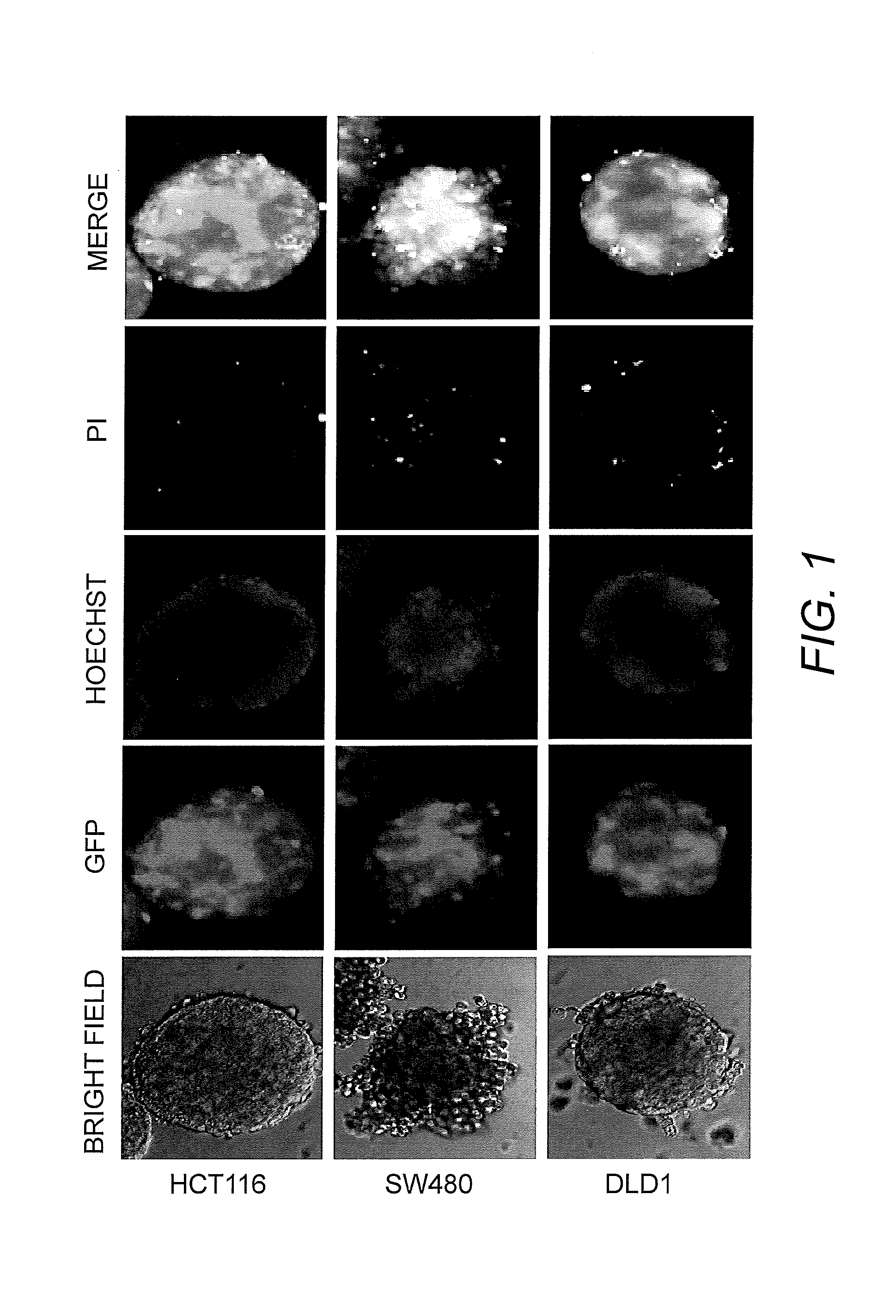
[0036]To prepare the Tumor in Dish model, Human Umbilical Vein Endothelial Cells (HUVEC), Human Lymphatic endothelial cells (HLEC), Human lung fibroblast cells (MRC-5), and Human lung epithelial cells (NL20) were combined. To the normal cells was added human colon cancer cells (DLD1 stably expressing Green Fluorescent Protein ((GFP)). The cells were cultured on the medium composed of the ingredients listed in Table 2 in ultra low attachment plates.

TABLE 2ComponentAmountEGM MV Microvascular Endothelial Cell Growth40%MediumAirway Epithelial Basal Medium 20%DMEM - Colosphere, NeurosphereorMEBM - Mammosphere 20%orRPMI-1640 Medium - Esophageal and GastricCancer SpheresFibroblast Growth Medium20%Hydrocortisone1.4μg / mlFGF6VEGF2ng / mlR3-IGF-13ng / mlAscorbic Acid20μg / mlhEGF6ng / mlHeparin calcium salt2.4μg / mlFBS1.2%P / S0.6%L-Alanyl-L-Glutamine0.48mMExtract P0.08% Plasma protein fraction0.1%Epinephrine0.2μMTransferrin1μg / mlT32nMInsulin1μg / mlB27 supp...

example 2

Exosome Analysis of Tumor in Dish Model

[0042]The Tumor in Dish model was exposed to a control and anti-cancer drug (compound 1). The media from the wells of control and drug-treated Tumor in Dish model were collected and exosomes were isolated using EXOQUICK exosome isolation kit (SBI, Mountain View, Calif.). RNA was isolated and real-time PCR analysis has conducted. This analysis indicated that the expression of DCLK1 and U6 mRNA decreased in the exosomes following compound 1 treatment. Exosomes are present in body fluids such as blood, milk, CSF, urine, etc. and have been shown to be released from the tumor to regulate tumor microenvironment. Therefore, regulation of tumor-released exosomes by anti-cancer agents can be analyzed using the Tumor in Dish model and system.

example 3

Analysis of Anti-Cancer Drug in Tumor in Dish Model

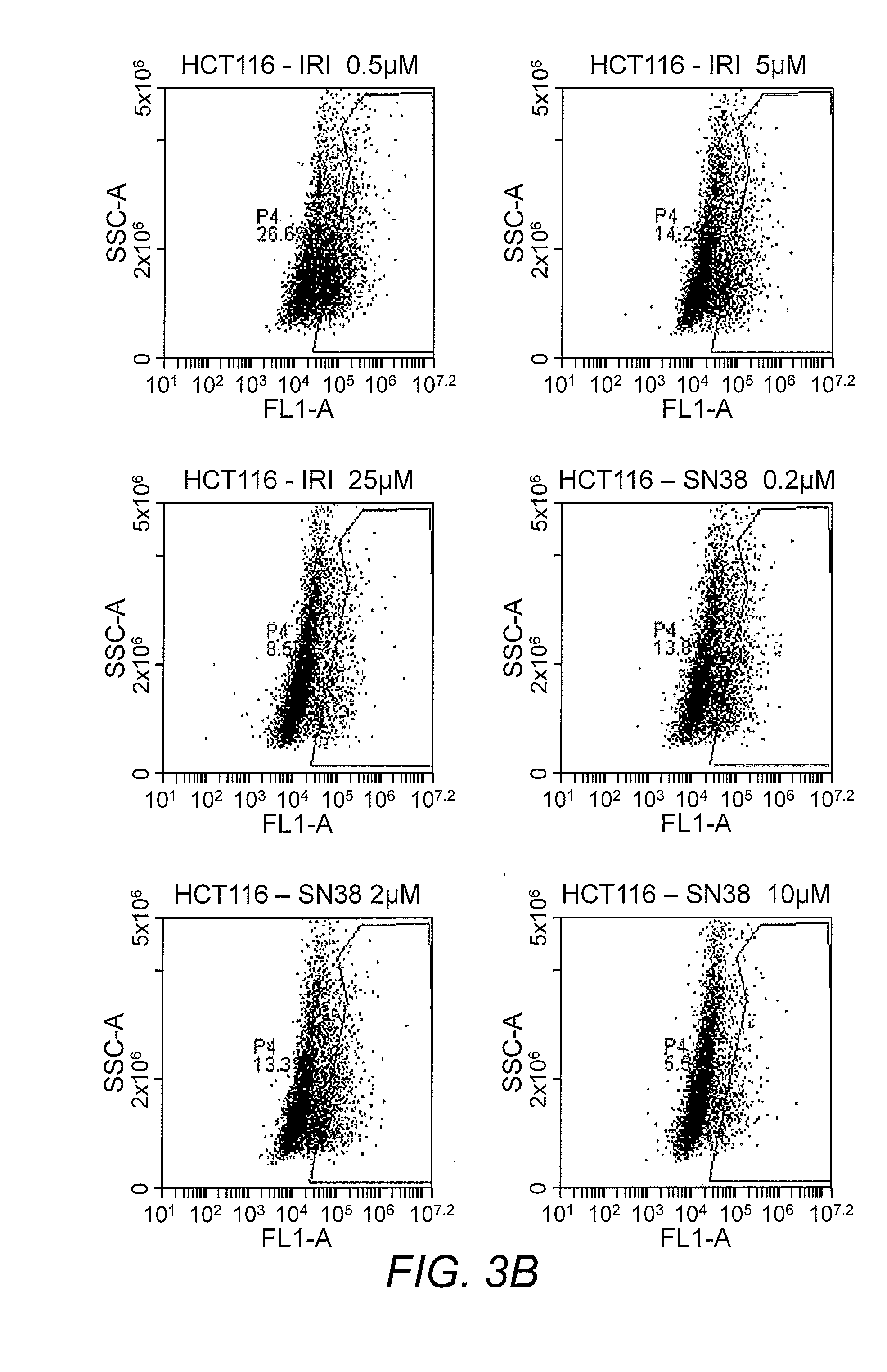
[0043]Cells from control and anticancer drug B-treated Tumor in Dish model were dissociated into single cell suspension using trypsin and were FACS sorted to determine the effect of the drug on different types of cells. The results indicated that the number of GFP expressing DLD1 cells was measurably decreased in the drug-treated group, while the number of red florescence protein expressing normal fibroblast cells, purple color-labeled endothelial cells, and the blue color-labeled lung epithelial cells (pseudo colored) were moderately decreased by drug B. Immunofluorescence imaging studies confirmed that drug B significantly decreased the number GFP-DLD1 cells. In a similar study with compound 1, no significant decrease in DLD1-GFP cells was observed; however, there was a significant inhibition of endothelial cell growth, indicating inhibition of angiogenesis.

[0044]The effect of conventional anti-cancer agents were also tested in th...


PUM


Abstract

This invention is a method and kit for preparing a Tumor in Dish model for use in screening anti-cancer agents and analyzing cancer growth and development.

Description

INTRODUCTION[0001]This application claims the benefit of U.S. Provisional Application Nos. 61 / 688,937 filed May 24, 2012 and 61 / 712,974 filed Oct. 12, 2012, whose contents are incorporated herein by reference in their entireties.BACKGROUND OF THE INVENTION[0002]Pharmacokinetics is the study of the fate of pharmaceuticals and other biologically active compounds from the time they are introduced into the body until they are eliminated. Pharmacokinetics provides a rational means of approaching the metabolism of a compound in a biological system. One of the fundamental challenges researchers face in drug, environmental, nutritional, consumer product safety, and toxicology studies is the extrapolation of metabolic data and risk assessment from in vitro cell culture assays to animals.[0003]To develop treatment options for patients diagnosed with tumors that are non-responsive or respond poorly to treatment with a particular anti-cancer agent, there is a need for reliable robust cell lines...

Claims


Application Information

Patent Timeline
IPC IPC(8): G01N33/50
CPCG01N33/5011C12N5/0693C12N2502/1323C12N2502/28C12N2503/02
Inventor ANANT, SHRIKANTRAMAMOORTHY, PRABHURAMALINGAM, SATISHSITTAMPALAM, GURUSINGHAM SITTA
Owner UNIVERSITY OF KANSAS
Why Eureka
Try Eureka