Method and system for providing versatile nirs sensors

a technology of nirs sensors and sensors, applied in the field of providing versatile nirs sensors, can solve the problems of increased pressure within the compartment, decreased size, and cast that is too tigh

Inactive Publication Date: 2013-11-28
J&M SHULER
View PDF16 Cites 39 Cited by
  • Summary
  • Abstract
  • Description
  • Claims
  • Application Information

AI Technical Summary

Benefits of technology

[0032]The sensor may further include a substrate for supporting the light source, portable energy source, the processing module, and wireless transceiver. The battery or capacitor may have a size which is substantially smaller than the substrate. The substrate may be part of a sterile bandage. The substrate of the wireless sensor may include absorbent materials to absorb any moisture or liquid adjacent to the light source or the light receiver. The wireless transceiver may include at least one of a radio-frequency transceiver, an optical transceiver, an acoustical transceiver, and a magnetic transceiver. The wireless sensor may include memory for storing the readings in connection with the light source. The wireless sensor may have multiple modes of operation.

Problems solved by technology

Bleeding or swelling within a compartment can cause an increase in pressure within that compartment.
A decrease in size results from a cast that is too tight, constrictive dressings, pneumatic anti-shock garments, and closure of fascial defects.
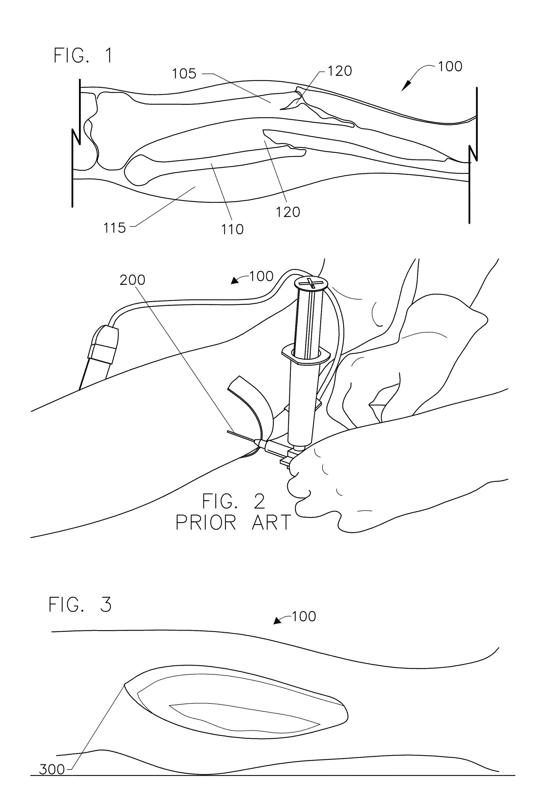
While compartment pressures can be measured with this conventional method, the method is highly invasive procedure which can cause tremendous pain to the patient.
In addition to causing tremendous pain to the patient, there are several more problems associated with the conventional needle measuring method.
First, it is very challenging for a medical practitioner to actually measure or read pressure of a compartment since the needle must be positioned at least within the interior of a compartment.
And it is very difficult for the medical practitioner to know if the needle has penetrated adequately through the intermediate layers to enter into the compartment.
This challenge significantly increases if the patient being measure is obese and has significant amounts of subcutaneous fat in which to penetrate with the needle.
Often, the medical practitioner may not have a needle accurately positioned inside a compartment which can yield a reading of the tissue adjacent to the compartment, such as muscle or skin.
Because of the challenge medical practitioners face with precisely positioning a needle within a compartment of interest and because of the numerous law suits associated with the diagnosis of compartment syndrome, many medical schools do not provide any formal training for medical practitioners to learn how to properly place a needle within a compartment of interest for reading a compartment's pressure.
Therefore, many medical practitioners are not equipped with the skills or experience to accurately measure compartment pressures with the needle measuring method.
Due to the legal climate regarding this condition, clinicians are forced to treat an elevated value for compartment pressures or expose themselves to legal ramifications with any complications.
As described later, the treatment of compartment syndrome can cause significant morbidity and increase the risk for infection.
Therefore inaccurate and elevated pressure readings are a very difficult and potential dangerous pitfall.
Another problem associated with the training and experience required for the needle measuring method is that, as noted above, compartment syndromes usually occur when tissues within the compartment are experiencing unusual levels of swelling and pressure.
A normal patient without any swelling would not provide a medical practitioner with the skills to accurately assess a size of a compartment when using the needle measuring method for determining compartment pressure.
In addition to the problem of entering a compartment that may have an abnormal size or anatomy, the needle measuring method has the problem of providing only a snap-shot of data at an instant of time.
This collection of single data points over long periods of time is usually not very helpful because pressures within a compartment as well as the patient's blood pressure can change abruptly, on the order of minutes.
Also, because of the pain associated with the needle measuring method noted above, the medical practitioner will seldom or rarely take pressure readings with within a few minutes of each other using a needle.
A further problem of the needle measuring method is that for certain regions of the body, such as the lower leg, there are four compartments to measure.
Another problem, besides pain that is associated with the needle pressure measuring method, is that there is a lack of consensus among medical practitioners over the compartment pressure ranges which are believed to indicate that a compartment syndrome may exist for a particular patient.
Other subjective methods for diagnosing compartment syndromes instead of the needle measuring method exist, however, they may have less accuracy than the needle measuring method because they rely on clinical symptoms of a patient.
One of the major drawbacks of these symptoms is that for many of them the patient must be conscious and must be able to respond to the medical practitioner.
For any inebriated patients or patients who are unconscious, the pain assessment and muscle power assessment cannot be used accurately by the medical practitioner.
In the setting of high energy trauma which is associated with compartment syndrome, many patients are not capable of cooperating with a good physical exam due to any number of causes including head trauma, medical treatment (including intubation), drug or alcohol ingestion, neurovascular compromise or critical and life threatening injuries to other body systems.
For the pain assessment, if a lower leg compartment syndrome exists in a patient, then the range of motion for a patient's foot or toes will be extremely limited and very painful when the patient's foot or toes are actively or passively moved.
Another drawback using pain to assess the likelihood of a compartment syndrome is that every human has a different threshold for pain.
This means that even if someone should be experiencing a high level of pain, he or she may have a high threshold for pain and therefore, not provide the medical practitioner with a normal reaction for the current level of pain.
Another problem with using pain to assess the likelihood of the existence of a compartment syndrome is that if the patient is experiencing trauma to other parts of their body, he or she may not feel the pain of a compartment syndrome as significantly, especially if the trauma to the other parts of the patient's body is more severe.
That initial trauma by definition will cause a baseline amount of pain that is often very difficult to separate from a potential compartment syndrome pain.
These initial injuries by themselves cause significant pain, so a patient that does not tolerate pain well may present similar to a compartment syndrome without having any increased pressures simply because they react vehemently to painful conditions.
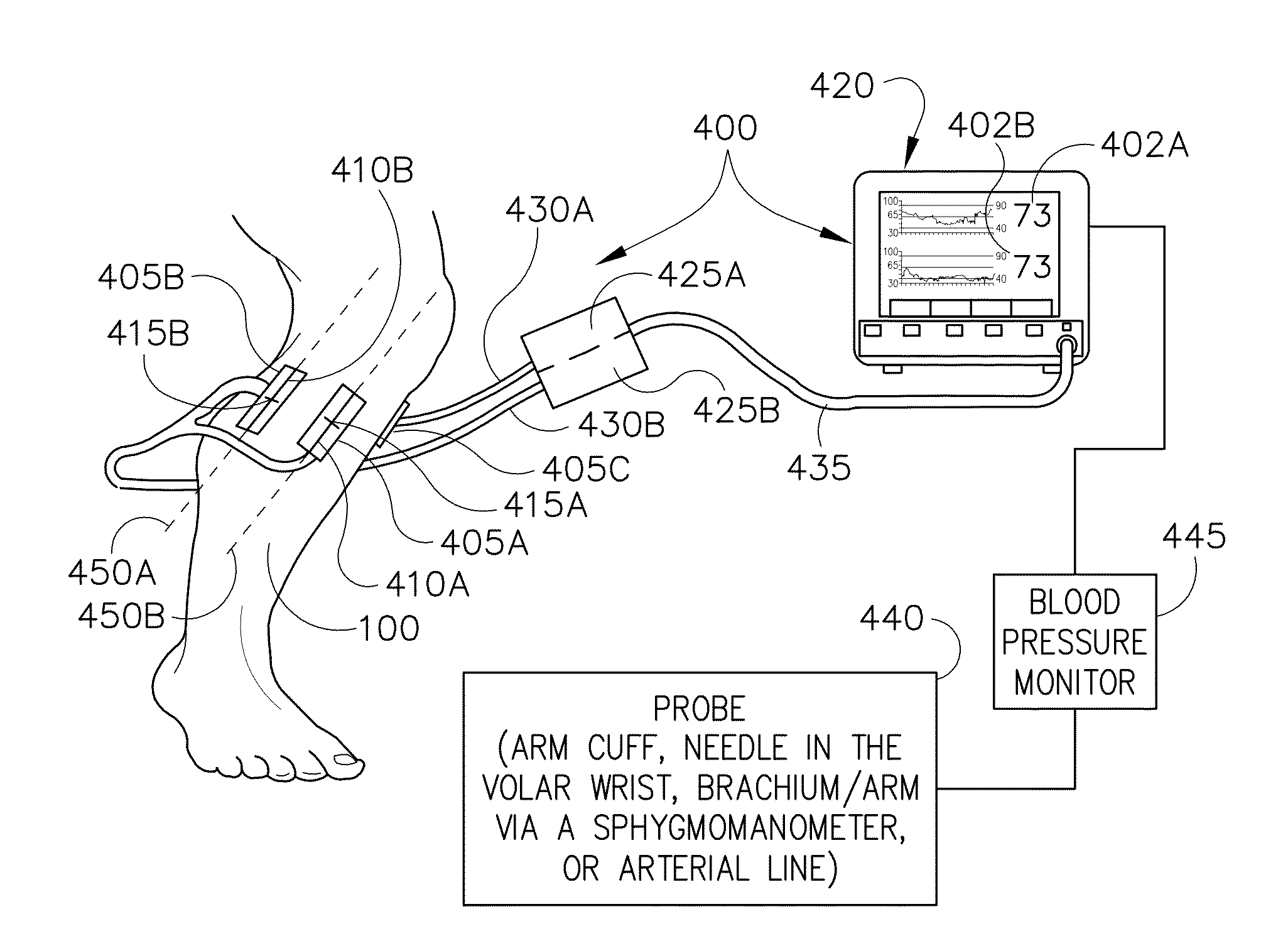
However, these conventional techniques have encountered the problem of a medical practitioner locating compartments of interest and accurately and precisely positioning a sensor over a compartment of interest.
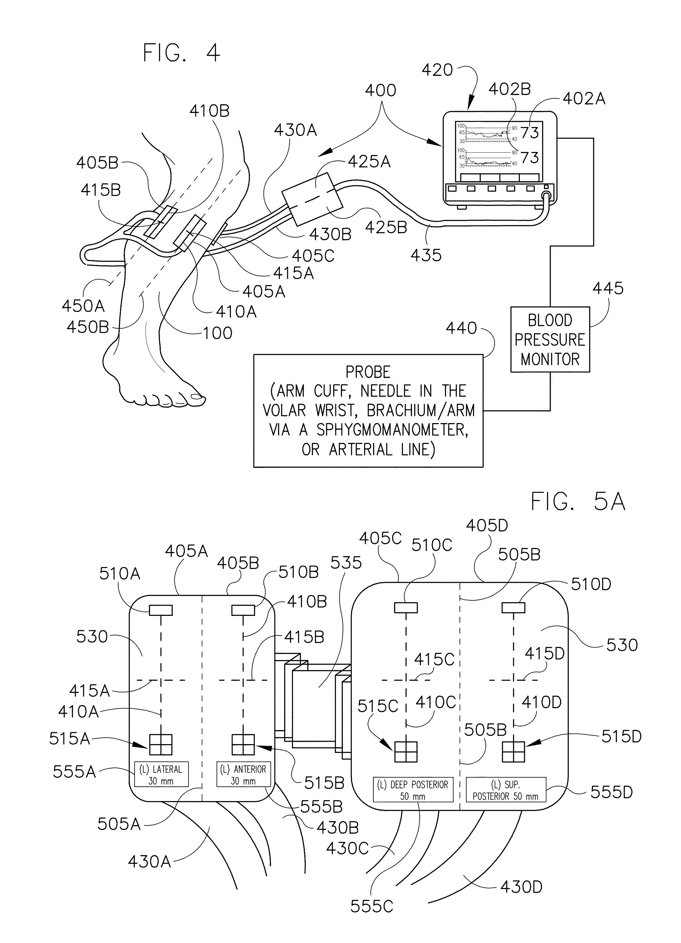
Often the orientation of the scan and the depth of the scan produced by a near infrared sensor as well as the orientation of a compartment can be challenging for a medical practitioner to determine because conventional sensors are not marked with any instructions or visual aids.
Another problem faced by the medical practitioner with conventional non-invasive techniques is determining how to assess the oxygenation level of compartments that lie underneath a particular neighboring compartment, such as with the deep posterior compartment of the human leg.
In trauma settings, near infrared sensors often do not work when they are placed over regions of the body that have hematomas or pools of blood.
Also, conventional near infrared sensors typically are not sterilized and cannot be used in surgical or operating environments.
Placement of the sensor can be difficult since an expanding hematoma can block a previously acceptable placement.
There is no product available at this time which will allow for multiple areas to be monitored in close proximity to one another without the potential for interference from other sensor light sources.
This procedure is very invasive and it often leaves the patient with severe scars and venous congestion once healed.
Open fractures have a much higher risk of bone infections which requires multiple surgical debridements and ultimately amputation in some cases in ordered to appropriately treat.
Additionally, some wound cannot be closed and require skin transfers, or skin grafts, from other parts of the body, usually from the anterior thigh.
Therefore, it is quite apparent that accurately diagnosing compartment syndrome is critical because any misdiagnosis can lead to significant morbidity.
A missed compartment syndrome can result in an insensate and contracted leg and foot.
Fasciotomies in the setting of dead muscle only increase the risk for severe infections and other complications.

Method used


Image

  • Method and system for providing versatile nirs sensors
  • Method and system for providing versatile nirs sensors
  • Method and system for providing versatile nirs sensors

Examples


case i

[0261]In 2007, a 44 year old Caucasian male fell 20 feet sustaining an isolated closed proximal tibia fracture with extension into the knee. Initial treatment included external fixation for stabilization on the day of injury. During surgery the compartments were firm but compressible. At post operative check revealed that the compartments were more firm. There was mild pain with passive stretch, though the patient was diffusely painful throughout both lower extremities. Intra-compartmental pressures were measured for all four compartments using a conventional needle method with a Striker device (Stryker Surgical, Kalamazoo, Mich.). The anterior and lateral pressures measured 50 mm Hg and the superficial and deep posterior compartments were 48 mm Hg. The diastolic pressure was 90 mm Hg resulting in a 40 mm Hg perfusion pressure.

[0262]Tissue oxygenation (StO2) or oxygenation levels were evaluated using two compartment sensors 405. The oxygenation levels were approximately 80% in all f...

case ii

[0267]Also in 2007, a 32 year old Hispanic male sustained an isolated, closed Schatzker VI tibial plateau fracture after falling from a scaffold. On initial evaluation, the patient had tight compartments, but there were no clinical symptoms of compartment syndrome. Active and passive range of motion resulted in no significant pain. Based on the concerns for the tense leg, intra-compartmental pressure measurements were obtained using a Stryker device.

[0268]All compartments were greater than 110 mm Hg. The patient's blood pressure was 170 / 112 mm Hg. The decision to perform a four compartment fasciotomy was made. The compartment sensors 405 were placed on the deep posterior compartment as well as the lateral compartment for continual monitoring. The lateral compartment was unable to give a consistent reading due to hematoma interference. The initial reading for the deep posterior was an oxygenation level of 65%. The deep posterior tissue oxygenation level steadily declined from 65% to ...

case iii

[0271]In 2007, a 62 year old Asian male suffered a closed midshaft tibia fracture in a motor vehicle crash. The patient was unresponsive and hypotensive at the scene of the accident and intubated prior to arrival. Upon presentation, the patient was hypotensive with a blood pressure of 90 / 55 mm Hg. The injured leg was clinically tight on examination.

[0272]Oxygenation levels were measured for all four compartments. The oxygenation levels were approximately at 50% for the anterior and lateral compartments while the two posterior compartments were approximately at 80%. The compartment sensors 405 were placed on the anterior and superficial posterior compartments for continued monitoring. Intra-compartmental pressures were measured at 50 mm Hg and 52 mm Hg in the anterior and lateral compartments respectively using the conventional Striker device (needle pressure measuring method). The superficial and deep compartment pressures were 19 mm Hg and 20 mm Hg respectively. After the patient w...


PUM


Abstract

A wireless near-infrared spectrometry sensor includes a light source for emitting near-infrared energy into tissue and a light receiver for receiving the near-infrared energy after it exits the tissue. The sensor may include a portable energy source for supplying energy to the light source. A processing module may control the light source and process readings in connection with the light source. A wireless transceiver may be coupled to the processing module for at least one of transmitting and receiving information, wherein the light source emits near-infrared energy at predetermined intervals in order to conserve energy in the portable energy source. The portable energy source may include at least one of a battery, a capacitor, a thermoelectric generator, a kinetic energy transducer, electricity derived from RF energy, and any combination thereof. The sensor may further include a substrate for support and which may be part of a sterile bandage.

Description

STATEMENT REGARDING RELATED APPLICATIONS AND PRIORITY CLAIMS[0001]This application is a continuation-in-part of U.S. Non-Provisional patent application Ser. No. 12 / 773,312 filed on May 4, 2010 entitled, “METHOD AND SYSTEM FOR MONITORING OXYGENATION LEVELS OF COMPARTMENTS AND TISSUE.” This application claims priority to this Non-Provisional patent application under 35 U.S.C. §120. This application also claims priority under 35 U.S.C. 119(e) to U.S. Provisional Patent Application Ser. No. 61 / 556,871 filed on Nov. 8, 2011 entitled, “METHOD AND SYSTEM FOR PROVIDING VERSATILE NIRS SENSORS.” The entire contents of both the provisional patent application and non-provisional patent application are hereby incorporated by reference.FIELD OF INVENTION[0002]The invention relates to a coordinated, continual and real-time method and system for monitoring oxygenation levels of a compartment and other tissue. More particularly, the invention relates to an orchestrated method and system that monitor...

Claims


Application Information

Patent Timeline
Patent Type & Authority Applications(United States)
IPC IPC(8): A61B5/00
CPCA61B5/6828A61B5/14552A61B5/4519A61B5/6824A61B5/0075A61B5/743A61B2562/0242A61B5/0017A61B5/021A61B5/0261A61B5/445A61B2560/0214A61B2562/0238A61B5/0002A61B5/02055A61B5/0215A61B5/022A61B5/024A61B5/0816A61B5/082A61B5/14539A61B5/412A61B5/413A61B5/416A61B5/4504A61B5/6831A61B5/742A61B5/746A61B2505/03A61B2505/05A61B2560/0242A61B2562/046
Inventor SHULER, MICHAEL SIMMS
Owner J&M SHULER
Why Eureka
Try Eureka