Methods for microvesicle isolation and selective removal

a technology of microvesicles and selective removal, applied in the field of cell biology, can solve the problems of unresolved size distinction, other technical challenges, and the inability to efficiently isolate microvesicles from various sources, and achieve the effect of rapid and inexpensive isolation of microvesicles

Active Publication Date: 2013-12-19
SYST BIOSCI
View PDF1 Cites 48 Cited by
  • Summary
  • Abstract
  • Description
  • Claims
  • Application Information

AI Technical Summary

Benefits of technology

[0029]The present invention provides methods for the rapid and inexpensive isolation of microvesicles, specifically circulating microvesicles such as exosomes. These methods utilize common laboratory reagents and apparatus, and do not require high speed centrifugation, such as ultracentrifugation. These methods of the invention also do not require the use of gradient sedimentation for the isolation of circulating microvesicles.

Problems solved by technology

However, various sources differ in their size-based definitions for exosomes, and this size distinction remains unresolved.
Although the study of microvesicles / exosomes had been greatly advanced with the development of analytical systems such as nanoparticle tracking analysis (NTA) and fluorescent nanoparticle tracking analysis (FNTA), other technical challenges remain.
One of the significant technical challenges in current research in microvesicles is the problem of how to efficiently isolate the microvesicles from various sources.
Current methodologies to isolate secreted microvesicles, including but not limited to exosomes, are constrained by technical limitations and other drawbacks.
These known methodologies are labor intensive, time-consuming and costly.
These ultracentrifugation procedures are time consuming and labor intensive, and furthermore, are constrained by the requirement for expensive ultracentrifugation equipment.
This approach is also time consuming and the yields are inconsistent.
This method has very low yields compared to other methods, and is costly due to the use of the immuno-reagents and magnetic beads, and further, these system reagents cannot be reused for subsequent isolations.
These endogenous microvesicles may impact the growth or differentiation of cells maintained in culture, and may skew or interfere with experimental results and experimental interpretation.
Although some cultured cell lines can be grown for a short time in minimal media in the absence of a serum supplement, this is not always possible.

Method used


Image

  • Methods for microvesicle isolation and selective removal
  • Methods for microvesicle isolation and selective removal
  • Methods for microvesicle isolation and selective removal

Examples


example 1

Development of Novel Systems for Isolation of Circulating Microvesicles

[0121]In an effort to develop new and improved methods for the isolation of circulating microvesicles, including exosomes, experiments were undertaken to test new approaches to microvesicle isolation.

[0122]Exosomes have been reported to have diameters ranging anywhere from about 40-200 nm. Lentivirus particles have an average diameter of about 80 nm, a size that is within the estimated range of exosome size. Formulations of polyethylene glycol (PEG-6000) had been previously used to precipitate and concentrate lentiviral particles from conditioned cell culture medium (Lewis and Metcalf, Applied and Environmental Microbiology, Vol. 54, No. 8 (1988). Because exosomes have similar diameters to a lentiviral particle, it was hypothesized that PEG formulations might be used to isolate exosomes from conditioned cell culture media that had been used to culture exosome-producing cell lines in vitro, and additionally, may a...

example 2

Isolation of Microvesicles from Urine and Western Blotting Verification

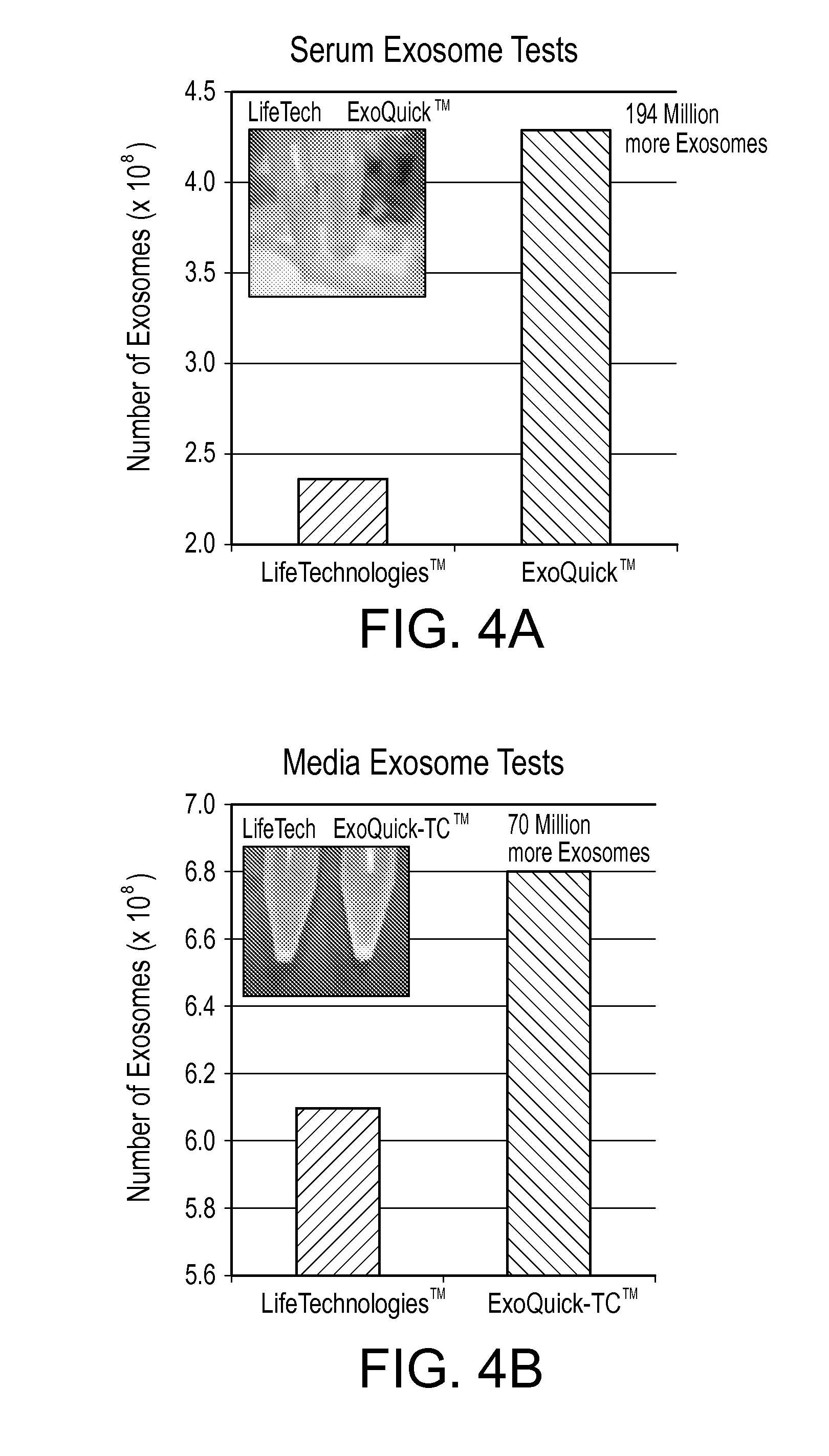
[0133]In this example, the ability of the a precipitation solution containing PEG-8,000 to precipitate and isolate microvesicles from human urine was tested, and the verified using western blotting using a known membrane marker.

[0134]Ten milliliters of normal human urine was combined with 2 ml PEG-8,000 precipitation solution, incubated for 16 hours at 4° C., and centrifuged at 1,500×g for 30 minutes. In some trials, a BECKMAN COULTER™ INC., Allegra™ 6R centrifuge with a swinging bucket rotor was used at a speed of 3,000 rpm producing 1,500×g relative centrifugal force. The resulting pellet was resuspended 175 uL PBS.

[0135]There are protein markers that are known to be abundantly associated with exosomes, including CD63, CD9, CD81 and Hsp70. The abundance of the CD9 protein marker in the resuspended pellet was analyzed by ELISA assay and Western blotting.

[0136]A) ELISA

[0137]Increasing amounts of the exosome suspe...

example 3

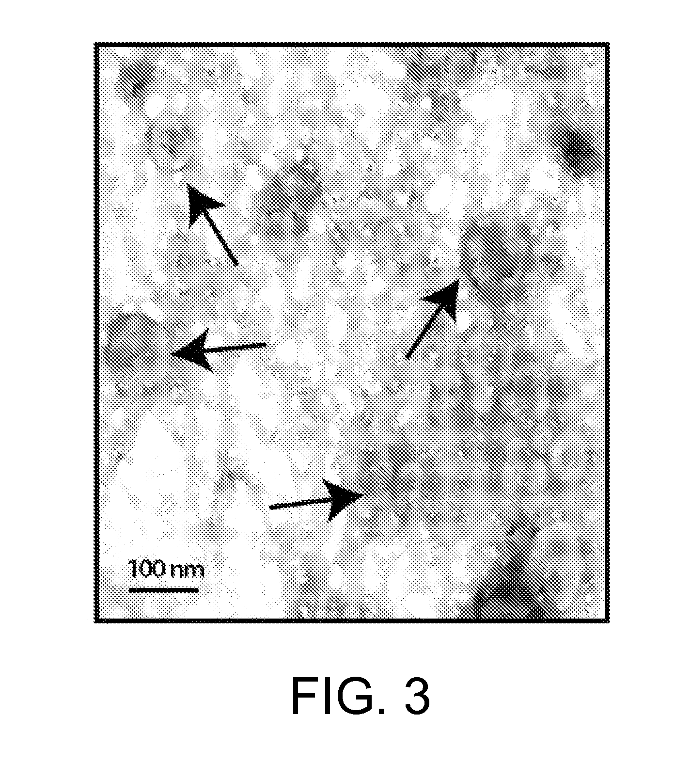
Exosomes Isolation from Urine and Analysis by Electron Microscopy (EM)

[0140]In this example, the ability of a PEG-8,000 precipitation solution to precipitate and isolate microvesicles from human urine was tested, and then verified using EM visualization.

[0141]Human urine containing exosomes was centrifuged at 3000×g and 10,000×g for 15 minutes. The supernatant was filtered through a 0.22 μm filter. PEG-8,000 precipitation solution was added to the urine supernatants, and the mixture was refrigerated at 5° C. overnight. Following incubation, the mixture was centrifuged at 1500×g, and a pellet was resuspended 1:10 in sterile PBS.

[0142]Formvar / carbon-coated nickel grids were incubated on drops of 0.1% poly-L-lysine for 5 minutes, rinsed by water and dried. Freshly prepared grids were placed on top of exosome drops and incubated for 10 minutes to adsorb exosomes. The grids were washed on several drops of PBS and fixed with 2.5% glutaraldehyde in 0.1 M cacodylate buffer for 15 minutes.

[0...


PUM

PropertyMeasurementUnit
average molecular weightaaaaaaaaaa
weight concentrationaaaaaaaaaa

Abstract

The invention relates to compositions and methods for isolation of microvesicles produced by mammalian cells. These microvesicles, known as extracellular microvesicles or circulating microvesicles, are isolated from sample materials such as body fluids, or from cell culture media that has been used to culture and maintain mammalian cells in vitro. The isolation of microvesicles as described herein results in purification and concentration of the microvesicles.The invention also provides related methods for producing blood serum and / or blood plasma that is free of detectable microvesicles, largely depleted of microvesicles, or has reduced concentration of microvesicles compared to the blood serum or blood plasma starting material (collectively termed “microvesicle-depleted”). The generation of microvesicle-depleted blood serum or plasma is critical for use in experimental systems where it is desirable to use a cell culture media that does not contain endogenous microvesicles, or has been depleted of endogenous microvesicles, from the source material.

Description

CROSS-REFERENCE TO RELATED APPLICATIONS[0001]This application claims the benefit of the U.S. Provisional Application Ser. No. 61 / 659,628, filed Jun. 14, 2012, and Ser. No. 61 / 800,213, filed Mar. 15, 2013.FIELD OF THE INVENTION[0002]The invention relates to the field of cell biology, and in particular, to the study of circulating microvesicle structures that are produced by cells. The invention relates to compositions and methods for the isolation of microvesicles produced by cells, finding use in biomedical research and potential therapeutic applications.BACKGROUND OF THE INVENTION[0003]The term microvesicles (also known as microparticles) refers to a heterogeneous in vivo collection of membrane bound (i.e., encapsulated) biological structures. These structures are formed from lipid bilayer, which is the same lipid bilayer that comprises eukaryotic cell membranes. Microvesicles can reside within the cell, or in the extracellular environment. Microvesicle structures (intracellular an...

Claims


Application Information

Patent Timeline
Patent Type & Authority Applications(United States)
IPC IPC(8): G01N1/34
CPCG01N1/34C12Q1/6895G01N33/5002G01N33/52C12M29/26C12Q2600/158C12Q2600/178C12Q1/6806C12Q2523/32C12Q2563/161
Inventor ANTES, TRAVIS JOHNKWEI, KEVIN
Owner SYST BIOSCI
Why Eureka
Try Eureka