Reduction of bioparticle levels of an organism

a bioparticle level and organism technology, applied in the field of cell biology, can solve the problems of unreliable fluids, unreliable methods, time-consuming, expensive, etc., and achieve the effects of reducing side effects, reducing costs, and reducing the number of stents

Pending Publication Date: 2019-08-22
YMIR GENOMICS
View PDF0 Cites 0 Cited by
  • Summary
  • Abstract
  • Description
  • Claims
  • Application Information

AI Technical Summary

Benefits of technology

The invention provides a way to quickly and easily isolate bioparticles from liquid samples using a method called matrix binding. This method can be used with almost any fluid, including in humans. The invention also includes a method for producing a complete cell culture media, which includes a defined minimal medium and a supplement that enhances cell growth. This media is used to culture cells in vitro for long periods of time. The technical effects of this invention are the improved isolation and enrichment of bioparticles, as well as a more efficient and cost-effective method for producing complete cell culture media.

Problems solved by technology

One of the significant technical challenges in current microvesicle research relates to the problem of how to isolate microvesicles efficiently from their various sources.
Current methodologies for isolating secreted microvesicles, including but not limited to exosomes, are constrained by technical limitations and other drawbacks.
These known methodologies are labor intensive, time-consuming, costly, and can be unreliable for different fluids; see Tauro et al., “Comparison of ultracentrifugation, density gradient separation, and immunoaffinity capture methods for isolating human colon cancer cell line LIM1863-derived exosomes,”Methods 56(2):293-304).
In particular, investigators and clinicians have previously possessed few choices for the selective, non-toxic “knock-out”, elimination and / or amelioration of EVs in vitro (i.e., in cell culture) or in vivo (i.e., in animal models or human subjects in clinical trials).
However, since many cells possess membrane proteins identical to those of EVs, it has been impossible to prevent such antibodies from affecting cells as well as exosomes.
Furthermore, antibodies are expensive and possess limited capacity.
However, Heparin has a very limited half-life and would thus require constant dosing to maintain any manner of blockade / chronic depletion of EVs.
Furthermore, Heparin is well known to affect cells and has been shown to possess significant side effects when used therapeutically in humans and animals (Smythe et al.
Two significant technical challenges in current research in extra-cellular vesicles are the problem of how to minimize contamination of cellular macromolecules from the cell-free biofluid sample and how to minimize the degradation of extra-cellular vesicle constituents during transport or storage.
Simply freezing the sample prior to removing the cells is not sufficient to ameliorate this problem, as it is well known that freeze-thaw cycles can actually lyse cells in fluids thus exacerbating the problem.
Furthermore, shipping a frozen sample increases the cost of sample collection and is not always possible for samples being sent from the developing world—where the cold-chain cannot always be maintained.
This issue is especially problematic for urine samples.
Furthermore, samples can be extremely dilute or extremely concentrated.

Method used


Image

  • Reduction of bioparticle levels of an organism
  • Reduction of bioparticle levels of an organism
  • Reduction of bioparticle levels of an organism

Examples


example 1

rous Beads to Reduce Levels of Bioparticles in Biofluids

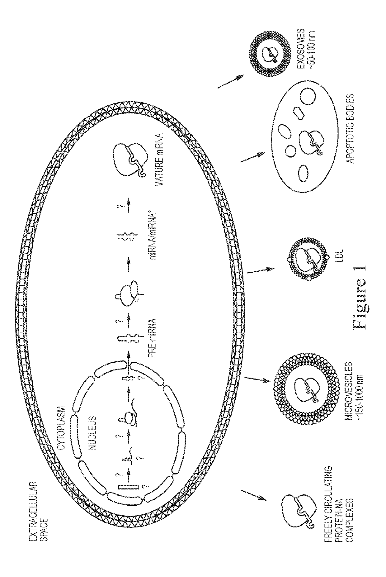
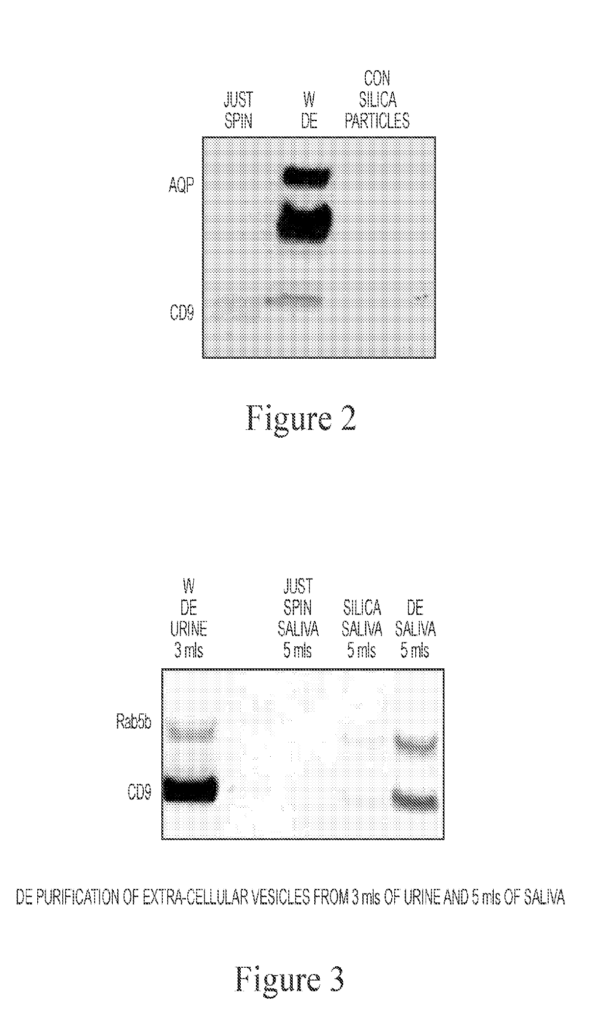
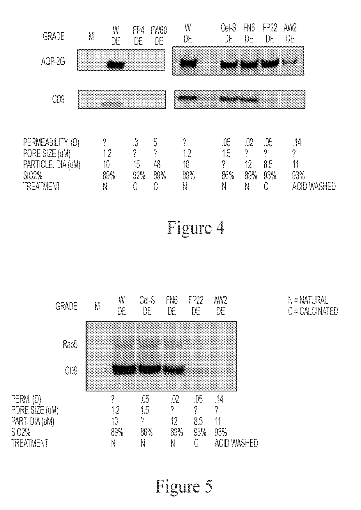
[0149]A broad range of biomarkers are secreted from cells, such as miRNA, proteins, lipids, glycoproteins, DNA, mRNA, tRNA, etc., which can exist in relatively stable form outside of cells, including but not limited to the following forms: protein-nucleic acid complexes, exosomes, microvesicles, LDL particles, and apoptotic bodies (FIG. 1). It was identified that Diatomaceous Earth (DE) isolated vesicle protein markers from urine, whereas control silica did not (FIG. 2). Diatomaceous Earth (DE) was also identified to isolate saliva exosomes (FIG. 3). DE (optionally non-calcinated (N) and low permeable / small pore size) also isolated EVs from urine (FIG. 4). Calcination and acid washing were identified to decrease DE's affinity for exosomes (FIG. 5). To examine the effect of pore size upon efficacy of isolation of bioparticles from biofluids, Perlite (Sil-Kleer) possessing smaller pore sizes / permeability that DE was examined, and...

example 2

in a Porous Container and Introduced to the Mouth of a Subject Isolated Exosomes

[0150]As shown in FIG. 9, Diatomaceous Earth placed in a porous cellulose bag, which was then held in the mouth of a subject for 30 minutes successfully and robustly sequestered exosomes, as evidenced by detection of the exosomal marker, Rab5b. A control Silica gel placed in the same type of container (cellulose bag) was meanwhile shown not to sequester this marker from saliva.

example 3

ic Reduction of Bioparticles in Urine of a Subject Having or at Risk of Developing Bladder Cancer

[0151]In an exemplary therapeutic method of the invention, a subject having or at risk of developing bladder cancer is identified. Porous beads, e.g., DE, are deposited in a porous, implantable membrane-bounded pouch or device. The implantable pouch or device is inserted into the bladder of the subject, optionally at or near a site of an existing tumor, and in contact with the urine of the subject. The device remains implanted in the subject for an appropriate period of time (e.g., ranging from a single day to a number of months or even years, noting the inert / non-toxic nature of both the implantable pouch or device and its contents (e.g., DE)). Growth, progression and / or metastasis of cancer (or markers of pro-cancer signaling) in the subject is assessed, using art-recognized methods, relative to an appropriate control subject or value, and the therapeutic efficacy of the implantable po...


PUM

PropertyMeasurementUnit
Timeaaaaaaaaaa
Timeaaaaaaaaaa
Timeaaaaaaaaaa

Abstract

Compositions and methods for the application of non-toxic bioparticle (e.g., extracellular vesicle (EV)) absorbing materials (e.g., non-toxic exosome reducing materials) for prophylactic, therapeutic, validation and / or experimental purposes are provided.

Description

CROSS-REFERENCE TO RELATED APPLICATION[0001]This application is an International Patent Application which claims the benefit of priority under 35 U.S.C. § 119(e) to U.S. Provisional Application No. 62 / 385,632, filed on Sep. 9, 2016, entitled, “Reduction of Bioparticle Levels of an Organism.” The entire contents of this patent application are hereby incorporated by reference herein.FIELD OF THE INVENTION[0002]The present invention relates to compositions and methods for the application of non-toxic bioparticle (e.g., extracellular vesicle (EV)) absorbing materials (e.g., non-toxic exosome reducing materials) for therapeutic, validation and / or experimental purposes. In addition, the invention relates to the field of cell biology, and in particular, to the study of circulating, cell-free, membrane-bound structures and protein-nucleic acid complexes that are produced and released by cells. The term “bioparticles” collectively describes these and other cell-free entities including protei...

Claims


Application Information

Patent Timeline
IPC IPC(8): A61M31/00G01N1/40
CPCA61M31/002G01N1/405G01N1/4077G01N2001/4088A61M2202/20A61M2210/0625A61M2210/1085B01D15/34C07K1/20A61B10/0051A61F5/453A61J1/05
Inventor PENDERGRAST, PATRICK SHANNONPENDERGRAST, ROBERT SCOTTPENDERGRAST, JOHN STEPHENMARKOWSKA, ANNA IRMINA
Owner YMIR GENOMICS
Why Eureka
Try Eureka