Catheter for Intravascular Ultrasound and Photoacoustic Imaging

a technology of intravascular ultrasound and photoacoustic imaging, which is applied in the field of intravascular catheter design and fabrication, can solve the problems that the catheter cannot be used for combined ivus, ivpa and elasticity imaging and radiation and/or acoustic therapy, and achieves the effects of reducing the intensity of the photoacoustic signal of the lipid based tissue, increasing the temperature, and modifying the contras

Inactive Publication Date: 2013-12-19
BOARD OF RGT THE UNIV OF TEXAS SYST
View PDF6 Cites 24 Cited by
  • Summary
  • Abstract
  • Description
  • Claims
  • Application Information

AI Technical Summary

Benefits of technology

[0017]In one embodiment, the present invention includes a method of imaging and treating a target tissue without the need to occlude or dilute luminal blood in a subject comprising the steps of: identifying a subject in need of treatment of a target tissue using an intravascular imaging and therapeutic device capable of combined intravascular ultrasound and photoacoustic imaging; irradiating the target tissue with electromagnetic radiation at a single wavelength from an intravascular imaging and therapeutic device comprising: a catheter with a proximal end and a distal end; one or more intravascular ultrasound units comprising a proximal end and a distal end, wherein the distal end comprises one or more single-element ultrasound transducer elements at a proximal end; one or more optical units comprising a proximal end and a distal end combination, wherein the distal end comprises at least one of one or more optical fibers or one or more optical bundles, wherein the proximal end comprises a port to couple at least one optical unit to a light source that irradiates at a single wavelength; an imager connected to the proximal end of the unit to capture one or more ultrasound and optical images; and performing at least one of an imaging or a therapy of the target tissue. In one aspect, the single wavelength is selected from one of 1060, 1064, 1070, 1200, 1210, 1700, 1710, 1715, 1720, 1725, 1730, or 1740 nm. In another aspect, the distribution of the ultrasound impedance is reconstructed by transmitting of short ultrasound waves into the target tissue with consequent detection of at least one of reflected or scattered ultrasound waves. In another aspect, the distribution of the optical absorption is reconstructed by transmitting of short light pulses of the same wavelength into the target tissue with a consequent detection of ultrasound waves generated in the tissue due to thermal expansion of the tissue due to absorbed light energy. In another aspect, the one or more ultrasound units irradiate an artery with long ultrasound pulses to perform an acoustic therapy of the artery. In another aspect, the one or more optical units can irradiate tissues by CW or long-pulse light to perform an optical therapy. In another aspect, the at least one of optical therapy, acoustic therapy, tissue target imaging, or therapy and imaging can be performed either simultaneously or separately. In another aspect, the imager is capable of providing imaging results or therapy results in a format determined by a user. In another aspect, the imager has a penetration depth of 1 to 5 mm. In another aspect, the imager has a resolution of 50 micrometers. In another aspect, the imager is able to detect the presence and spatially resolved location of lipid in the arterial wall. In another aspect, the imager modifies the contrast between lipid tissue and water-based tissue by increasing the temperature at the target tissue, wherein photoacoustic signal intensity of the lipid based tissue decreases and the water-based tissue increases. In another aspect, the method further comprises the step of mechanically or electrically steering an ultrasound beam to generate a two-dimensional cross-sectional image of the target. In another aspect, the method further comprises the step of using a contrast agent in the target tissue.
[0018]In another embodiment, the present invention includes a method of imaging and treating a target tissue in vivo without the need to occlude or dilute luminal blood in a subject comprising the steps of: identifying a subject in need of treatment of a target tissue using an intravascular imaging and therapeutic device capable of combined intravascular ultrasound and photoacoustic imaging; irradiating the target tissue with electromagnetic radiation at a single wavelength selected from wavelengths in which lipid and water have a different index of refraction comprising: a catheter with a proximal end and a distal end; one or more intravascular ultrasound units comprising a proximal end and a distal end, wherein the distal end comprises one or more single-element ultrasound transducer elements at a proximal end; one or more optical units comprising a proximal end and a distal end combination, wherein the distal end comprises at least one of one or more optical fibers or one or more optical bundles, wherein the proximal end comprises a port to couple at least one optical unit to a light source that irradiates at a single wavelength; an imager connected to the proximal end of the unit to capture one or more ultrasound and optical images; and performing at least one of an imaging or a therapy of the target tissue in vivo. In one aspect, the single wavelength is selected from one of 1060, 1064, 1070, 1200, 1210, 1700, 1710, 1715, 1720, 1725, 1730, or 1740 nm. In another aspect, the distribution of the ultrasound impedance is reconstructed by transmitting of short ultrasound waves into the target tissue with consequent detection of at least one of reflected or scattered ultrasound waves. In another aspect, the distribution of the optical absorption is reconstructed by transmitting of short light pulses of the same wavelength into the target tissue with a consequent detection of ultrasound waves generated in the tissue due to thermal expansion of the tissue due to absorbed light energy. In another aspect, the one or more ultrasound units irradiate an artery with long ultrasound pulses to perform an acoustic therapy of the artery. In another aspect, the one or more optical units can irradiate tissues by CW or long-pulse light to perform an optical therapy. In another aspect, the at least one of optical therapy, acoustic therapy, tissue target imaging, or therapy and imaging can be performed either simultaneously or separately. In another aspect, the imager is capable of providing imaging results or therapy results in a format determined by a user. In another aspect, the imager has a penetration depth of 1 to 5 mm. In another aspect, the imager has a resolution of 50 micrometers. In another aspect, the imager is able to detect the presence and spatially resolved location of lipid in the arterial wall. In another aspect, the imager modifies the contrast between lipid tissue and water-based tissue by increasing the temperature at the target tissue, wherein photoacoustic signal intensity of the lipid based tissue decreases and the water-based tissue increases. In another aspect, the method further comprises the step of mechanically or electrically steering an ultrasound beam to generate a two-dimensional cross-sectional image of the target. In another aspect, the method further comprises the step of using a contrast agent in the target tissue.

Problems solved by technology

Currently available catheters cannot be used both for combined IVUS, IVPA and elasticity imaging and for radiation and / or acoustic therapy.

Method used


Image

  • Catheter for Intravascular Ultrasound and Photoacoustic Imaging
  • Catheter for Intravascular Ultrasound and Photoacoustic Imaging
  • Catheter for Intravascular Ultrasound and Photoacoustic Imaging

Examples


example 1

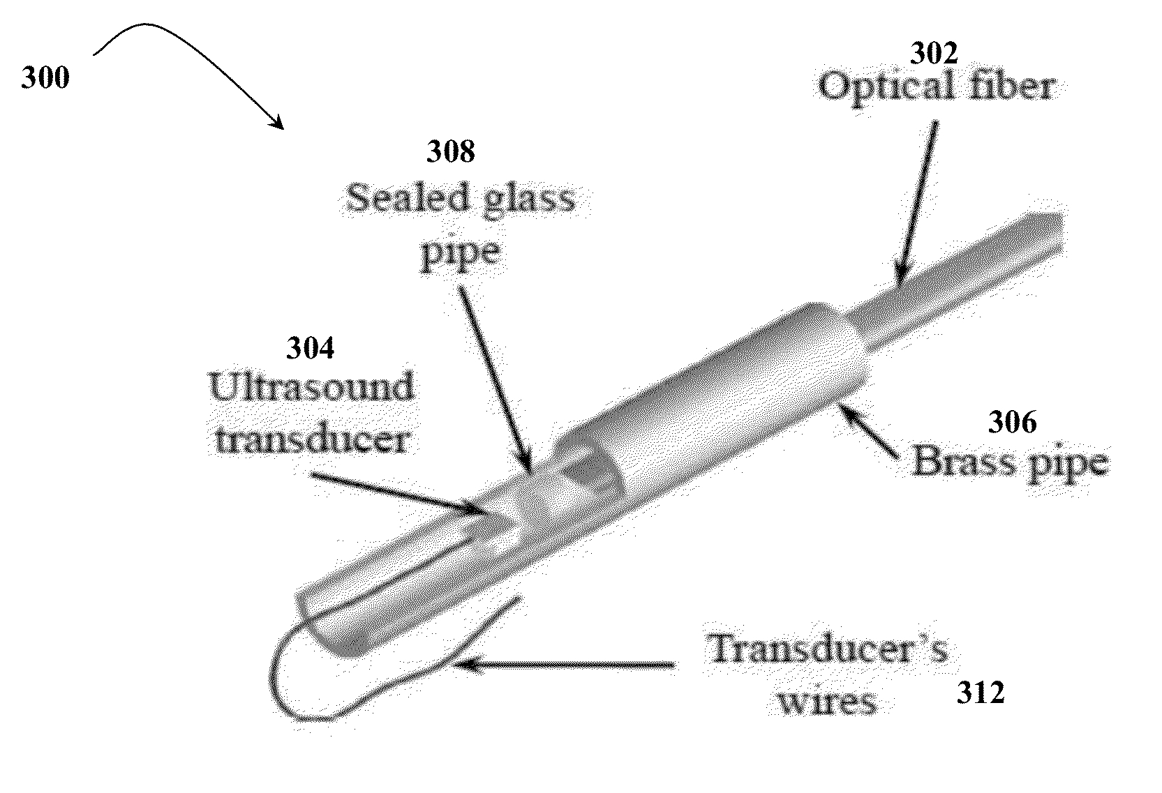
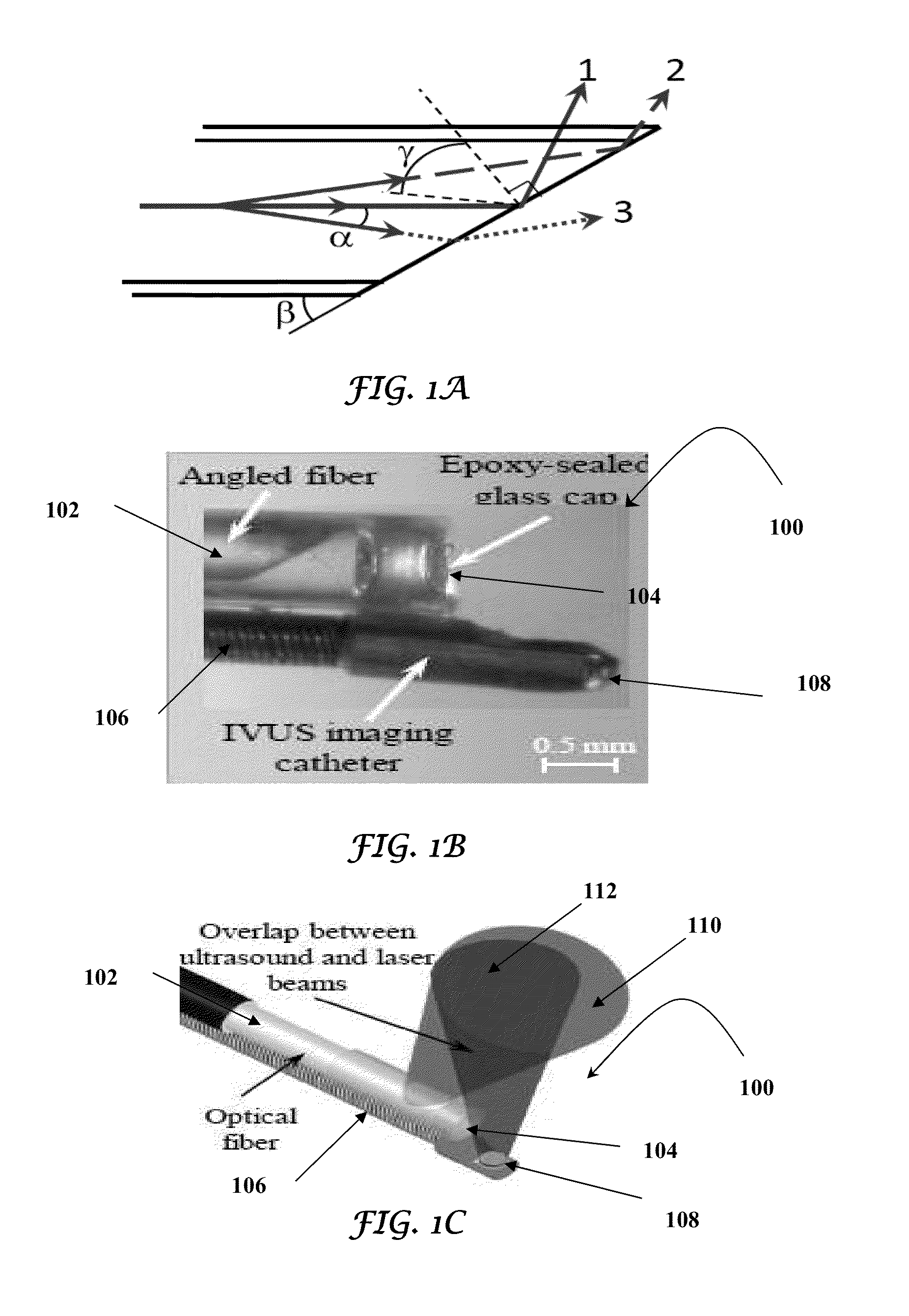
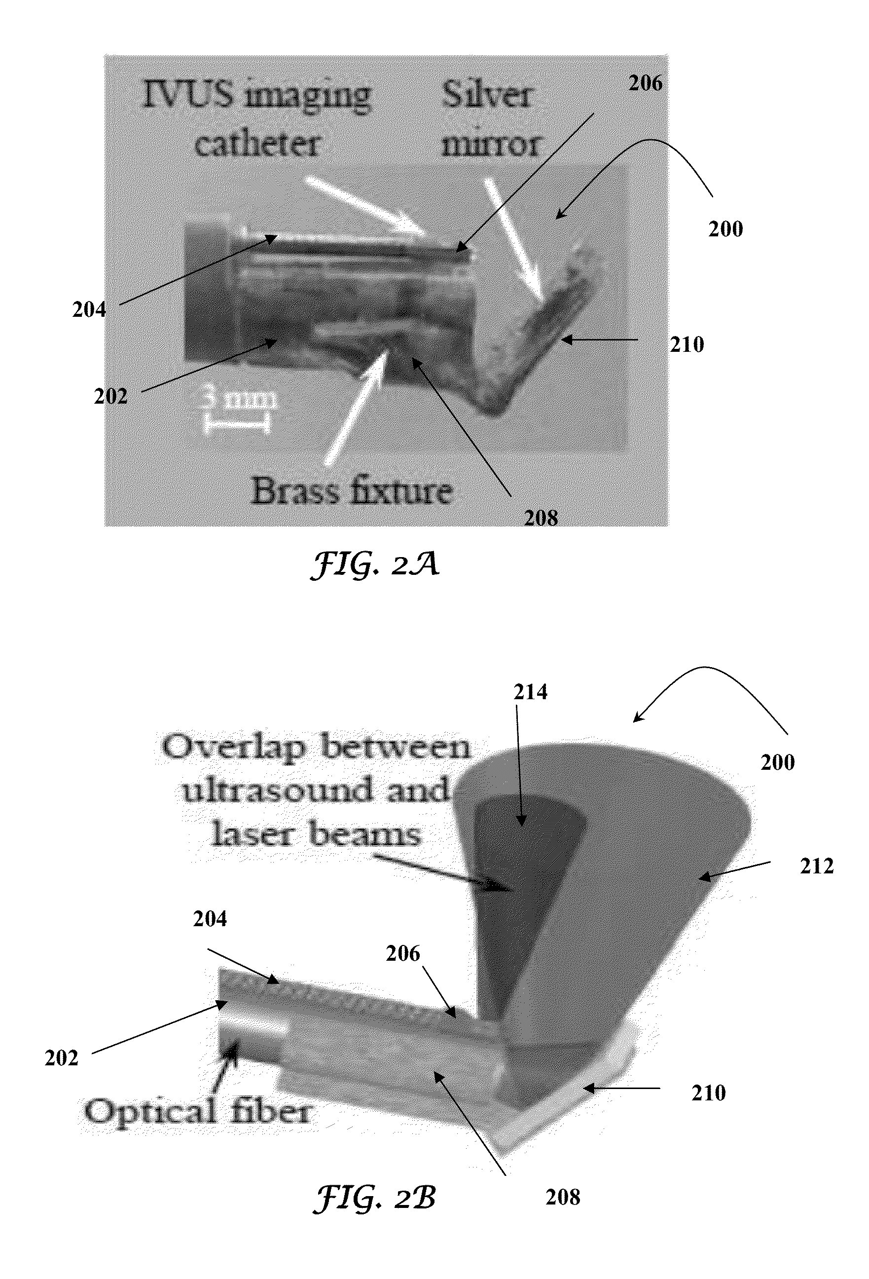
[0050]The present invention describes two designs of an integrated intravascular ultrasound, photoacoustic (IVUS / IVPA) and elasticity imaging catheter capable both of combined intravascular ultrasound, photoacoustic, and elasticity imaging and of radiation and / or acoustic therapy on an artery and / or nearby tissues. Such catheter include of one or more ultrasound units that are either a single element ultrasound transducer or an ultrasound array transducer or a combination thereof, and one or more optical units that comprise one or more optical fibers, one or more optical bundles or a combination thereof. A light delivery system is mounted on one or more optical units. The one or more ultrasound units and one or more optical units are assembled into a single device such that ultrasound and optical beams propagate orthogonally to the longitudinal axis of the catheter with maximum overlap with each other.

[0051]The elasticity imaging of an artery is performed by one or more ultrasound u...

example 2

[0090]The location and size of the necrotic lipid core is critical for analyzing the stability of atherosclerotic plaques (Falk 2006). Identification of vulnerable plaques depends on the distribution of lipid (Virmani 2011). Unfortunately, current invasive imaging modalities cannot reliably delineate spatially resolved lipid distribution (Vancraeynest et al. 2011). Intravascular optical coherence tomography (OCT) can detect lipid, but it lacks the imaging depth to assess the area of the lipid-rich plaques and requires temporal removal of luminal blood during imaging. Intravascular magnetic resonance imaging (MRI) has a better imaging depth, but it requires an occluding balloon to stabilize the catheter. Intravascular near-infrared spectroscopy (NIRS) can be performed in presence of blood, but the signals are not depth-resolved. Thus, a catheter-based imaging modality—ultrasound-guided intravascular photoacoustic imaging—was used to detect the depth-resolved distribution of lipid in ...


PUM


Abstract

The present invention includes a method of imaging and treating a target tissue without the need to occlude or dilute luminal blood in a subject by a combination of intravascular ultrasound and photoacoustic imaging by irradiating the target tissue with electromagnetic radiation at a single wavelength.

Description

CROSS-REFERENCE TO RELATED APPLICATIONS[0001]This application is a Continuation-in-Part application of U.S. patent application Ser. No. 13 / 505,345, filed Jul. 10, 2012, which is a 35 U.S.C. 371 National Stage application of International Application No. PCT / US2010 / 055006, filed Nov. 1, 2010, which claims the benefit of U.S. Provisional Application No. 61 / 257,390, filed Nov. 2, 2009. The contents of each of which are incorporated by reference in their entirety.STATEMENT OF FEDERALLY FUNDED RESEARCH[0002]This invention was made with U.S. Government support by the NIH grant number HL096981. The government has certain rights in this invention.TECHNICAL FIELD OF THE INVENTION[0003]The present invention relates in general to the field of combined intravascular ultrasound, photoacoustic and elasticity imaging and intravascular radiation and / or acoustic therapy, and more particularly, to the design and fabrication of an intravascular catheter for combined intravascular ultrasound, photoacou...

Claims


Application Information

Patent Timeline
Patent Type & Authority Applications(United States)
IPC IPC(8): A61B8/12
CPCA61B8/12A61B5/0035A61B5/0084A61B5/0095A61B8/4416A61B8/445A61B8/485A61B8/587
Inventor EMELIANOV, STANISLAVWANG, BOKARPIOUK, ANDREIYEAGER, DOUGLAS
Owner BOARD OF RGT THE UNIV OF TEXAS SYST
Why Eureka
Try Eureka