Methods and Compositions for Diagnosis of Ovarian Cancer

a technology for ovarian cancer and compositions, applied in the direction of instruments, nucleotide libraries, tumor rejection antigen precursors, etc., can solve the problems of poor protein biomarker for early detection, poor sensitivity and specificity, and ineffective early detection

Inactive Publication Date: 2014-09-18
THE WISTAR INST OF ANATOMY & BIOLOGY
View PDF0 Cites 6 Cited by
  • Summary
  • Abstract
  • Description
  • Claims
  • Application Information

AI Technical Summary

Benefits of technology

[0006]In another aspect, a diagnostic device include a kit, panel or microarray comprising at least two diagnostic reagents, each reagent identifying a different biomarker of Table 1, or molecular form, modified molecular form, isoform, pro-form, or peptide fragment thereof. In another embodiment, the kit, panel or microarray includes diagnostic reagents that bind or complex individually to 2, 3, 4, 5, 6, 7, 8, 9, 10 and 11 or more different biomarkers, molecular forms or fragments thereof, desirably in a protein level profile or panel. In another embodiment, the kit, panel or microarray includes diagnostic reagents that bind or complex to different molecular forms or fragments of the same biomarker, desirably in a protein level profile or panel.
[0007]In another aspect, a method for diagnosing or detecting the existence or absence of, or monitoring the progress of, ovarian cancer in a subject is provided. This method includes contacting a sample obtained from a test subject with a composition or reagent as described herein; and detecting or measuring in the sample or from a protein level profile generated from the sample, the protein levels of one or more of the biomarkers of Table 1, or a molecular form, modified molecular form, isoform, pro-form, or peptide fragment thereof. The protein level(s) of the biomarker(s) in the subject's sample or from a protein level profile or ratio of multiple biomarkers is then compared with the level or ratio of the same biomarker or biomarkers in a reference standard. A sig

Problems solved by technology

When ovarian cancer is diagnosed at an early stage (stages I or II), treatment is highly effective, with a five-year survival rate of up to 90%, whereas the five-year survival rate for patients with advanced disease (stages III and IV) is reduced to 30% or less.4, 5 Unfortunately, most ovarian cancers are not diagnosed until after the cancer has spread, primarily because earlier-stage diseases are asymptomatic and the ovaries are buried deep within the body.
Current screening methods for ovarian cancer typically use a combination of pelvic examination, transvaginal ultrasonography, and serum CA125, but these methods are not effective in detecting early-stage ova

Method used


Image

  • Methods and Compositions for Diagnosis of Ovarian Cancer
  • Methods and Compositions for Diagnosis of Ovarian Cancer
  • Methods and Compositions for Diagnosis of Ovarian Cancer

Examples


example 1

Materials and Methods

[0187]Cell Culture.

[0188]The human epithelial ovarian cancer cell line TOV-112D was obtained from the American Type Culture Collection (ATCC, Manassas, Va.). The TOV-112D cells were maintained in a 37° C. incubator with a 5% CO2-95% air atmosphere in a 1:1 mixture of MCDB 105 media and Medium 199 (Sigma-Aldrich, St. Louis, Mo.) supplemented with 15% fetal calf serum, as described previously.40

[0189]Ovarian Cancer Growth In Vivo and Mouse Serum Collection.

[0190]Approximately 3 million TOV-112D cells in 100 μl PBS were injected subcutaneously into the flanks of nine severe combined immunodeficient (SOD) female mice. Tumor volumes were monitored by caliper measurements. When tumors were approximately 1 cm in length, blood was collected from mice by cardiac puncture under anesthesia, animals were immediately euthanized, and the tumor at the injection site and other internal organs were collected.

[0191]The collected blood was allowed to clot at room temperature and ...

example 2

Overview of the Ovarian Cancer Biomarker Discovery and Verification / Validation Strategies

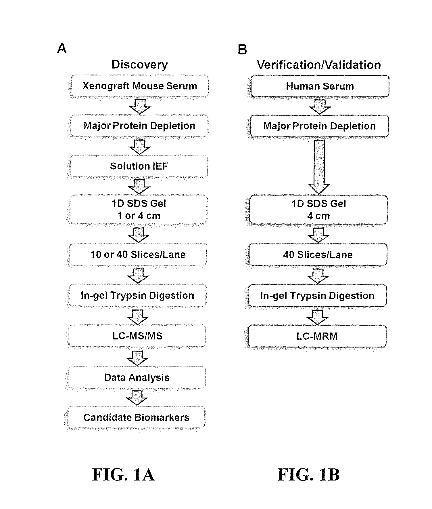
[0213]The general experimental workflow we used for discovery and verification of candidate ovarian cancer protein biomarkers is shown in FIGS. 1A and 1B. For discovery of candidate human biomarkers, serum proteins obtained from SCID mice harboring human ovarian cancer tumors were subjected to a 4-D separation consisting of three sequential, and substantially orthogonal, protein separations, i.e., major protein depletion, solution IEF, and 1-D SDS-PAGE, followed by online, reversed-phase LC peptide separation prior to MS / MS analysis. We previously developed this 4-D protein profiling method for comprehensive analysis of human serum and plasma proteomes, which resulted in the most comprehensive coverage of a serum sample in the pilot phase of the Human Proteome Organization Plasma Proteome Project (HUPO PPP).22, 44 That study also demonstrated that 14 of the 20 proteins known at that time to be i...

example 3

Xenograft Mouse Model of Human Ovarian Endometrioid Cancer

[0215]To identify candidate serum biomarkers using the xenograft mouse model, TOV-112D ovarian endometrioid tumor cells were injected subcutaneously. This cell line was originally derived from a patient with a malignant ovarian tumor that had never been exposed to radiation or chemotherapy.46 This cell line was chosen because it has a fast growth rate and has been demonstrated to form tumors readily in immune-compromised mice. More importantly, the in vitro growth characteristics of the cell line mimic the aggressive clinical behavior of the cancer.46 Previous proteomics biomarker discovery studies used human SKOV-3 serous ovarian cancer cells.36, 38 Certain sets of biomarkers likely characterize different types of ovarian cancer, where some proteins are common to all or multiple cancer subtypes and other proteins are specific to a single subtype.


PUM

PropertyMeasurementUnit
Ratioaaaaaaaaaa
Levelaaaaaaaaaa

Abstract

Methods and compositions are provided for diagnosing ovarian cancer in a mammalian subject, preferably in a serum or plasma sample of a human subject. The methods and compositions enable the detection or measurement in the sample or from a protein level profile generated from the sample, the protein level of one or more specified biomarkers. Comparing the protein level(s) of the biomarker(s) in the subject's sample or from protein abundance profile of multiple biomarkers, with the level of the same biomarker(s) or profile in a reference standard, permits the determination of a diagnosis of ovarian cancer, or the identification of a risk of developing ovarian cancer, or enables the monitoring of the status of progression or remission of ovarian cancer in the subject followed during a therapeutic protocol.

Description

STATEMENT REGARDING FEDERALLY SPONSORED RESEARCH OR DEVELOPMENT[0001]This invention was made with government support under Grant Nos. CA131582, CAl20393 and CA10815 awarded by the National Institutes of Health. The government has certain rights in this invention.BACKGROUND OF THE INVENTION[0002]Ovarian cancer is the fifth-leading cause of cancer-related death in women in the United States, and is the most lethal of all gynecological malignancies.1 In 2010, an estimated 21,880 women were diagnosed with ovarian cancer, and 13,850 deaths occurred in the United States alone.1 The most common and deadly form of ovarian cancer is epithelial ovarian cancer, which further can be divided into four major histopathological groups: serous, endometrioid, mucinous and clear cell tumors.2, 3 The high mortality rate of ovarian cancer is due largely to the lack of effective screening strategies for early detection. When ovarian cancer is diagnosed at an early stage (stages I or II), treatment is hig...

Claims


Application Information

Patent Timeline
IPC IPC(8): C12Q1/68G01N33/574
CPCG01N33/57449C12Q1/6886G01N2800/50G01N2800/54G01N2800/56C07K14/4748
Inventor SPEICHER, DAVID W.TANG, HSIN-YAOBEER, LYNN A.
Owner THE WISTAR INST OF ANATOMY & BIOLOGY
Why Eureka
Try Eureka