Soft tissue repair allografts and methods for preparing same

a soft tissue repair and allograft technology, applied in the field of allografts, can solve the problems of uniform microstructure of the papillary dermis pd, and achieve the effect of uniform density and porosity

Inactive Publication Date: 2014-09-18
MUSCULOSKELETAL TRANSPLANT FOUND INC
View PDF3 Cites 14 Cited by
  • Summary
  • Abstract
  • Description
  • Claims
  • Application Information

AI Technical Summary

Benefits of technology

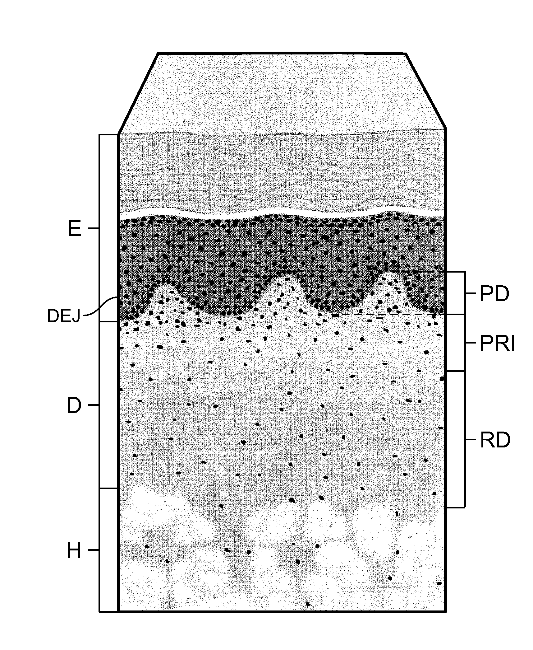
[0008]The present invention relates to a soft tissue repair allograft, and more particularly to an allograft dermal tissue-derived ACDM, and its use in plastic surgery procedures, including breast reconstruction. The ACDM of the present invention is derived from deeper-cut dermal tissue, which constitutes a collagen matrix having substantially uniform density and porosity, and therefore possesses the foregoing structural and biomechanical properties that make it well-suited for use in breast reconstruction procedures, e.g., as a sling, as well as other plastic surgery applications. The allograft dermal tissue form, or ACDM, includes a portion of dermal tissue having a first exposed surface formed by a first cut and a second exposed surface formed by a second cut opposite the first exposed surface, wherein the portion of dermal tissue constitutes a collagen matrix having substantially uniform density and porosity between the first and second exposed surfaces.
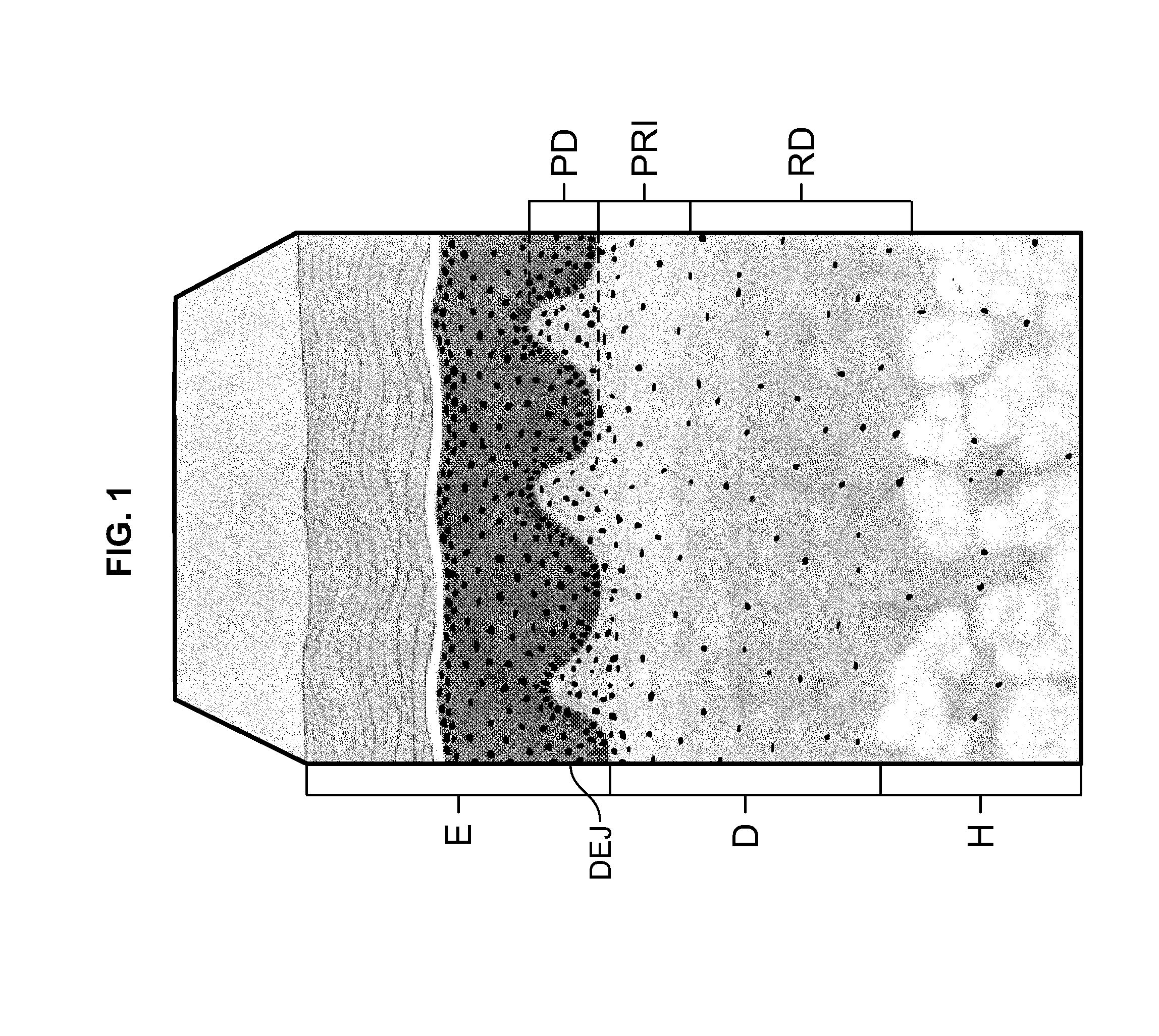
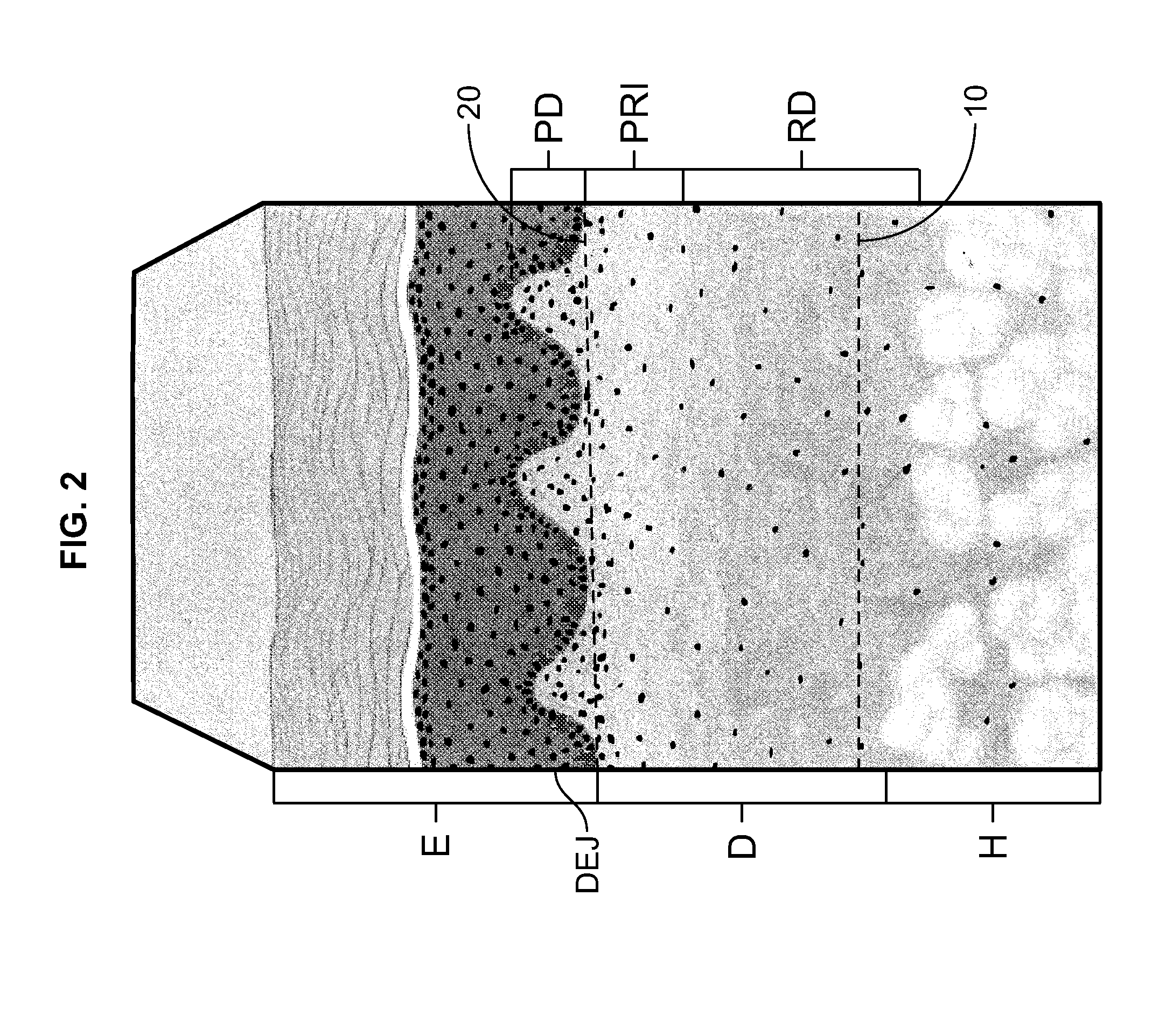
[0009]The present invention also relates to a method for preparing an ACDM, i.e., an allograft dermal tissue form, from donor tissue. The method involves (1) making a first cut into the reticular dermis RD at a first location distal the papillary-reticular dermis interface PRI, and along a first plane substantially parallel to the papillary-reticular dermis interface PRI; (2) removing the hypodermis H from the reticular dermis RD along the first cut to form a first exposed surface on a remaining portion of the donor tissue; (3) making a second cut into the papillary dermis PD at a second location proximate the DEJ, and along a second plane substantially parallel to the papillary-reticular dermis interface PRI and the first plane; and (4) removing the epidermis E, DEJ and a portion of the papillary dermis PD from the remaining portion of the donor tissue to form a second exposed surface on a remaining portion of the dermis D opposite the first exposed surface. The first and second locations are selected such that the remaining portion of the dermis D constitutes a collagen matrix having substantially uniform density and porosity between the first exposed surface and the second exposed surface.
[0010]The present invention further relates to an allograft hybrid bilayer tissue form having a dermal side and an adipose side for use in surgical procedures, as well as a method for forming the allograft hybrid bilayer tissue form. The method involves (1) providing donor tissue including skin having (a) an epidermis E and (b) a dermis D underlying the epidermis E, the dermis D including a papillary dermis PD adjacent the epidermis E, a reticular dermis RD distal to the epidermis E, and a papillary-reticular dermis interface PRI between the papillary dermis PD and reticular dermis RD; and a hypodermis H adipose tissue underlying the reticular dermis RD; (2) making a cut into the reticular dermis RD at a location proximate the hypodermis H, and along a plane substantially parallel to the papillary-reticular dermis interface PRI; and (3) removing the hypodermis H and a portion of the reticular dermis RD attached to the hypodermis H to form the allograft hybrid bilayer tissue form such that the allograft hybrid bilayer tissue form includes both a dermal side and an adipose side.

Problems solved by technology

The microstructure of the papillary dermis PD is not uniform.
Nevertheless, this dual structure, which may only be visible on a microscopic scale, presents concerns about identifying and maintaining the side orientation of the ACDM, i.e., during a surgical procedure.

Method used


Image

  • Soft tissue repair allografts and methods for preparing same
  • Soft tissue repair allografts and methods for preparing same
  • Soft tissue repair allografts and methods for preparing same

Examples


example 1

In Vitro Fibroblast Attachment to the ACDMs

Materials and Methods

[0037]7 mm punches of each tissue sample (i.e., each ACDM) were prepared and seeded with 1×10⁵ BJ neonatal human foreskin fibroblasts (ATCC, Manassa, Va.) on both sides in Eagles Minimum Essential Medium +10% fetal bovine serum. After 30 minutes, the tissue sections were washed to remove any non-adherent cells and incubated at 37° C. for 1 hour in complete growth medium. Attached cells were quantified using CyQuant Cell Proliferation Assay (Invitrogen, Carlsbad, Calif.) according to the manufacturer's instructions. Non-adherent seeded controls were measured for all samples. The test was replicated with each sample set. Values for cell fluorescence were reported. Tissue from multiple donor lots were collected, processed as described and tested. In addition, five lots of AlloDerm® RTU thick tissue were obtained and tested as commercial controls.

Results

[0038]

TABLE 1In vitro fibroblast attachmentNo. ofSamplesCells*Grouping*...

example 2

Tensile Properties of the ACDMs

Materials and Methods

[0046]Tissue samples (i.e., for each ACDM) were tested on an MTS 858 Mini Bionix System. Sample thickness was first measured with a laser micrometer (Z Mike, Benchmike 4050S). Samples in dogbone configuration (1 cm×7 cm; ASTM 638) were positioned in pneumatic action grips set at 29 psi pressure at a gage length of 26 mm. Tissue was pulled to break at a strain rate of 50.6 mm / min. Ultimate tensile strength, elongation-at-break and elastic modulus were recorded. Statistical analysis included both tests of the means and the estimates of variability for tensile strength, elongation-at-break, and modulus.

Results

[0047]As a result of the more open structure and greater porosity of the Disclosed ACDM, as contrasted with the FlexHD Structural ACDM, the Disclosed ACDM has reduced tensile strength as compared to the FlexHD Structural ACDM; 10.97 vs. 15.36 MPa.

[0048]As can be seen from the data in Table 2 and the graph illustrated in FIG. 7, t...

example 3

Surface Characterization of the ACDMs by Scanning Electron Microscopy (SEM)

Materials and Methods

[0055]Tissue samples (i.e., for the Disclosed ACDM and the FlexHD Structural ACDM) were lyophilized and coated with a 10 nm layer of gold. Images were taken using a Field Emission Zeiss Scanning Microscope (Carl Zeiss, Inc., Thornwood, N.Y.) with a working distance of 5-10 mm and voltage range of 30-200 kV. All images were taken at the Department of Ceramics and Material Science at Rutgers University, New Brunswick, N.J.

Results

[0056]Scanning electron micrographs of the epidermal side and the dermal side of both the FlexHD Structural ACDM and the Disclosed ACDM are presented in FIGS. 8a and 8b, respectively. Representative images were taken at 250× for all samples. For both ACDMs, the micrographs of the epidermal side of the ACDMs are shown on the left, and the micrographs of the dermal side are shown on the right.

Discussion

[0057]The deeper cut method of the present invention that was used...


PUM


Abstract

Allografts for soft tissue repair, including breast reconstruction and other plastic surgery procedures, are disclosed. One allograft is made from decellularized dermal tissue and constitutes a collagen matrix having substantially uniform density and porosity. Another allograft is a hybrid bilayer tissue form that is made from decellularized dermal and adipose tissues. Methods for making both allografts are also disclosed.

Description

RELATED APPLICATION[0001]This application claims the benefit of U.S. Provisional Patent Application No. 61 / 783,237, filed Mar. 14, 2013, the disclosure of which is incorporated in its entirety herein.FIELD OF THE INVENTION[0002]The present invention relates generally to allografts made from decellularized dermal tissues, and in particular, to the use of such allografts for soft tissue repair, including breast reconstruction and other plastic surgery procedures.BACKGROUND OF THE INVENTION[0003]Human allograft dermal tissue has been widely accepted for use in various surgical procedures for decades. For example, acellular dermal matrices (“ACDMs”) derived from allograft dermal tissue are used in the repair of ventral abdominal hernias and other abdominal wall defects. Commercially available ACDMs include FlexHD® Structural™ ACDM, which is marketed by Musculoskeletal Transplant Foundation (Edison, N.J.), as well as AlloDerm® ACDM and AlloDerm® Ready to Use (“RTU”) ACDM, both of which a...

Claims


Application Information

Patent Timeline
Patent Type & Authority Applications(United States)
IPC IPC(8): A61F2/10
CPCA61F2/105A61L27/362A61K35/36C12N5/0698A61L27/3683A61L27/38A61L27/60
Inventor LOCARNO, MICHAELCHOI, BRYAN J.NGO, MAHN-DAN
Owner MUSCULOSKELETAL TRANSPLANT FOUND INC
Why Eureka
Try Eureka