Decellularized porcine cornea tissue and preparation method and application thereof

A decellularization and corneal technology, applied in medical science, prosthesis, etc., can solve the problems of large damage to collagen lamellar structure, disorderly arrangement of collagen fibers, and inability to store in wet state for a long time, achieving no cell residue and biocompatibility. and good biological safety, the effect of retaining the integrity of the laminate structure

Active Publication Date: 2015-04-15
QINGDAO CHUNGHAO TISSUE ENG
View PDF4 Cites 14 Cited by
  • Summary
  • Abstract
  • Description
  • Claims
  • Application Information

AI Technical Summary

Problems solved by technology

[0006] The purpose of the present invention is to overcome the large damage to the collagen lamellar structure of the corneal stroma substitute prepared by the existing method, which makes the arrangement of collagen fibers messy, the pores are uneven, and the light transmittance is low, which cannot fully meet the needs of clinical transplantation. To provide a decellularized collagen fiber that is neatly arranged, uniform in pores, intact in lamellar structure, high in light transmittance, and capable of long-term wet storage under the premise of invariant corneal stroma. Corneal tissue and its preparation method and application

Method used


Image

  • Decellularized porcine cornea tissue and preparation method and application thereof
  • Decellularized porcine cornea tissue and preparation method and application thereof
  • Decellularized porcine cornea tissue and preparation method and application thereof

Examples


preparation example Construction

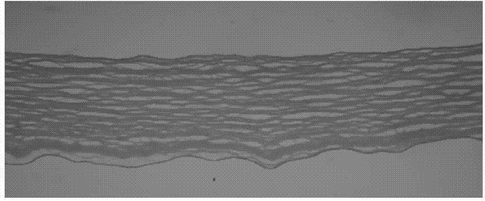
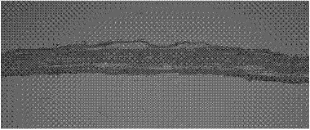
[0026] In the first aspect, the present invention provides a method for preparing decellularized corneal tissue, the method comprising: performing hypotonic swelling, repeated freezing and thawing, and enzymatic digestion of the corneal stroma slices of fresh animal eyeballs cut under sterile conditions , sonication, sterilization and wet storage, the enzymatic digestion is treated with a buffer solution containing DNase and RNase.

[0027] According to the method of the present invention, those skilled in the art can understand that, before cutting out the corneal stromal piece under aseptic conditions, generally the fresh animal eyeballs are soaked in alcohol or PBS solution containing tobramycin. The PBS buffer can be 1×PBS buffer.

[0028] There are no special requirements for the concentration and soaking time of the alcohol and tobramycin, which may be commonly used concentrations and soaking times in the art. Preferably, the volume percent concentration of alcohol is 7...

Embodiment 1

[0052] This example is used to illustrate the decellularized corneal tissue and its preparation method of the present invention.

[0053] (1) Take the pig eyeball, and soak the fresh pig eyeball with 75% alcohol by volume for 20 seconds, and directly cut out a corneal stroma sheet with a diameter of 9 mm and a thickness of 400 μm in an ultra-clean bench;

[0054] (2) Soak the cut corneal stroma sheet in triple distilled water for 10 hours;

[0055] (3) Place the hypoosmotically swollen corneal stroma sheet obtained in step (2) in a sterile cryopreservation tube, freeze and thaw repeatedly, the freezing time is 40 minutes, the freezing temperature is -80 ° C, the melting time is 20 minutes, and the melting temperature is 37°C, the number of cycles is 3 times;

[0056] (4) Put the corneal stromal slices that have been repeatedly frozen and thawed in Tris-Hcl buffer containing DNase and RNase for enzymatic digestion. The volume ratio of DNase to Tris-Hcl buffer is 1:1000, and RN...
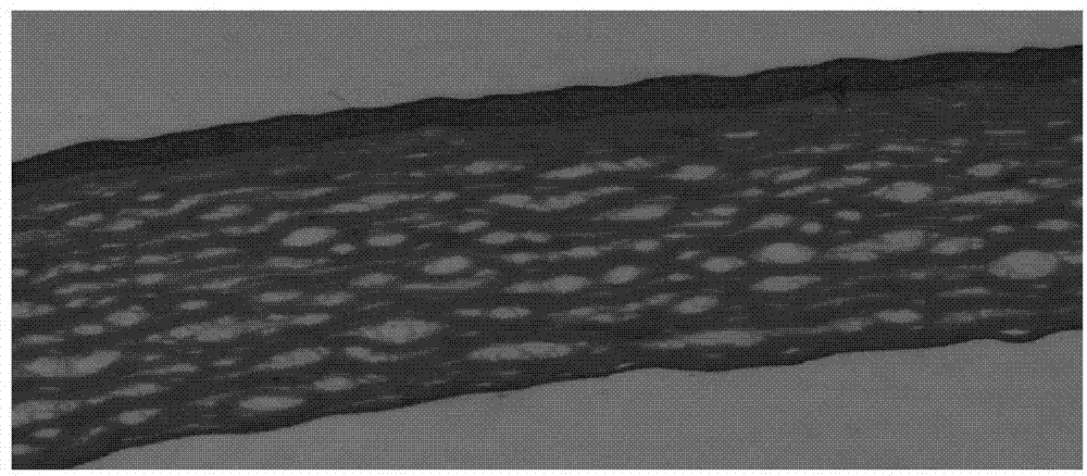
Embodiment 2

[0065] This example is used to illustrate the decellularized corneal tissue and its preparation method of the present invention.

[0066] (1) Take the bull's eyeball, soak the fresh bull's eyeball in 1×PBS solution containing tobramycin (the concentration of tobramycin is 40000U / L) for 30s, and cut it directly with a keratome in an ultra-clean bench Corneal stroma sheet with a diameter of 8 mm and a thickness of 200 μm;

[0067] (2) Place the cut corneal stroma piece in triple distilled water and soak for 8 hours;

[0068] (3) Place the hypotonic and swollen corneal stroma sheet obtained in step (2) in a sterile cryopreservation tube, freeze and thaw repeatedly, the freezing time is 30min, the freezing temperature is -200°C, the melting time is 30min, and the melting temperature is 40°C, the number of cycles is 2;

[0069] (4) Put the corneal stromal slices that have been repeatedly frozen and thawed in 1×PBS buffer containing DNase and RNase for enzymatic digestion. The vol...


PUM

PropertyMeasurementUnit
molecular weightaaaaaaaaaa
diameteraaaaaaaaaa
thicknessaaaaaaaaaa

Abstract

The invention relates to the field of corneal stroma substitutions, and discloses a decellularized porcine cornea tissue and a preparation method thereof. The method comprises the steps: performing low-permeability swelling, repeated freeze thawing, enzymic digestion, ultrasonic treatment, sterilization and wet-state sealed storage in sequence on a corneal stroma sheet, which is cut under a germfree condition, of a fresh animal eyeball, wherein the enzymic digestion is implemented by a buffering solution containing DNA enzyme and RNA enzyme. The invention further provides the application of the decellularized porcine cornea tissue in serving as the corneal stroma substitution. According to the method, damage to the collagen structure of the corneal stroma is reduced to the maximum extent; collagen fibers are tidily arrayed, and the porosity is uniform and regular; no cell is retained; the lamellar structure is kept intact; the biocompatibility of a bracket material is improved; furthermore, on the premise that the corneal stroma is not modified, the decellularized porcine cornea tissue can be stored in a wet state for a long time.

Description

technical field [0001] The invention relates to the field of corneal stroma substitutes, in particular to a decellularized corneal tissue and its preparation method and application. Background technique [0002] The cornea is located on the outermost surface of the eyeball and is in direct contact with the outside world. It is vulnerable to damage (physical, chemical) and infection. Once a lesion occurs, it will become cloudy and affect vision, or even cause blindness. Common corneal lesions include keratitis, corneal ulcer, keratoconus, keratomacia, and corneal degeneration. According to the statistics of the World Health Organization (WHO), there are nearly 50 million people with corneal disease blindness in the world at present, and there are about 4 million people in my country. Corneal disease has seriously affected the patients' normal life, work and study. They need family and social care, which increases the financial burden of the family and the burden of the whole...

Claims


Application Information

Patent Timeline
Patent Type & Authority Applications(China)
IPC IPC(8): A61L27/38
Inventor 王宝泉李青
Owner QINGDAO CHUNGHAO TISSUE ENG
Why Eureka
Try Eureka