Intraocular shunt manufacture

a technology of intraocular shunts and gelatin, which is applied in the field of system and method of making gelatin shunts, can solve the problems of affecting the ability of the shunt, affecting the shunt, and blindness, and achieves the effect of removing the skin

Active Publication Date: 2014-09-25
AQUESYS INC
View PDF5 Cites 31 Cited by
  • Summary
  • Abstract
  • Description
  • Claims
  • Application Information

AI Technical Summary

Benefits of technology

[0011]Certain aspects of the invention address the problems of controlling and manipulating liquid gelatin. The invention recognizes that simply routing a wire through a temperature controlled gelatin bath is not sufficient to produce a gelatin shunt with a uniform cross-section and uniform shape along a length of the implant. Heated gelatin alone forms a skin layer on top of the gelatin. Pulling a wire through a gelatin bath alone results in the wire passing through the skin layer, which makes it impractical to control the gelatin uptake on the wire. The invention solves this aspect of the problem by adding a water layer on top of the gelatin. The water layer eliminates the skin effect, and allows for production of a uniform cone of gelatin upon pulling a wire through the gelatin and then the water layer. Where the gelatin cone intersects the water-air boundary, a spot forms. This spot is exposed to air, and gelatin from this spot is taken up the wire.
[0012]However, the invention also recognizes that more gelatin reaches the top of the water than can be taken up by the wire. This results in cast-off, which renders the cone, and thus the spot at the boundary layer, unstable, i.e., the gelatin deposit is inconsistent in diameter. To solve this aspect of the problem, the systems and methods of the invention use a plate having an aperture, which aperture controls the gelatin spot. The plate having the aperture is situated in the water layer, and with the aperture plate in place, the cone of gelatin that feeds the spot is consistent and yields a uniform uptake of gelatin onto the wire.

Problems solved by technology

Build-up of aqueous humor and resulting intraocular pressure may result in irreversible damage to the optic nerve and the retina, which may lead to irreversible retinal damage and blindness.
A problem with implantable shunts is that they are composed of a rigid material, e.g., stainless steel, that does not allow the shunt to react to movement of tissue surrounding the eye.
Consequently, existing shunts have a tendency to move after implantation, affecting ability of the shunt to conduct fluid away from the anterior chamber of the eye.
Such anchors result in irritation and inflammation of the surrounding tissue.
Further, implanting a rigid shunt may result in the shunt causing blunt trauma upon insertion into an eye, such as producing a cyclodialysis cleft, or separation of the ciliary body from the scleral spur, creating hypotony by allowing the uncontrolled escape of aqueous humor through the cleft into the suprachoroidal space.
However, there are numerous issues associated with making gelatin shunts.
Additionally, there are challenges associated with the drying process that also make it difficult to produce a gelatin shunt with a uniform cross-section and uniform shape along a length of the implant.
Pulling a wire through a gelatin bath alone results in the wire passing through the skin layer, which makes it impractical to control the gelatin uptake on the wire.
This results in cast-off, which renders the cone, and thus the spot at the boundary layer, unstable, i.e., the gelatin deposit is inconsistent in diameter.
However, the wire constrains the gelatin from shrinking axially.
If humidity is left uncontrolled, an outer skin of the gelatin dries and hardens before the gelatin has completed shrinking This results in non-uniform cross sections and shapes along the implant length.

Method used


Image

  • Intraocular shunt manufacture
  • Intraocular shunt manufacture
  • Intraocular shunt manufacture

Examples


example 1

Gelatin Preparation

[0049]Into a 600 mL beaker was added 98±1 grams of porcine gelatin. An amount of about 172±1 grams of USP sterile water was measured and poured into the beaker containing the porcine gelatin. The beaker was sealed with parafilm, and the covered beaker was placed in a water bath at 55±1° C. for a minimum of 8 hours (maximum 36 hours). Ensure water level is higher than mixture in beaker. The lid of the water bath was checked to ensure that it was closed and after the minimum time period had elapsed, the beaker was removed from bath. The beaker was visually observed to verify that all gelatin in the mixture was dissolved and that mixture appeared homogeneous.

example 2

Gelatin Transfer

[0050]The water circulator was checked to make sure that it was at a sufficient level (between the high and low marks). The circulator was set and run at 40.5° C. and allowed to come to temperature before proceeding to the next step. The gelatin mixture from Example 1 was poured into the jacketed beaker on a fixture so that the meniscus was about 20 mm from the top. Within 1 minute of adding the gelatin mixture, 60 cc's of USP sterile water was added above the gelatin surface using a syringe. The water was added slowly so as not to disturb the gelatin. The mixture was allowed to settle for minimum 30 min.

example 3

System Set-Up

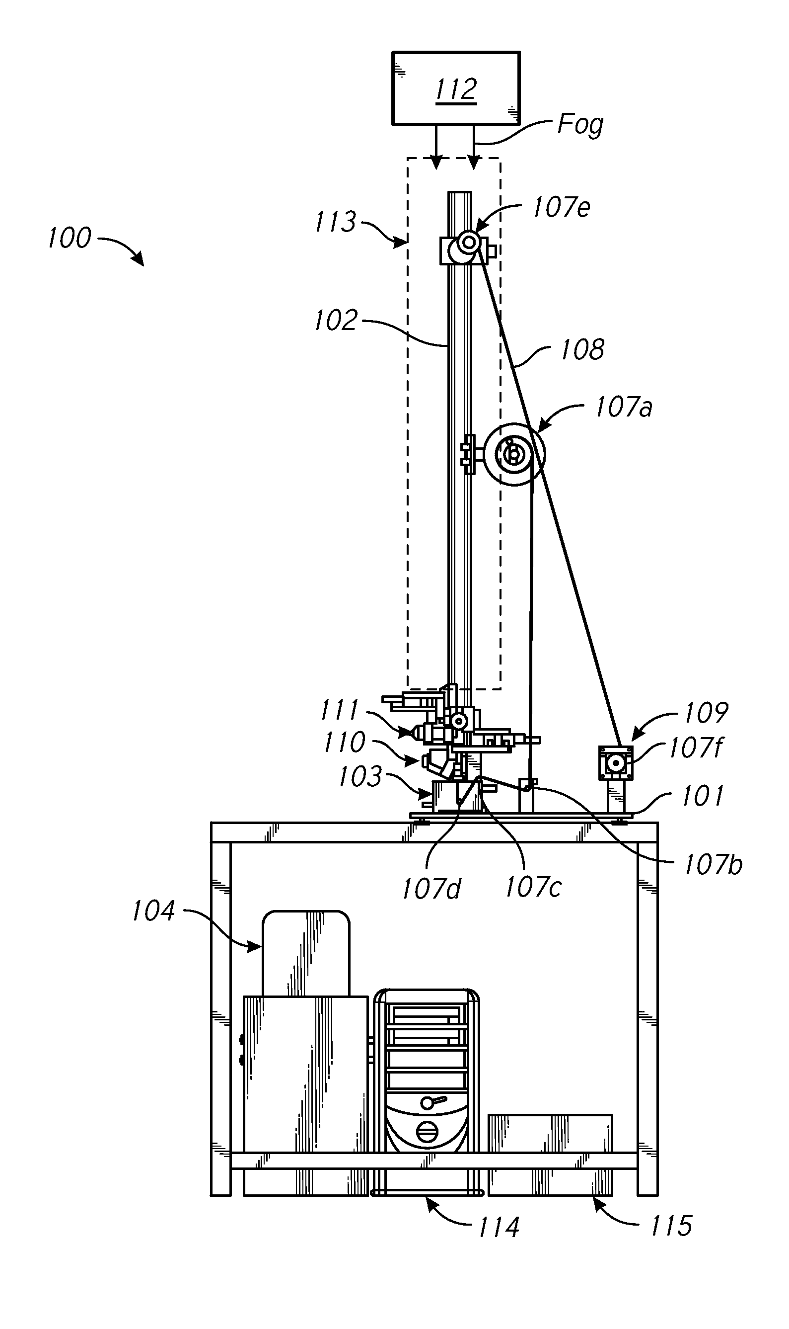
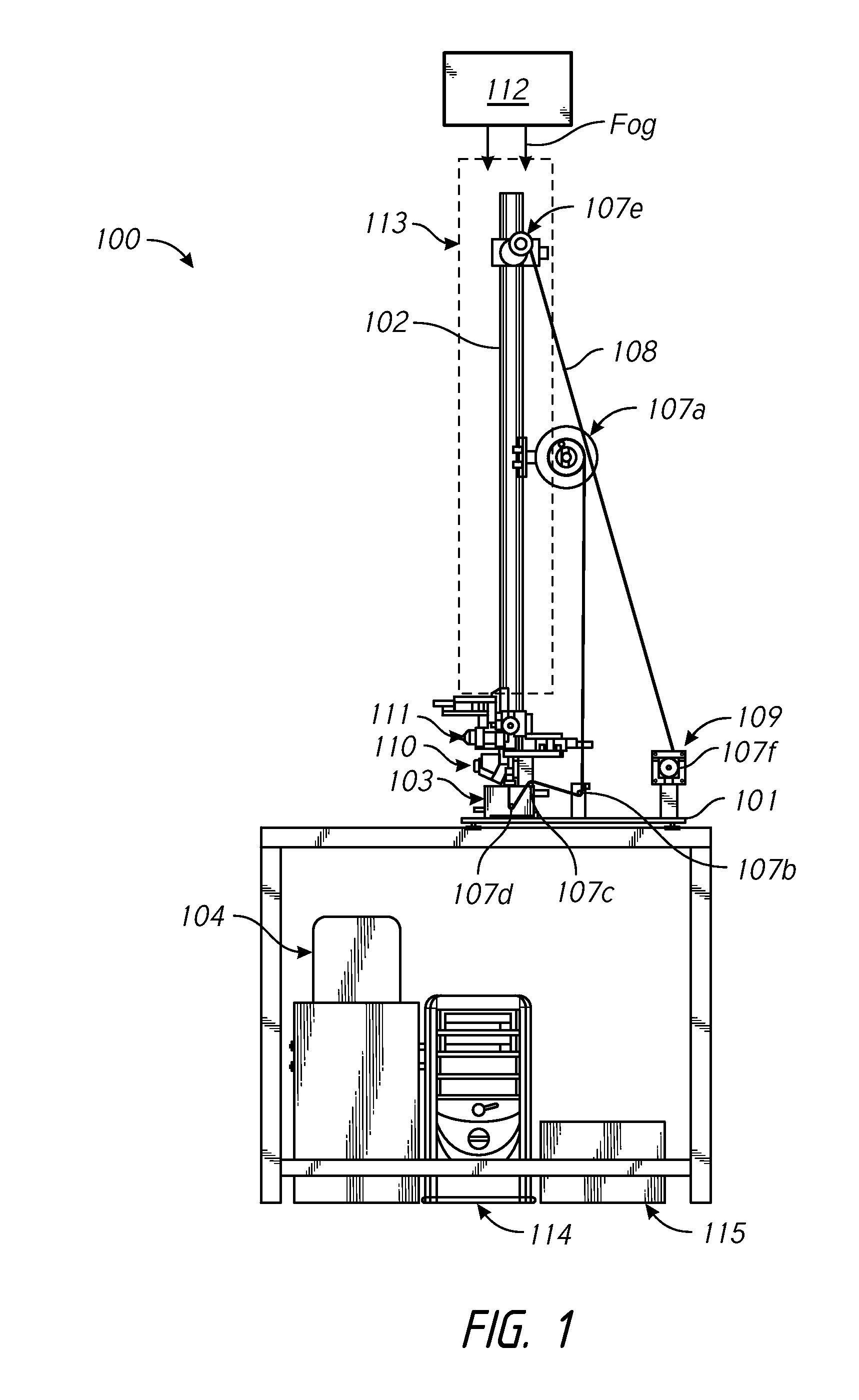
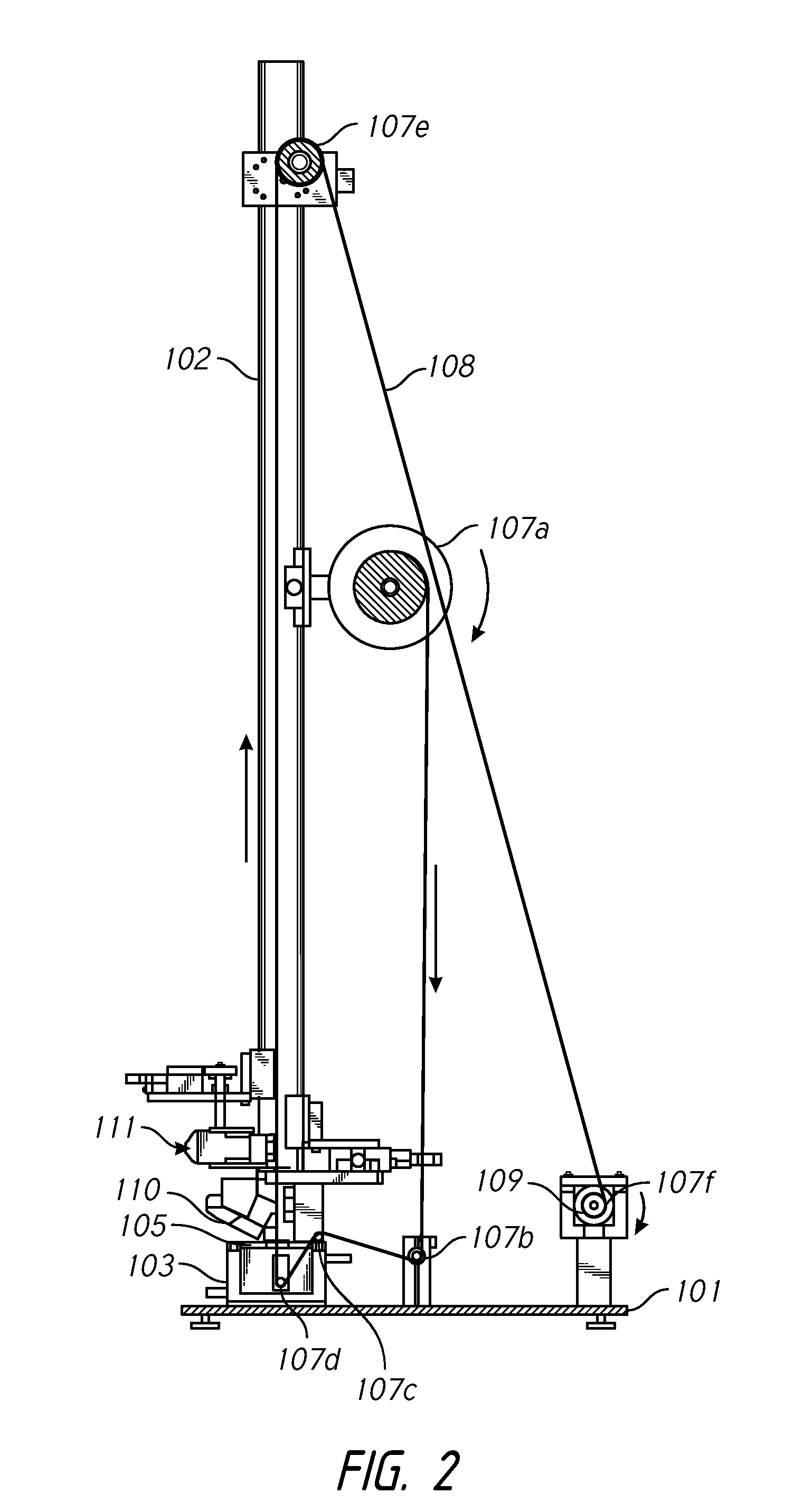
[0051]A spool was assembled onto an axle using parts as shown in FIG. 7. The final assembled spool is shown in FIG. 8. The M6 screw (Item 8) was finger tightened, and pinch bolt (Item 7) was fastened onto the mount. In order to increase the friction on the spool, the M6 screw (Item 8) was advanced approximately a ¼ turn. The wire was then threaded onto the spool, and the spool was slid up the shaft. The spindle assembly was lowered into the gelatin mixture, about 5 mm from the bottom of flask. The aperture plate was lowered into the water layer under a top surface was submerged about 1 mm below the surface of water. The bottom surface of the aperture plate was above the gelatin layer.

[0052]The first and second cameras were positioned on the shaft so as to properly view the wire as it emerged from the aperture in the aperture plate. The tube of the fogger assembly was lowered over the shaft until a bottom of the tube is positioned just above the second camera. In this po...


PUM

PropertyMeasurementUnit
inner diameteraaaaaaaaaa

Abstract

An intraocular shunt can be manufactured using a system that includes a liquid bath and a wire, which is moved through the bath. When moved through the bath, the wire is coated with a material, such as gelatin. For example, the liquid bath can have a top layer, including water, and a bottom layer, including gelatin. The coated wire passes through an aperture formed in a plate component of the system. The gelatin can be dried on the wire in a humidity-controlled space, thereby forming the shunt.

Description

CROSS-REFERENCE TO RELATED APPLICATIONS[0001]This application is a divisional of U.S. patent application Ser. No. 13 / 314,950, filed on Dec. 8, 2011, the entirety of which is incorporated herein by reference.BACKGROUND[0002]1. Field of the Invention[0003]The invention generally relates to systems and methods for making gelatin shunts.[0004]2. Description of the Related Art[0005]Glaucoma is a disease of the eye that affects millions of people. Glaucoma is associated with an increase in intraocular pressure resulting either from a failure of a drainage system of an eye to adequately remove aqueous humor from an anterior chamber of the eye or overproduction of aqueous humor by a ciliary body in the eye. Build-up of aqueous humor and resulting intraocular pressure may result in irreversible damage to the optic nerve and the retina, which may lead to irreversible retinal damage and blindness.[0006]Glaucoma may be treated in a number of different ways. One manner of treatment involves deli...

Claims


Application Information

Patent Timeline
Patent Type & Authority Applications(United States)
IPC IPC(8): A61F9/007
CPCB05C3/12A61F9/00781A61F2240/001A61F9/0017
Inventor ROMODA, LASZLO O.HAMSTROM, BRIANCHANDRAKANT, PARTHAHORVATH, CHRISTOPHER
Owner AQUESYS INC
Why Eureka
Try Eureka