Metabolic flux measurement, imaging and microscopy

a metabolic flux and microscopy technology, applied in the field of metabolic flux determination, microscopy and functional histopathology, can solve the problems of loss of spatial information, all current histological analyses are ‘blind’ to the spatial order, and altered metabolism may limit the efficacy of standard anti-cancer therapy, etc., to achieve the effect of allowing or increasing the energy-dependent volatilization of molecules

Active Publication Date: 2014-11-06
RGT UNIV OF CALIFORNIA
View PDF5 Cites 36 Cited by
  • Summary
  • Abstract
  • Description
  • Claims
  • Application Information

AI Technical Summary

Problems solved by technology

Thus altered metabolism may limit efficacy of standard anti-cancer therapy, but this feature may also be used to identify and characterize subtypes of neoplastic tissue.
However, all current histological analyses are ‘blind’ to the spatially ordered metabolic dynamics of the tumor.
However, the requisite sample homogenization and preparation results in a loss of spatial information, with averaging of metabolite concentrations, and loss of information of critical tumor subpopulations.
Major approaches include: Time-of-Flight Secondary Ionization Mass Spectrometry (TOF-SIMS) [17, 18], which has the highest spatial resolution (˜100 nm), but the extensive fragmentation which often complicates molecular characterization and the lack of tandem MS capabilities on commercial instruments limits identification.
Unfortunately, tissue imaging by DESI shows relatively low spatial resolution (˜100 um) and is currently incapable of imaging tissues at the cellular level.

Method used


Image

  • Metabolic flux measurement, imaging and microscopy
  • Metabolic flux measurement, imaging and microscopy
  • Metabolic flux measurement, imaging and microscopy

Examples


example 1

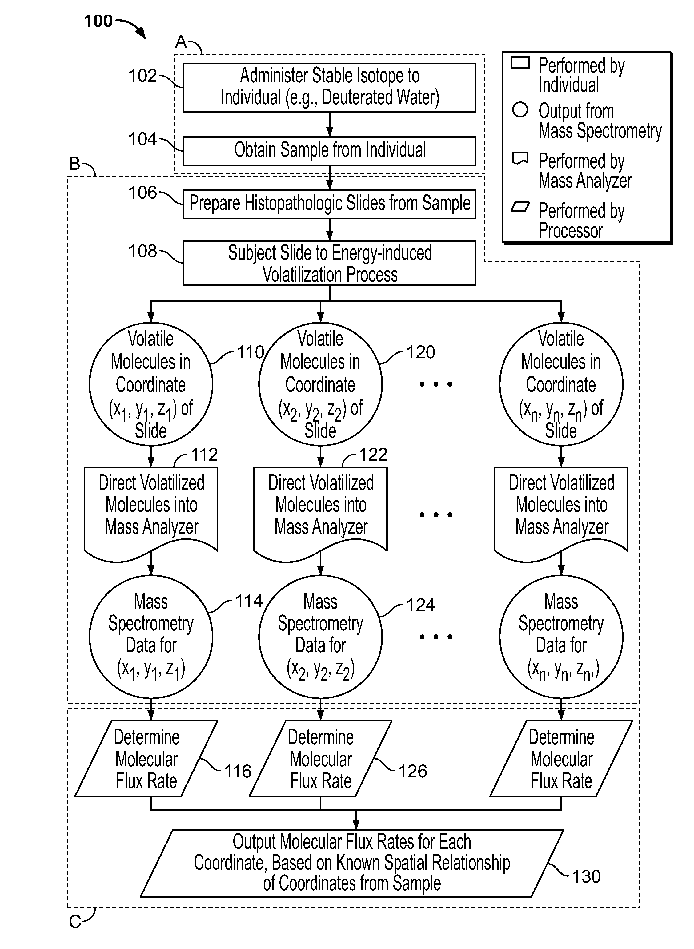
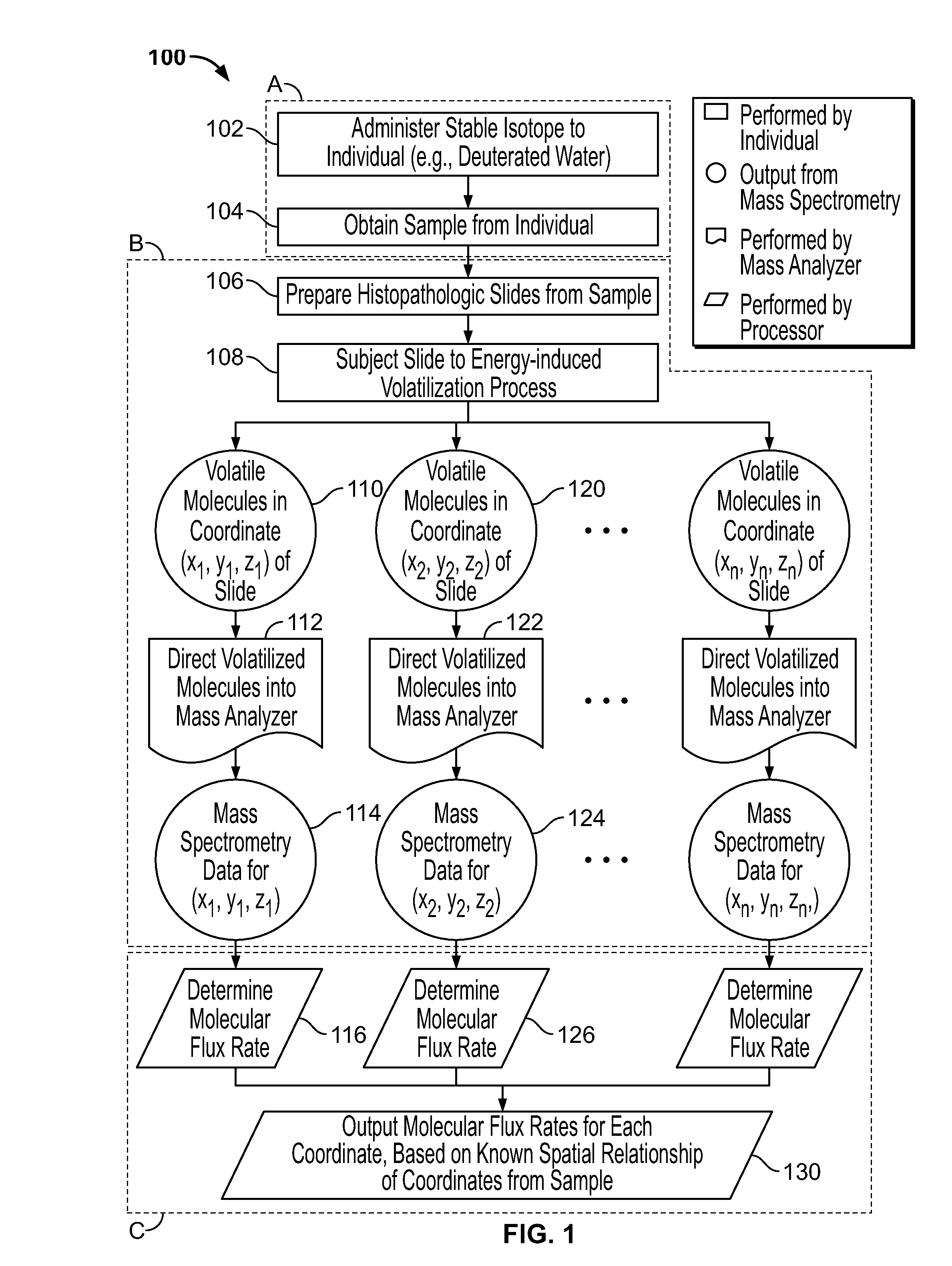
Integration of Deuterium Isotopic Labeling with Nanostructure-Initiator Mass Spectrometry (NIMS)-Based Metabolite Imaging Technology to Generate Histochemical Flux Images

[0182]Here we propose a novel integration of nanotechnology based mass spectrometric tumor imaging, with mass isotopomer quantitation and kinetic analysis after deuterium label incorporation, with the goal of quantifying fluxes directly within the 3D architecture of the tumor and the microenvironment. This overcomes critical challenges in the field by directly measuring metabolic phenotypes within the physiological context.

[0183]In vivo approaches for characterizing the molecular dynamics of complex biochemical networks, based on the internal patterns of incorporated labels have been developed, tested and extensively validated. See Hellerstein, M. K. and R. A. Neese, Mass isotopomer distribution analysis at eight years: theoretical, analytic, and experimental considerations. American Journal of Physiology—Endocrinol...

example 2

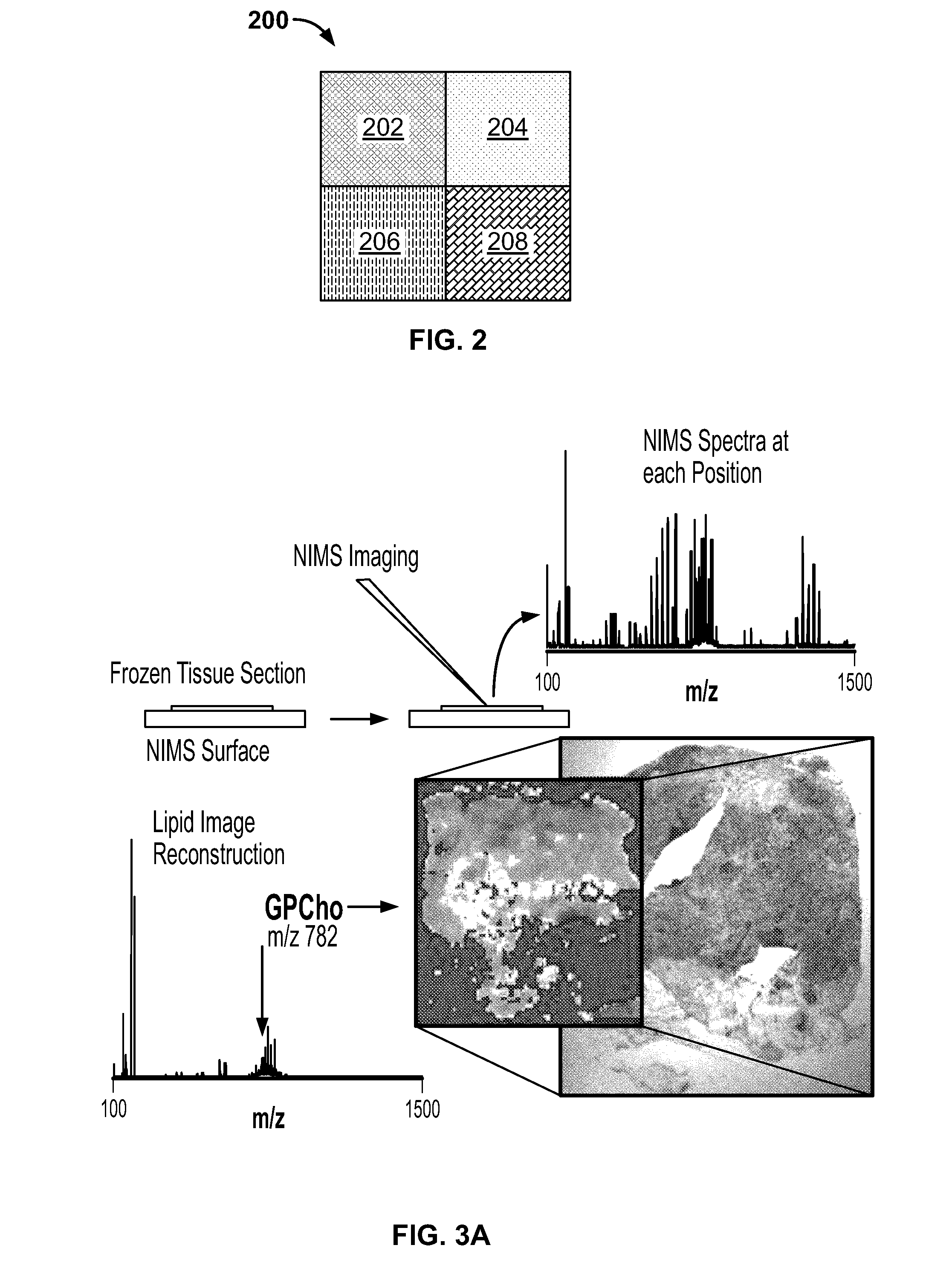
Kinetic Histology of Mouse Tissue with NIMS

[0220]Whole mouse imaging has shown that there are complex metabolite patterns that can be imaged using NIMS over large areas (FIG. 7). To determine the ability to image isotopomer abundance patterns and consequently determine the flux, dissected tissues from adult mice that were given 5% 2H2O for three days were sectioned and imaged with NIMS (FIG. 7A). For each pixel, numerous labeled metabolites could be observed. For example, phosphocholine was one of the dominant signals[37] in the TOF-SIMS imaging spectra. In the case of phosphocholine, there are 15 hydrogen atoms in C—H bonds. As can be seen in FIG. 7D, the ratio of M1 / (M1+M0), or relative flux, increases over the course of 3 days of administration of 2H2O. It can be calculated that there are 3 hydrogens that could be exchanged with deuterium in cellular water for phosphocholine. All of the other hydrogens are non-labile metabolically and come from nutritional-sources such as exogeno...

example 3

Flux Microscopy for Healthy Human Tissue

[0223]Determine Spatial Turnover Kinetics of Lipid Metabolites (Kinetic Histochemistry) in Healthy Human Tissue.

[0224]Here, we will validate existing clinical protocols and determine the clinically feasible time points for tissue collection that give useful lipid flux distributions using NIMS tissue imaging. Specifically, we will define the clinical 2H2O administration, sample handling, and analysis protocols by using relatively accessible human tissue samples to identify and overcome procedural and technical challenges. Compared to preclinical experimentation, human studies have a number of distinct challenges that affect the quantification of newly-synthesized metabolites in heavy water labeling studies. Different isotopic enrichments in the metabolic products compared to mouse tissues will almost surely result due to slower basal turnover rates, lower precursor pool enrichments (2H2O % in body water), and time-varying labeling enrichment cu...


PUM

PropertyMeasurementUnit
massaaaaaaaaaa
pixel sizeaaaaaaaaaa
thicknessaaaaaaaaaa

Abstract

Provided herein are methods for measuring molecular flux rates of molecules of interest in a tissue sample in spatially-organized manner and generating output (e.g., an image, a heat map, a contour map, a table or a database) representing the molecular flux rates of each spatially-defined location of the sample. Provided herein are also the output, as well as systems and computer-readable medium with computer-executable instructions for determining molecular flux rates of molecules of interest in the sample.

Description

CROSS-REFERENCE TO RELATED APPLICATION[0001]This application claims the benefit of U.S. Provisional Patent Application Ser. No. 61 / 532,522, filed Sep. 8, 2011, which is incorporated herein by reference in its entirety.STATEMENT REGARDING GOVERNMENTAL SUPPORT[0002]This invention was made with Government support under Contract No. DE-AC03-05CH11231 awarded by the Department of Energy. The Government has certain rights in the invention.BACKGROUND[0003]1. Field[0004]The present disclosure relates generally to the fields of metabolic flux determination, microscopy and functional histopathology, and more specifically to in situ spatial imaging, mapping and display of dynamic metabolic processes in histopathology specimens.[0005]2. Related Art[0006]Breast cancer development involves dynamic and reciprocal interactions between neoplastic cells, activated stromal cells, extracellular matrix (ECM) and soluble molecules in their vicinity. Together these environmental factors foster the maligna...

Claims


Application Information

Patent Timeline
Patent Type & Authority Applications(United States)
IPC IPC(8): G01N33/58
CPCG01N33/58G06F19/18G01N2570/00G01N2458/15G01N33/6848G01N2800/7028G16B20/00Y10T436/24
Inventor BOWEN, BENJAMIN P.LOUIE, KATHERINE B.NORTHEN, TRENT R.HELLERSTEIN, MARC K.
Owner RGT UNIV OF CALIFORNIA
Why Eureka
Try Eureka